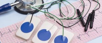
What is "bradycardia"?
A decrease in heart rate (HR) below the established norm (60 beats per minute) is called “bradycardia”.
It can occur both in healthy people and in patients with various pathologies. Compared to tachycardia (increased heart rate more than 90 beats per minute), bradycardia is less common. It is typical for people over 60 years of age, since heart disease is more common at this age. Heart rate usually corresponds to the pulse, since with each release of blood into the aorta under pressure, rhythmic vibrations of the arterial walls occur.
What is the normal heart rate?
In adults, the heart rate should be 60–90 beats per minute (bpm); for children, normal heart rates depend on age. Thus, in babies under one year old, heart rate fluctuates between 100–170 beats/min. As the child grows up, normal heart rate values gradually decrease. By age 12–15, your resting heart rate should approach 70–90 beats per minute.
How to measure pulse?
The pulse can be measured by feeling the arteries that are located shallow to the surface of the skin and where they can be pressed against the bones. Then you can feel the movement of the vessel wall with your fingertips (Fig. 1). It occurs with every heartbeat. The pulse is measured in the following arteries: temporal, carotid, brachial, radial (at the wrist), femoral, popliteal, dorsalis pedis, posterior tibial (at the ankle of the foot).
Figure 1. How to measure pulse Source: MedPortal
Causes of bradycardia
A decrease in heart rate can occur not only with cardiac lesions, but also with non-cardiac pathologies. Common reasons for a decrease in heart rate include:
- coronary heart disease, heart defects, atherosclerotic cardiosclerosis, heart attack, myocarditis, cardiomyopathies, pericarditis;
- jaundice and kidney failure, in which toxins slow the heart's impulses;
- connective tissue diseases (rheumatism, rheumatoid arthritis, scleroderma, lupus erythematosus);
- hypothyroidism - decreased function of the thyroid gland;
- infections (diphtheria, borreliosis, syphilis, toxoplasmosis, viral hepatitis, etc.);
- disturbance of the electrolyte composition of the blood - decreased potassium levels and increased calcium;
- increased intracranial pressure due to brain tumors, stroke, trauma, developmental defects;
- lymphogranulomatosis, multiple myeloma, amyloidosis, sarcoidosis, hemochromatosis are conditions in which myocardial damage is possible.
Drug-induced bradycardia often occurs. This occurs when taking or overdosing on beta blockers, cardiac glycosides, calcium channel blockers, and antiarrhythmic drugs.
The heart rate is affected by the work of the nervous system: the parasympathetic system reduces the heart rate, and the sympathetic system increases it. Reflex bradycardia occurs as a result of the influence of the parasympathetic nervous system on the sinus node. The causes of such bradycardia may be:
- diseases of the nervous system;
- gastrointestinal pathology (peptic ulcer of the stomach and duodenum, cholecystitis, cholelithiasis);
- diseases of the esophagus and diaphragm;
- low body temperature, hypothermia;
- blow to the upper abdomen (up to cardiac arrest);
- pressure on the eyeballs or neck in the area of the carotid artery;
- cough, vomiting.
When the heart rate slows, blood pressure (BP) can be normal, high or low.
Bradycardia with high blood pressure is an uncommon occurrence. It is dangerous due to respiratory arrest, loss of consciousness, risk of blood clots, insufficient blood supply to the brain and heart with the development of stroke and heart attack. The combination of hypertension and bradycardia requires special treatment approaches so as not to aggravate the slowing of the rhythm.
For newborns (especially premature ones), bradycardia can occur against the background of apnea - stopping breathing for more than 20 seconds (Fig. 2). This occurs due to the immaturity of the nervous and muscle tissue of the newborn.
Figure 2. Bradycardia and apnea in infants. Source: MedPortal
Clinical tests
From the given answer options, select those that you think are correct. 1. Disease in which nephrotic syndrome develops:
a) amyloidosis; b) pneumonia; c) viral pericarditis; d) trichinosis; d) chronic hepatitis.
2. A 45-year-old patient has chronic osteomyelitis of the right femur. In urine: protein - 1.66 g/l, leukocytes - 4-6 in the field of vision, erythrocytes - single in the field of vision, there are hyaline casts. Establish a probable diagnosis:
a) chronic pyelonephritis; b) chronic glomerulonephritis; c) renal amyloidosis; d) renal vein thrombosis; d) everything is possible.
3. Clinical manifestations of Rendu-Osler disease:
a) the presence of telangiectasia; b) vasculitic purpuric type of bleeding; c) development of iron deficiency anemia; d) recurrent nosebleeds; e) hemarthrosis.
4. Criteria for diagnosing myeloma:
a) reduced content of plasma cells in the bone marrow; b) increased content of plasma cells in the bone marrow; c) the presence of paraproteins in the blood serum; d) the presence of paraproteins in the urine.
5. Specify the main diagnostic sign in the differential diagnosis of Minkowski–Choffard disease and Gilbert syndrome:
a) bilirubin level; b) the general condition of the patient; c) age of the patient; d) morphology of erythrocytes; d) hemoglobin level.
6. In case of hypoglycemic coma, the skin:
a) hyperemic; b) wet; c) icteric; d) dry.
7. Characteristic odor from the mouth during hyperglycemic coma:
a) alcohol; b) ammonia; c) acetone; d) rotten eggs.
8. Decreased memory, constipation, bradycardia are observed with:
a) hypothyroidism; b) diffuse toxic goiter; c) diabetes mellitus; d) pheochromocytoma.
9. Dry skin, itching, thirst and polyuria are observed with:
a) hypothyroidism; b) diffuse toxic goiter; c) diabetes mellitus; d) endemic goiter.
10. In acute glomerulonephritis, the following develops:
a) glucosuria; b) dysuria; c) oliguria; d) polyuria.
11. With pyelonephritis, the kidneys are predominantly affected:
a) calyxes; b) tubules; c) glomeruli; d) glomeruli and tubules.
12. 64-year-old woman for the last 3 months. I began to notice a decrease in the frequency of bowel movements to 1-2 times a week, and I was worried about the feeling of incomplete bowel movements. Several times I noticed streaks of blood in my stool. What is the most likely cause of these symptoms?
a) colorectal cancer; b) recurrent hemorrhoids; c) ulcerative colitis; d) irritable bowel syndrome; e) colonic localization of Crohn's disease.
13. In a 61-year-old man for 3 months. progressive constipation is observed (from 3 times a week at the beginning to 1 time every 7–10 days); often, after the absence of bowel movements, the patient notes the appearance of loose stools. Often notes tenesmus and discharge of scarlet blood with feces. During this time I lost 6 kg. The most likely diagnosis for the patient is:
a) cancer of the descending colon; b) Crohn's disease localized in the colon; c) rectal cancer; d) anal fissure; e) ulcerative colitis.
14. A patient with gout consults a doctor about diet. He lists the foods he loves. Select from its list the product containing the highest concentration of purines and to be excluded from the diet:
a) cucumbers; b) shish kebab; c) boiled potatoes; d) tangerines; d) pasta.
15. In what pathological condition does the following occur: pleural effusion-exudate is serous or hemorrhagic, with a high content of eosinophils, the amount of exudate is small, but after puncture it can accumulate again; Characterized by pain in the chest, more on the left, the presence of effusion not only in the pleura, but also in the pericardium, increased temperature, in the blood test - leukocytosis, increased ESR:
a) Mallory-Weiss syndrome; b) Kohn syndrome; c) Dressler syndrome; d) nephrotic syndrome; e) Itsenko-Cushing syndrome.
16. What disease is characterized by pleural effusion containing 10–40% protein, rel. density 1008–1017, unilateral, hemorrhagic, non-renewable after evacuation:
a) pulmonary infarction; b) myocardial infarction; c) splenic infarction; d) pancreatic cancer; d) lung cancer.
17. Exudate is considered hemorrhagic if the number of red blood cells in the effusion contains:
a) 500–1000 per µl; b) 1000–2000 in µl; c) more than 3000 per µl; d) more than 5000 in µl; e) more than 10,000 in µl.
18. Tuberculosis infection can be suspected if the pleural effusion contains:
a) 10–15% eosinophils; b) 30–50% eosinophils; c) 20–30% lymphocytes; d) more than 30% lymphocytes; e) more than 50% lymphocytes.
19. Chylothorax is formed when:
a) injury; b) pneumonia; c) abscess formation of the lung; d) metastatic damage to the pleura; e) pericarditis.
20. A patient with severe nephrotic syndrome suddenly developed abdominal pain without precise localization, nausea, vomiting, the temperature increased to 39°C, and erythema on the skin of the anterior abdominal wall and thighs. Most likely reason:
a) bacterial peritonitis; b) abdominal nephrotic crisis; c) renal colic; d) apostematous pyelonephritis; d) intestinal colic.
21. The patient is 27 years old. Denies any history of pulmonary diseases. 1 hour ago, against the background of complete health, sudden severe pain in the left half of the chest and lack of air appeared. The temperature is normal. Breathing over the left lung cannot be heard; upon percussion there is a “box” sound. The mediastinum is shifted to the right by percussion. He needs to suspect:
a) fibrinous pleurisy; b) myocardial infarction; c) pulmonary tuberculosis; d) spontaneous pneumothorax; e) strangulated diaphragmatic hernia.
22. A 45-year-old patient, a mechanic by profession, was found to have hypertrophy of the parotid and salivary glands, Dupuytren’s contracture, proteinuria (2.5 g/l), hematuria (40–60 in p.s.). The level of IgA in the blood is increased. Most likely diagnosis:
a) idiopathic IgA nephritis; b) glomerulonephritis with hemorrhagic vasculitis; c) glomerulonephritis of alcoholic etiology; d) lupus glomerulonephritis; e) chronic pyelonephritis.
23. A 16-year-old patient suffering from osteomyelitis of the left leg developed edema, ascites, and hydrothorax. The examination revealed nephrotic syndrome and hepatosplenomegaly. The level of fibrinogen in the blood is sharply increased. Probable diagnosis:
a) post-infectious glomerulonephritis; b) decompensated cirrhosis of the liver; c) hepatorenal syndrome; d) secondary amyloidosis with kidney damage; e) lupus glomerulonephritis.
24. A 50-year-old patient complains of loss of strength and pain in the spine, the level of hemoglobin in the blood is 65 g/l, proteinuria is 22 g, the level of serum albumin is 40 g/l. Most likely diagnosis:
a) chronic glomerulonephritis in the stage of uremia; b) multiple myeloma; c) secondary amyloidosis with kidney damage; d) chronic pyelonephritis; e) polycystic kidney disease.
25. In a 45-year-old patient for 4 months. Fever and episodes of painless gross hematuria are noted. The level of hemoglobin in the blood is 160 g/l, ESR is 60 mm/h. Most likely diagnosis:
a) chronic glomerulonephritis of hematuric type; b) lupus nephritis; c) kidney cancer; d) urate nephrolithiasis; e) amyloidosis.
Answers
1 – a. 2 – c. 3 – a, c, d. 4 – b, c, d. 5 – d. 6 – b. 7 – c. 8 – a. 9 – c. 10 – c. 11 – a. 12 – a. 13 – a. 14 – b. 15th century 16 – a. 17 – d. 18 – d. 19 – a. 20 – b. 21 – g. 22 – c. 23 – d. 24 – b. 25 – c.
Classification of bradycardia
The work of the heart is impossible without the normal functioning of the conduction system. These are special muscle fibers that form nodes and bundles and provide coordinated contraction of the atria and ventricles of the heart. A normal rhythm is generated with a frequency of 60–90 per minute in the sinus node (SU), which is located in the right atrium. Along the internodal bundles, the electrical impulse is conducted to the atrioventricular node (atrioventricular - AV), located in the interatrial septum. From the AV node, the impulse goes directly to the muscles that provide contraction of the ventricles of the heart.
Depending on the mechanism of occurrence, bradycardia occurs:
- Sinus. Associated with a slowdown in impulse generation in the sinus node itself (Fig. 3).
- Sinoatrial, in which the transmission of impulses from the sinus to the atria is blocked.
- Atrioventricular, when there is a blockade of excitation transmission in the AV node.
Figure 3. Development of sinus bradycardia.
Source: mayoclinic.org Bradycardia can be pathological and physiological - a decrease in heart rate is typical for young athletes and during sleep. Pathological bradycardia occurs:
- acute and chronic;
- with symptoms and without clinical manifestations;
- cardiac and extracardiac (depending on the reasons).
Pacemaker
When a person experiences severe bradycardia and heart failure develops against this background, they resort to implanting a pacemaker in the heart. This device independently generates electrical impulses. A stable preset heart rhythm favors the restoration of adequate hemodynamics.
The operation is performed under general anesthesia and lasts about an hour. Through the subclavian vein, under the control of an X-ray machine, a double electrode is inserted into the right ventricle and atrium. The stimulator is sutured in the subclavian area or under the skin on the abdomen.
The patient spends no more than a week in the surgical department.
Why is bradycardia dangerous?
A decrease in heart rate can cause heart failure, chronic attacks of bradycardia, and the formation of blood clots.
Heart failure
It rarely develops when the decrease in heart rate is significant (less than 40 per minute). Bradycardia results in insufficient cardiac output, which means that the heart can no longer supply oxygen to all organs and systems of the body at the proper level. In this case, the brain and the heart itself suffer first. The risk of developing coronary artery disease and myocardial infarction also increases, and fainting and cardiac arrest may occur.
Blood clots
With prolonged or frequently occurring bradycardia with an irregular rhythm, as a result of slowing down the movement of blood flow in the chambers of the heart, blood thickens and the gradual formation of blood clots occurs. When they enter the vessels of the brain or heart, strokes and heart attacks occur.
Chronic attacks of bradycardia
When the cause of bradycardia cannot be identified and eliminated, attacks occur that reduce the quality of life and are difficult to correct. During an attack, the patient experiences weakness, dizziness, and decreased performance. Sometimes fainting is possible.
Forecast
The presence of organic heart lesions has an adverse effect on the prognosis of bradycardia. The possible consequences of bradycardia are significantly aggravated by the occurrence of Morgagni-Adams-Stokes attacks without solving the issue of electrical stimulation. The combination of bradycardia with heterotopic tachyarrhythmias increases the likelihood of thromboembolic complications. Against the background of a persistent decrease in rhythm, the patient may develop disability.
With a physiological form of bradycardia or its moderate nature, the prognosis is satisfactory.
Symptoms of bradycardia
Manifestations of bradycardia are caused by a lack of blood supply to vital organs: the brain, heart and lungs. Bradycardia can manifest itself (Fig. 4):
- decrease in blood pressure;
- presyncope and loss of consciousness;
- dizziness, headache;
- rapid fatigue;
- chest pain;
- shortness of breath and poor tolerance to any physical activity;
- pale skin.
Figure 4. Symptoms of bradycardia. Source: MedPortal
When to see a doctor?
If you have symptoms of bradycardia and a pulse less than 60 beats per minute, you should consult a physician or cardiologist. The doctor will determine the causes of this condition and prescribe effective treatment.
Not all bradycardia causes symptoms. However, low heart rates cannot be ignored. A repeated decrease in heart rate is the “first bell” that will force the timely diagnosis of heart disease and non-cardiac pathology, and prevent the development of dangerous complications.
Diagnosis of bradycardia
Only a doctor can differentiate physiological or pathological bradycardia. To do this, he uses several diagnostic methods.
Auscultation
A simple diagnostic method is to listen to the heart using a phonendoscope. Auscultation of the heart allows not only to determine a rare rhythm, but also to suspect certain diseases of the cardiovascular system.
Electrocardiography
ECG is a method of instrumental diagnostics of the heart, which records not only heart rate, but also helps to diagnose cardiac causes of bradycardia (myocardial ischemia and infarction, sinus node pathology, AV block). This is important for effective therapy. In some cases, daily ECG monitoring is necessary - a Holter study.
Phonocardiography
Phonocardiography (PCG) is a method of hardware diagnostics of sound phenomena that are created during the work of the heart. The study is carried out in conjunction with an ECG. At the same time, the heart sounds that are created during the operation of the valves, as well as additional noise, are assessed.
To diagnose the causes of bradycardia, the doctor may also prescribe:
- EchoCG - ultrasound examination of the heart;
- blood test for markers of damage to the heart muscle (troponins, creatine kinase);
- study of inflammation indicators (C-reactive protein, rheumatic tests, markers of collagenosis);
- blood test for thyroid function indicators (TSH, T4, T3);
- study of blood electrolytes (potassium, magnesium, calcium, sodium);
- biochemical blood test (glucose, bilirubin, cholesterol, creatinine, etc.).
Do they join the army if they have bradycardia?
In the list of diseases when a conscript is considered unfit for military service, bradycardia is absent, since it is not a disease, but a diagnostic sign of heart pathologies.
When diagnosing bradycardia, a young man must undergo a cardiovascular examination, and only on the basis of the identified/undetected disease is the issue of suitability for service decided. According to Art. 42-48 young men with diseases such as AV block and sick sinus syndrome are considered unfit for service. In the absence of these pathologies, the conscript is not exempt from military service.
Treatment of bradycardia
Not all bradycardia requires treatment. Only a doctor can determine the need for treatment by excluding physiological causes of a decrease in heart rate.
Conservative treatment
For conservative treatment use:
- anticholinergics - drugs that prevent the influence of the parasympathetic nervous system on the heart;
- adrenaline analogues - drugs that stimulate heart rate;
- antiarrhythmic drugs.
Treatment is carried out with tablets and injections. Their doctor can prescribe them occasionally during attacks and for a long time.
Surgery
In cases where conservative treatment is ineffective or there is damage to the conduction system of the heart, surgical treatment is indicated - installation of an electrical pacemaker (pacemaker). This is an electronic device that is implanted subcutaneously in the left subclavian region and sets the necessary rhythm to the heart through electrodes (Fig. 5). The pacemaker not only stimulates impulses to contract the heart, but also controls its natural electrical activity.
Figure 5. Installation of a pacemaker for bradycardia. Source: MedPortal
Treatment with folk remedies
To treat bradycardia at home, infusions of herbs are mainly used, which reduce the tone of the parasympathetic nervous system, increase myocardial contractions, and maintain blood pressure. These are Chinese lemongrass, tartar, immortelle, yarrow, walnuts, a mixture of lemon, honey and garlic and others.
Important! Treatment with folk remedies should be carried out very carefully and only after consultation with a doctor. It is possible with mild degrees of bradycardia (heart rate of at least 40), in the absence of complications and manifestations such as fainting, shortness of breath, dizziness, chest pain.
How to treat bradycardia with folk remedies?
Currently, the following traditional methods have proven effectiveness in the treatment of bradycardia:
- A mixture of honey, lemon and garlic. To prepare it, wash the lemons and scald them with boiling water, then squeeze the juice out of them. Then peel 10 medium heads of garlic and chop them to a paste. Mix the prepared garlic pulp with lemon juice until a homogeneous mass is obtained. Then add one liter of honey to the garlic-lemon mixture and mix the whole mixture well. Place the finished mixture in a sealed container in the refrigerator and leave for 10 days. Then eat 4 teaspoons every day before meals.
- Yarrow decoction. To prepare it, pour 50 g of dry grass into 500 ml of warm water, then bring it to a boil. Boil for 10 minutes, then leave for an hour. Strain the finished broth and take one tablespoon three times a day.
- Walnuts that you should eat every day. Nuts should be present in the human diet every day. The best time to eat nuts is for breakfast.
Typically, treatment for bradycardia is long-term, and traditional methods can be used for as long as desired.
Prevention
To prevent complications of bradycardia, you need to consult a specialist as soon as possible if your pulse decreases and heart rate is less than 60, and undergo a full examination to identify the causes of the condition. It is equally important to strictly follow your doctor's recommendations for treating episodic or prolonged bradycardia.
To prevent bradycardia, it is necessary to adhere to a lifestyle that reduces the risk of heart disease:
- stop smoking and drinking alcohol;
- eat well: limit animal fats, salt, caloric content of food should correspond to energy costs;
- reduce excess weight if you are overweight;
- avoid nervous stress;
- normalize blood pressure.
