Magnetic resonance imaging combines high information content, safety and non-invasiveness. MRI of cerebral vessels is the most optimal radiation method for studying the vascular system. The contrast agent used during the study practically does not cause allergic reactions and is well tolerated.
In the absence of contraindications, the procedure is not associated with risks to the patient’s health. Electromagnetic radiation, which provides reading of information about the vascular system, has indicators that do not exceed standard values for humans in any research mode.
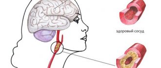
MRI of cerebral vessels provides detailed information for making a diagnosis in complex cases and with a confusing clinical picture - many diseases have similar symptoms. The blood supply to the head and brain comes from branches of the aorta passing through the neck. At this level, diseases or damage to the vascular system are often noted that affect cerebral circulation. To accurately determine the cause of painful conditions, an MRI of the vessels and arteries of the neck is performed in conjunction with a study of the brain vessels.
The examination allows you to study the condition of the main groups of cerebral vessels:
- internal carotid artery and its branches (ophthalmic, anterior and middle cerebral arteries);
- vertebral arteries located in the cervical spine, basilar, posterior cerebral arteries;
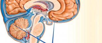
- Circle of Willis - a system of connected arteries at the base of the brain that increases the reliability of the blood supply to the brain;
- veins of the brain and venous sinuses of the dura mater.
Carrying out MRI of the vessels of the head and neck with a contrast agent allows you to visualize the smallest vessels and determine disorders of microcirculation and metabolic processes.
When is the examination carried out?
Before undergoing an MRI of the head and neck vessels, it is advisable to consult a neurologist. The doctor will assess the patient’s symptoms and condition and refer him for the most appropriate study.
The examination is usually carried out if the patient observes the following phenomena for a long time:
- prolonged headaches of unknown cause that cannot be treated with known drugs;
- periodic dizziness, loss of consciousness, fainting, darkening of the eyes, difficulty in spatial orientation;
- unstable blood pressure;
- decreased vision;
- numbness of the arms, shoulder girdle;
- actively developing osteochondrosis of the cervical spine with corresponding symptoms;
- herniated disc in the cervical region;
- subluxation, instability of the cervical vertebrae;
- traumatic brain injury, spinal injury;
- suspicion of cancer or mass formations in the brain.
The value of MRI of the head and neck vessels lies in the possibility of making an early diagnosis. The examination helps to start therapy in a timely manner and prevent the progression of the disease. Having received the necessary treatment, the patient can undergo the procedure again an unlimited number of times without risk to health. The doctor monitors the effectiveness of therapy and, if necessary, makes adjustments or changes tactics.
MRI of cerebral vessels is actively used by neurosurgeons to plan operations. Information about the location and structural features of the vascular system allows you to avoid unforeseen situations and make precise interventions. MRI is important in the postoperative period to monitor recovery processes.
What is the difference between CT and MRI of the brain?
Computed tomography is the only method that can compete with MRI. The principles of their operation differ, first of all, in the nature of the radiation: magnetic for MRI and x-ray for CT. For this reason, the first can be performed repeatedly without harm to the patient’s health, and the second can be performed no more often than a regular x-ray. At the same time, CT, unlike MRI, is not performed in a confined space, which makes it more preferable for claustrophobics.
In addition, MRI shows the chemical structure of the tissues being studied, and CT will tell us about their physical state.
Another important difference is that MRI is more effective for examining soft tissue, while CT is more effective for examining bones and joints.
The price of a brain MRI is not much different from the cost of a CT scan.
Sign up for diagnostics Do not self-medicate. Contact our specialists who will correctly diagnose and prescribe treatment.
What will the examination show?
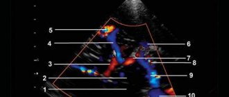
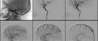
MRI of the vessels of the head and neck is performed in angiography mode - in this case, only areas of the vascular system are visible in the images, without surrounding tissues. The examination makes it possible not only to assess the condition of the arteries and veins, but also to determine the speed of blood flow in the vessels in real time. Based on the scanning results, a three-dimensional 3-D model of the area under study is constructed.
Signs of which diseases will be shown by MRI of cerebral vessels:
- stroke - hemorrhagic (leakage of blood from the vascular system into the brain tissue or meninges) or ischemic (cessation of blood supply to a part of the brain, more common);
- arterial aneurysm - pathological expansion and thinning of the vascular wall with the risk of rupture;
- arterial dissection as a result of congenital weakness of the arterial wall or damage by a pathological process;
- thrombosis, thromboembolism;
- inflammatory diseases of the vascular system – phlebitis, arthritis, systemic vasculitis;
- vascular tumors – hemangioma, hemangioblastoma, angioreticuloma;
- atherosclerosis;
- narrowing of the lumen of the vessel due to structural features or compression from the outside by a space-occupying formation;
- congenital developmental anomalies and structural features - absence, decrease or increase in diameter, bifurcation of some parts of the vascular system, arteriovenous malformation.
MRI of the vessels and arteries of the neck will show structural defects and pathological processes leading to impaired cerebral circulation:
- dissection of the carotid and vertebral arteries - damage to the artery wall as a result of injury, bending, with further formation of a hematoma between the walls of the vessel, narrowing of the lumen of the artery and disruption of the blood supply to the brain;
- pathological tortuosity of the carotid artery;
- developmental anomalies and acquired lesions of the vertebral artery;
- neoplasms;
- aneurysm of a section of the vascular system;
- pathologies in the area of the carotid sinus.
What is the essence of the EEG procedure?
An electroencephalogram records electrical signals from brain cells and allows us to identify epilepsy, trauma, neoplasms, inflammatory processes, and changes in blood vessels. Pathology is indicated by disturbances in the electrical activity of neurons, which are recorded using special sensors placed on the patient’s head.
Using an electroencephalogram, the doctor can:
- analyze the performance of the brain;
- identify foci of pathologies;
- assess the nature and extent of damage;
- confirm or clarify the diagnosis;
- monitor the effectiveness of the treatment.
Contraindications for magnetic resonance imaging of the vascular system of the head and neck
The ban on MRI of head and neck vessels is due to the peculiarities of the interaction of electromagnetic radiation with certain metals and magnets. The examination is prescribed with caution in certain patient health conditions.
MRI of cerebral vessels is contraindicated if the patient has the following devices in the body:
- pacemaker and other stimulating implants and pacemakers, cochlear implant - implants may fail under the influence of a magnet;
- metal intravascular stents, artificial heart valves;
- endoprostheses, foreign metal bodies in the body;
- metal clips installed in the vessels of the brain after surgery - under the influence of a magnetic field they can move and cause bleeding.
Not every material prohibits MRI of vessels and arteries of the neck and head. Metals that react to a magnetic field by displacement and heating: stainless steel, iron, nickel, cobalt. In other cases, the procedure is not prohibited.
In some patient conditions, examination is allowed by the doctor’s decision on an individual basis:
- pregnancy up to 12 weeks - the fetus is especially sensitive to external factors;
- serious condition of the patient, severe pain;
- epilepsy, seizures, mental illness;
- body weight should not be more than 130 kg - the devices have restrictions on the patient’s weight and waist circumference.
The procedure can be performed in patients who find it difficult to maintain a stationary body position under general anesthesia or in an open-type apparatus. During the entire period of using MRI, there have been no cases of fetal pathology developing after the procedure in the first trimester of pregnancy, however, doctors are careful. If the patient suffers from a mild form of claustrophobia, he may take sedatives before the examination.
MRI of the vessels of the head and neck with the use of contrast agents is contraindicated in pregnant women at any stage, as well as in patients with renal failure.
Diagnostics
At the beginning of development, atherosclerosis does not manifest itself in any way. Later, as the disease progresses, several visible manifestations of atherosclerosis appear, which can be identified during a routine examination by a neurologist. This is weight loss, swelling, fatty tissue, and trophic disorders. The doctor will listen to the heart, measure blood pressure, and prescribe clinical tests. The following diagnostic measures are used:
- Ultrasound Dopplerography. Allows you to identify diseases at an early stage, assess the size of plaques and blood flow.
- Coronary angiography is used to assess the condition of the heart vessels.
Preparing for the study
First of all, you should make sure that there are no contraindications, and if there are any, collect all the technical documentation describing the devices and endoprostheses implanted into the body. The examination becomes possible after analyzing the composition of metal implants.
It is advisable to have with you a doctor’s referral, extracts from medical documents, expert advice, as well as the results of previous studies, if any. A patient who is scheduled to receive a contrast agent should undergo a kidney function test in advance. Women should be sure that they are not pregnant.
There is no need for special preparation for MRI of vessels and arteries of the neck and head. If an examination with contrast is prescribed, you must arrive on an empty stomach.
Do I need to prepare for an ultrasound of the neck vessels?
No special preparation is required, but in 1-2 days it is advisable to minimize the impact of factors that may affect the state of the cardiovascular system. Try not to drink a lot of tonic drinks - strong tea, coffee, energy drinks, give up alcohol. It is better not to smoke before the test. If you take any medications regularly, talk to your doctor about taking them on the day of the test - some medications can affect blood flow, and your doctor may advise stopping them for the day or delaying taking them.
How is the examination carried out?
First of all, you need to get rid of metal items of clothing, the neck and ears should be free of jewelry. Bags, money, plastic cards, telephones, and electronic devices must be left outside the office. The patient receives disposable comfortable clothing for a comfortable procedure.
The examination without contrast agent lasts about 20 minutes; the introduction of contrast agents doubles the examination time. The patient is positioned on a table, which smoothly slides into the body of the tomograph, where the scanning will take place. During the examination, the neck and head should not make any movements. Fixation is ensured by a special coil.
When performing MRI of vessels and arteries of the neck and head, the tomograph operation is accompanied by unusual sounds, so the patient receives earplugs or headphones before the examination. The body of the tomograph is equipped with a fresh air supply system, as well as an intercom through which the doctor can give the command to hold your breath or slightly change the position of your head. If the patient becomes ill, it is possible to urgently contact the staff and stop the scan. After completing the examination, the patient changes clothes and can await the result.
How is an EEG performed?
The examination takes place in several stages.
Preparatory stage
- the patient enters the office, protected from light and sound;
- an encephalograph “cap” consisting of special sensors is put on it;
- The sensor wires are connected to a device that records the bioelectric impulses of the brain.
Diagnostic stage
- the encephalograph transmits data to the monitor in the form of a graph;
- the power of electric fields and its distribution in different parts of the brain are recorded;
- Functional tests are carried out: the patient is asked to blink, look at flashes of light, breathe less often or deeper, listen to a sharp sound.
Final stage
- the electrodes are removed from the patient;
- print out the results.
What the results look like
After performing an MRI of the vessels and arteries of the neck and head, the doctor receives layer-by-layer images of the vascular pattern with the ability to construct a three-dimensional model. The patient can pick up the images on electronic media or in printed form. The photographs are accompanied by a report from a radiologist.
Normally, the internal carotid arteries are located symmetrically, have clear contours, are not displaced, and are not compressed. The cerebral arteries arise in a typical place, the diameter is not changed, and there is no pathological tortuosity of the vessels. The cerebral veins and sinuses should be of normal diameter, without areas of blood flow disturbance, filling defects or deformations.
On MRI images of the vessels and arteries of the neck you can see signs inherent in certain diseases:
- an aneurysm is a round formation with a high signal intensity; when a thrombus attaches, a layered structure appears;
- vascular malformations appear as linear or tortuous structures with dilated vessels;
- dissection of the arterial wall is characterized by the appearance of a crescent-shaped hematoma located along the vessel, while the lumen of the artery is narrowed evenly or in the form of a rosary;
- atherosclerosis of the vascular system looks like an area of increased intensity on the vessel wall, the lumen is narrowed;
- hemangioma is a high-intensity formation with clear edges and a lobular structure.
Despite a detailed description of all identified pathological formations, the conclusion of a radiologist is not a diagnosis. The MRI result of the area of the cerebral vascular system should be shown to the doctor who ordered the study. Clinics offer the opportunity to consult with the radiologist who performed the procedure. If necessary, he will recommend contacting specialized specialists - a neurologist, oncologist or surgeon.
How to do an MRI of the brain
Only the patient’s head enters the tomograph chamber, so the feeling of enclosed space is minimized
There is no need to prepare specially for the study. The only requirement is not to wear metal or jewelry, hairpins, or watches when going for an examination (or be prepared to remove them or take them out of your pockets before placing them in the tomograph). Also, during the procedure you cannot have magnetic media (flash drives, bank cards, etc.) with you, since the information stored on them will be destroyed.
The diagnosis will take 30–40 minutes. You cannot move your head during the process, but this restriction does not apply to the facial muscles. You can even talk to your doctor while he takes sequential pictures. From time to time, the doctor himself will contact the patient to inquire about his well-being.
If you suffer from claustrophobia, but an MRI of the brain is necessary, you need to say this in advance. Then the doctor will suggest taking a sedative before starting the procedure.
Where to get a magnetic resonance imaging scan
The search for a clinical diagnostic center can be significantly speeded up if you use the help of our website. The service contains information about a large number of clinics, ranging from contact details to discount offers. If necessary, our specialist will advise you free of charge over the phone about operating hours, the availability of tomographs of the required type and the time available for making an appointment.
A patient who wants to undergo magnetic resonance imaging of the vascular system of the head and neck can independently filter the list of medical institutions by rating, cost and features of the procedure (24-hour clinics, pediatric MRI).
Advantages and disadvantages of the procedure
Today, almost every medical institution has the necessary equipment to conduct ultrasound of the neck vessels. Let's consider the advantages of this technique:
- This method is absolutely safe;
- The procedure can be performed at any age;
- She has no direct contraindications;
- The process is absolutely painless and does not cause discomfort.
- The only disadvantage we can note is the lack of information content - this is not the main method of diagnosing diseases, but only an auxiliary one.
Treatment
Treatment depends on the stage of the process. This may be a conservative approach or surgery. In the first case, the focus is on normalizing the patient's lifestyle. Nutrition is adjusted, a special diet is prescribed, and a weight loss program is developed, if necessary. As drug therapy, drugs are used that normalize blood pressure, lower cholesterol levels, normalize blood counts, and replenish the supply of vitamins and microelements.
If the process has gone far enough and medications do not achieve the desired result, surgery is used. This can be open or modern endovascular intervention. A gentle method is angioplasty, during which the lumen of the vessel expands; if necessary, a stent can be installed to maintain the lumen of the vessel in the desired position. The operation is aimed at removing plaques that are blocking normal blood flow. In some cases, a severely damaged vessel may be replaced or a new blood flow path created.
Get diagnosed with atherosclerosis at Clinic No. 1
- Neurologist appointment
- Doppler ultrasound
- Coronary angiography
For one-time payment for services - 20% discount
Call