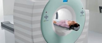
Disorders of the venous circulation of the brain
Classification. The following chronic and acute variants of venous circulation in the brain are distinguished. Chronic include venous congestion and venous encephalopathy, acute include venous hemorrhage, thrombosis of the veins and venous sinuses, thrombophlebitis.
Venous stagnation. The most common form of venous circulation disorder is due to various reasons: cardiac and cardiopulmonary failure, respiratory diseases (bronchitis, bronchiectasis, bronchial asthma, emphysema, etc.); compression of extracranial veins (internal jugular, innominate, superior vena cava), struma, arterial aneurysm, tumor in the neck; neoplasms of the brain, membranes and skull, arachnoiditis, traumatic brain injury, thrombosis of the veins and sinuses of the dura mater, compression of the veins with cerebral edema and craniostenosis. With venous stagnation, metabolic changes and brain hypoxia occur, venous and intracranial pressure increases, and cerebral edema develops. More often, milder disorders occur in the form of changes in the tone of the cerebral veins, which is detected using orbital plethysmography and rheography.
Clinical manifestations. A dull headache, more pronounced in the morning, increases with movements of the head to the sides, changes in atmospheric pressure, changes in ambient temperature, after excitement, drinking alcohol, etc., there is a hum or noise in the head, cyanotic lips, cheeks, ears, nose , mucous membranes of the oral cavity, swelling of the lower eyelids, especially in the morning, dilation of the veins in the fundus. Venous pressure ranges from 55 to 80 mmH2O, arterial pressure is usually within normal limits. Stupefaction, dizziness, darkening of the eyes, fainting, and numbness of the extremities are observed. Epileptic seizures and mental disorders are possible. With severe venous stagnation, patients are unable to lower their heads and remain in a horizontal position.
Measurements of pressure in the ulnar vein, radiography of the skull (increased development of diploic veins, graduates and veins of the dura mater), and phlebography are of diagnostic value in venous pathology.
Venous encephalopathy. With venous encephalopathy, the following syndromes are distinguished: hypertensive (pseudotumorous), diffuse small-focal brain damage, bettolepsy and asthenic.
Bettolepsy, or cough epilepsy, develops with chronic bronchitis and emphysema, pneumosclerosis, bronchial asthma, especially with cardiopulmonary failure. A persistent cough may result in sudden loss of consciousness (syncope).
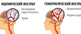
Venous hemorrhages. Capillary-venous hemorrhages in the brain and capillary-venous stasis are observed in hypertension. Venous stroke occurs in patients with heart failure, traumatic brain injury, brain tumor, infectious and toxic brain lesions. Clinical manifestations develop slowly: confusion, speech disorders, diplopia, pyramidal reflexes, hemiparesis, hemihypesthesia, damage to cranial nerves.
Thrombosis of cerebral veins. It occurs in the practice of clinicians of many specialties as a complication of various inflammatory processes, infectious diseases, operations, abortions, pregnancy, childbirth, skull trauma, “blue” heart defects, etc. Changes in the walls of the veins, a slowdown in the speed of blood flow and increased blood clotting play a role in the pathogenesis, as well as a change in the colloidal properties of endothelial cells, which contributes to the aggregation of blood cells. Often, thrombosis of the cerebral veins is combined with thrombosis of the sinuses of the brain, as well as the veins of the lower extremities.
Clinical manifestations. Cerebral vein thrombosis usually develops gradually. Headache, nausea, vomiting, meningeal symptoms, congestive optic discs, increased body temperature, and increased ESR appear. In the cerebrospinal fluid, mild pleocytosis and an increase in protein content, sometimes blood, are detected. Characterized by confusion, partial motor-type seizures, and less commonly generalized convulsions. Depending on the location of the venous lesion, focal symptoms occur: aphasia, alexia, hemianopsia, flaccid or spastic paresis or paralysis, and sensory disturbances. The outcome is often favorable, focal symptoms often undergo significant or even complete regression, but there are relapses of the disease. A slow chronic course over many months and even years is possible. Sometimes there are consequences in the form of mental disorders, aphasia, convulsive seizures and paresis of the limbs.
Thrombosis of the dural sinuses. Usually develops when infection penetrates into them from a nearby focus (furuncles or carbuncles of the scalp, face, erysipelas, etc., purulent osteomyelitis of the skull bones, purulent acute and chronic otitis media, mastoiditis, purulent processes in the orbit, paranasal sinuses) along the brain and diploic veins. In addition, phlebitis and thrombosis of the dural sinuses can occur hematogenously with thrombophlebitis of the veins of the extremities or pelvis and during septic processes. Thrombosis of the cerebral sinuses is sometimes accompanied by thrombophlebitis of the retinal veins, purulent meningitis, brain abscesses, etc. Sinus thrombosis can also occur with chronic infections (tuberculosis), malignant tumors and other diseases that occur with cachexia, in debilitated patients and in old age.
Clinical manifestations. Low-grade or sometimes very high, stable or fluctuating body temperature, headache, vomiting, leukocytosis in the blood, increased intracranial pressure. With thrombosis of the sinuses of the convexital surface of the brain, general cerebral symptoms predominate; in the sinuses of the base of the brain, signs of damage to the cranial nerves predominate. Drowsiness develops, sometimes, on the contrary, motor restlessness, insomnia, delirium, epileptic seizures, rigidity of the neck muscles, Kernig's symptom, hyperesthesia to visual, auditory and skin stimuli, and sometimes trismus. Focal symptoms of brain damage correspond to the location of the sinus. Swelling and cyanosis of the face or mastoid area are noted. In the fundus, dilated veins and swelling of the optic discs are detected. The cerebrospinal fluid is clear or xanthochromic, sometimes with an admixture of red blood cells; moderate pleocytosis is noted. Septic thrombosis of the dural sinuses is manifested by chills and very high remitting temperature. With thrombosis of the superior sagittal sinus, epileptic seizures of the motor type, hemi- and paraplegia or paresis occur.
Symptoms of thrombosis of the transverse or sigmoid sinus: headache, bradycardia, sometimes double vision, septic temperature, chills, stupor, turning into a soporous and even comatose state, sometimes delirium and agitation, anti-pain position of the head with an inclination to the painful side, meningeal phenomena, leukocytosis in blood. The jugular vein may be involved in the process. In this case, swelling of the tissue surrounding the vein and signs of damage to the glossopharyngeal, vagus, accessory and hypoglossal nerves occur.
Symptoms of cavernous sinus thrombosis: exophthalmos, swelling and venous hyperemia of the eyelids, orbits, forehead, root of the nose, dilation of the fundus veins (congestion), pain and hyperesthesia in the area of innervation of the superior branch of the trigeminal nerve, chemosis of the conjunctiva, ophthalmoplegia - paralysis or paresis of muscles, innervated by the III, IV, VI cranial nerves, stupor, delirium, sometimes coma, metabolic and endocrine dysfunction.
Complications: purulent meningitis, metastatic abscesses in the lungs, septic pneumonia.
Thrombophlebitis of the cerebral veins. With thrombophlebitis of the cerebral veins, the temperature rises to subfebrile levels with periodic rises to 38–39 °C. Patients complain of headache, nausea, and vomiting. Observed stupor, stuporous state, epileptic seizures, paresis of the limbs; in the fundus – swelling and dilation of veins; in the blood - leukocytosis; in the cerebrospinal fluid - slight pleocytosis, an increase in the amount of protein and positive protein reactions, sometimes an admixture of erythrocytes.
Venous discirculation in childhood and adolescence
Introduction
Vascular lesions of the nervous system are an important problem in modern clinical neurology.
The study of cerebral venous circulation disorders remains one of the urgent tasks of modern medicine. The improvement of ultrasound equipment, as well as its software, has led to the fact that when studying blood flow in the arteries of the brain, it is possible to assess the state of venous blood flow at a fairly good level.
However, the main problem is that the data on the normative values of velocities in the venous system of the brain are extremely scattered, fragmentary and not always unambiguous.
In this regard, you often have to rely on your own experience, taking as a basis data from a number of literary sources (Table 1), which are more consistent with the features of this device, the quality of the resulting image and the age of the patient. The small number of ultrasound studies, which would contain data on the state of venous blood flow at the extra- and even more so at the intracranial levels, is explained primarily by hardware features, and only then by the insufficient amount of information on this issue in the periodical literature, the complexity of the spatial-anatomical three-dimensional perception of the intracranial venous system by diagnosticians, low need for such studies on the part of neurologists. Table 1
. Blood flow indicators (Vmax, cm/s) in the internal jugular veins and in the main intracranial veins/sines of the brain.
| Author | Internal jugular vein | Middle cerebral vein | Vienna Rosenthal | Vienna Galena | Direct sine | Internal cerebral vein | Vertebral veins |
| V.G. Lelyuk, S.E. Lelyuk, 2003 [1] | 7-45 right\ 12-33 left | 9 | 11 | 15 | |||
| J. Valdueza et al., 1996 [2] | 6-18 | 4-17 | |||||
| BG Schoser, 1999 [3] | 5-12 | ||||||
| E. Stolz et al., 1997 [4] | 13,8 ± 8,9 | 13,7 ± 4,7 | 31,7 ±15,6 | ||||
| R. Baumgartner et al., 1997 [5] | 4-15 | 7-19 | 12-39 | 10-18 | |||
| R. Aaslid, 1991 [6] | 23 ± 3 | ||||||
| V.A. Shakhnovich, 1998 [7] | 14-28 | ||||||
| M.L. Dicheskul et al., 2008 [8] | 9,8-20,9 | ||||||
| G.A. Ivanichev, G.B. Dolgikh, 2007 [9] | 22,0 ± 4,6* 20,1 ± 3,2** | 17,7 ± 3,3* 16,2 ± 2,2** | |||||
| G.B. Dolgikh, G.A. Ivanichev, 2008 [10] | 13,6 ± 0,3 | 23,4 ± 0,9 | 18,7 ± 0,9 |
Note. * - in children aged 1-12 months; ** - in children aged 1-3 years.
The purpose of this study was to assess correlation dependencies in patients with signs of venous discirculation at the intra- and extracranial levels, cerebral venous hemodynamics in children and adolescents with a clinical picture of “cranialgia”, with clarification of the cause-and-effect relationships that determine the formation of venous discirculation.
Material and methods
The study included 106 children aged from 2 to 18 years, average age 9.87 ± 3.9 years (from 2 to 6 years - 18 people, average age 3.8 ± 1.43 years; from 7 to 18 years - 88 people, average age 11.1 ± 2.99 years), sent for examination to the diagnostic center of Kaliningrad with a headache clinic or symptoms of vertebrobasilar insufficiency. During the examination, signs of dysgemia at the intra- and extracranial levels were identified in all of them. Doppler ultrasound studies of arterial and venous blood flow at the level of the neck and base of the brain were performed on a Medison Accuvix V10 device (South Korea), in B-, C-, PW-modes, linear (L5-12 MHz) and sector phased (P2- 4 MHz) sensors. Correlation dependencies were assessed between 94 clinical and instrumental indicators.
results
As a result of the study, it was found that discirculation in the vertebral vein (PV) system, as a rule, is a consequence of pronounced extravasal influences (vascular compression) on blood flow in the internal jugular vein (IJV) on the side of registration of dyshemia (r = + 0.67; p 0.05).
Dysgemia in the vein of Galen on the right is often accompanied by an increase in the tone of the VA, ICA and MCA on the ipsilateral side (as a consequence of reflex changes), just as the former is associated with kinks and S-shaped tortuosity of the ICA on the right. The effect of ICA tortuosity on venous outflow may be due to extravasal compression of venous vessels by tortuous arterial trunks with significantly higher intravascular pressure in the places of their closest abutment.
The relationship between “headache syndrome” and accelerated venous blood flow through the veins of Galen turned out to be extremely low (r = +0.22; p
In addition, a fairly clear picture of pronounced unilateral disturbances of both arterial blood flow and venous outflow was noted, which is probably due to the unity of the autonomic innervation of the arteriovenous system “head-neck”, the presence of complex compensatory mechanisms (reflex), as well as the indirect influence of the tone of the striated muscles neck and cervical spine, which is confirmed in a number of studies.
Discussion
From our own experience of working with devices from leading manufacturers of ultrasound equipment, it should be admitted that the best quality images of the cerebral venous network were often obtained using Medison Acuvix devices. At the same time, the images were of high quality in both C and PW modes, with a minimum of artifacts and interference.
Assessment of venous blood flow at the extracranial level (jugular veins, vertebral veins) causes fewer difficulties and is available on almost any device, except, perhaps, assessment of blood flow through the vertebral veins in persons with excess body weight, widespread osteochondrosis and a short neck, when even visualization vertebral arteries is carried out with great difficulty.
The structure of the cerebral venous system is presented in Figure 1.
Rice. 1.
Arteries and veins/venous plexuses at the base of the brain [11]. 1 - sphenoid-parietal venous sinus, 2 - middle cerebral artery (MCA), 3 - middle cerebral vein (deep), 4 - posterior cerebral artery (segment P1) (PCA), 5 - basilar venous plexus, 6 - basilar (main ) artery, 7 - vein of Rosenthal (right) and branch of the posterior cerebral artery (right), 8 - vertebral artery (segment V1), 9 - marginal venous sinus, 10 - vein of Galen (great cerebral vein), 11 - straight sinus, 12 - branch of the posterior cerebral artery (left), 13 - vein of Rosenthal (left), 14 - inferior ventricular vein.
Variants of normal and pathological venous blood flow are shown in Table 2 and Figures 2-18.
table 2
. Variants of normal and impaired venous blood flow in C- and PW-regimes. Causes of venous discirculation in the main venous pools. *
| Blood Flow Options | Causes of venous discirculation |
| Rosenthal's Vienna Fig. 2, 3, 4 |
|
| Inferior ventricular vein (tributary vein of Rosenthal) Fig. 5 | |
| Middle cerebral vein Fig. 6, 7, 8 |
|
| Vienna Galena Fig. 9, 10, 11 |
|
| Vertebral vein Fig. 12, 13 |
|
| Internal jugular vein Fig. 14, 15 |
|
| External jugular vein Fig. 16 | |
| Sphenoparietal venous sinus Fig. 17, 18 |
Note
*
The following should also be considered as possible causes of venous discirculation:
- violation of the central regulatory mechanisms of vascular tone;
- hereditary constitutional predisposition, manifested by undifferentiated connective tissue dysplasia (including in the form of bone deformities and hypermobility of the joints of the cervical spine);
- consequences of perinatal pathology (cervical spine injuries [12]; perinatal hypoxic-ischemic processes [13]);
- extravasal causes of venous stagnation: tumors of the mediastinum and neck; osteochondrosis of the cervical spine; traumatic compression of the chest and abdomen, leading to compression of the superior vena cava, jugular and vertebral veins; early osteochondrosis; deforming arthrosis; spondylosis; craniovertebral anomalies (basilar compression; defects of the odontoid process of the second cervical vertebra, Kimmerle anomalies; Arnold-Chiari anomalies);
- disorders of exclusively myogenic nature; compression of the vertebral artery by the inferior oblique muscle of the capitis during tonic tension with subsequent contracture or compression by the anterior scalene muscle;
- vasculitis of cerebral vessels, rheumovasculitis, bacterial meningitis, thrombosis of intracranial venous sinuses [10].
**
The literature covers various methods for invasive and non-invasive assessment of intracranial pressure (ICP) [7, 14]. In the specialized literature, the possibility of assessing ICP by displacement of the tympanic membrane is discussed [15]. However, this technique is described only for patients with hydrocephalus. The development of non-invasive methods for measuring ICP is still relevant, with various ultrasound and telemetric measurement methods taking the leading place. However, the question of the accuracy of the data obtained with non-invasive methods remains open and requires further clarification. None of the non-invasive techniques allows you to measure the absolute value of ICP, but only extrapolates its dynamics.
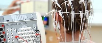
The only possible method for identifying ICH in patients with chronic venous insufficiency remains a comprehensive clinical and instrumental diagnosis, including assessment of the neurological status, condition of the fundus, Echo-encephalography (EchoEG), Dopplerography of the vessels of the neck (USDG BCS) and brain (TG USDG). Only the combination of these methods can bring the researcher closer to the expected conclusion. According to H. Bode [16], it is almost impossible to detect an increase in ICP in a child with hydrocephalus, based only on Doppler data.
In the studies of Yu.A. Rosin [17] proved the presence of a pressure gradient between the vein of Galen and the straight sinus. During transoccipital examination in the oral part of the straight sinus, in the area where the vein of Galen flows into the straight sinus, high venous outflow (more than 50 cm/s) is detected in children, which significantly exceeds the blood flow in the veins of Rosenthal, internal cerebral veins and the inferior sagittal sinus.
At the same time, a number of authors note that an increase in the tone of the main cerebral arteries should be considered as a compensatory mechanism for facilitating venous outflow [18].
The main hemodynamic signs of benign intracranial hypertension with transcranial Dopplerography (TCD) are considered to be an increase in maximum velocity and increased pseudopulsation of blood flow in the cerebral veins and sinuses (veins of Rosenthal > 15 cm/s, vein of Galen > 20 cm/s and straight sinus > 30 cm/s )
[1]. It is assumed that during chronically ongoing processes in the cranial cavity, venous circulation suffers more significantly.
It has been noted that a sharp increase in the venous signal and a change in the physiological direction of blood flow along the internal ophthalmic vein to retrograde are detected on the side of the “focus” of brain damage in cases of cerebrovascular accidents and traumatic brain injury, accompanied by an increase in intracranial pressure [13].
Rice. 2.
Middle cerebral artery (MCA). Transtemporal access. Color Doppler mapping mode at the level of the anterior cerebral artery (ACA) (3), MCA (5), first (7) and second (8) segments of the PCA, vein of Rosenthal (9), vein of Galen (10), middle cerebral vein (4), inferior ventricular vein (tributary vein of Rosenthal) (6). Brain peduncles (pedunculi cerebri) (1; 2).
Rice. 3.
Middle cerebral vein (deep). Right there. Color flow control, PW mode. Flow scanning in the vein of Rosenthal. Vmax 15.88 cm/s.
Rice. 4.
Posterior cerebral artery (segment P1) (PCA). Transtemporal access. Color flow control, PW mode. Scanning of pathological accelerated flow in the vein of Rosenthal. Vmax 28.59 cm/s.
Rice. 5.
Basilar venous plexus. Transtemporal access. Color flow control, PW mode. MCA (1), PCA segment P1 (4), cerebral peduncles (6; 7), middle cerebral vein (2), vein of Rosenthal (5). Flow scanning in the inferior ventricular vein (inflow of the vein of Rosenthal) (3).
Rice. 6.
Basilar (main) artery. Transtemporal access. Color flow mode at the level of the ACA (1), MCA (2), first segment of the PCA (4), middle cerebral vein (3). Brain peduncles (pedunculi cerebri) (5; 6).
Rice. 7.
Vein of Rosenthal (right) and branch of the posterior cerebral artery (right). Right there. Color flow control, PW mode. Flow scanning in the middle cerebral vein (proximal segment).
Rice. 8.
Vertebral artery (segment V1). Transtemporal access. Color flow control, PW mode. Scanning of pathological accelerated flow in the middle cerebral vein (proximal segment). Vmax 24.62 cm/s
Rice. 9.
Marginal venous sinus. Transtemporal access. Color circulation mode at the level of the first segment of the PCA (3), vein of Rosenthal (4), vein of Galen (5). Brain peduncles (pedunculi cerebri) (1; 2).
Rice. 10.
Vein of Galena (great vein of the brain). Right there. Color flow control, PW mode. Flow scanning in the vein of Gallen. Vmax 21.18 cm/s
Rice. eleven.
Direct sine. Transtemporal access. Color flow control, PW mode. Scanning of pathological accelerated flow in the vein of Galen. Vmax 50 cm/s
Rice. 12.
Branch of the posterior cerebral artery (left). Longitudinal scanning in the projection of the V2 segment of the vertebral artery (1) and vertebral vein (2). Color flow control and PW mode. Vmax in the vertebral vein is 34.69 cm/s.
Rice. 13.
Vienna of Rosenthal (left). Longitudinal scanning in the projection of the V1 segment of the vertebral artery (1). Color flow control and PW mode. Pathological accelerated flow in the vertebral vein (2). Vmax 83.73 cm/s.
Rice. 14.
Inferior ventricular vein. Transverse scanning in the projection of the internal carotid artery (3), external carotid artery (2) and internal jugular vein (1). Color flow control and PW mode. Vmax in the internal jugular vein is 41.49 cm/s.
Rice. 15.
Transverse scanning in the projection of the internal carotid artery (1) and the tortuous internal jugular vein (2). Color flow control and PW mode. Pathologically accelerated turbulent flow in the internal jugular vein up to 80 cm/s.
Rice. 16.
Transverse scanning in the projection of the internal (1) and external (2) carotid arteries, external jugular vein (3). Color flow control and PW mode. Vmax in the external jugular vein is 22.88 cm/s.
Rice. 17.
Transtemporal access. Mode of color circulation at the level of the MCA (2), and the sphenoparietal venous sinus (1). Brain peduncles (pedunculi cerebri) (3).
Rice. 18.
Ibid (Fig. 17). Color flow control, PW mode. Flow scanning in the sphenoparietal venous sinus (1). Vmax 19.19 cm/s.
Another problem facing the researcher, even in the case when it is possible to assess the nature of venous blood flow at the intra- and extracranial level, is the correct interpretation of the results obtained. Since the available literature data does not provide a holistic picture of the causes of venous discirculation, and in some cases, increased ICP or connective tissue dysplasia is indicated as the main reason for its occurrence, without indicating the possible mechanisms of the formation of venous discirculation, the benefit of such conclusions is extremely small. It is also impossible to influence the tactics of further treatment, since the points of possible application of efforts by doctors of different specialties are unknown or not indicated.
An increase in ICP as a probable cause of venous discirculation should not be forgotten that due to its low prevalence in the population (0.025-0.05% among children and adolescents), this pathology cannot be considered as the leading etiological cause of dysgemia and is most likely a diagnosis of exclusion.
Functional disorders of the musculoskeletal system with the formation of blocks in small joints of the spine with the appearance of reflex musculoskeletal pain syndromes are unreasonably rarely diagnosed, and the role of myofascial pain syndromes, in which the muscle suffers primarily, is underestimated. An important role in this in children is played by certain injuries to the cervical spine in the anamnesis (mainly during childbirth). The literature describes the mosaic nature of emerging pathogenetic factors in the obstruction of the outflow of venous blood from the skull. At the same time, the leading place in the genesis of dynamic disorders of venous circulation belongs to myofascial pain syndrome of cervical localization. When myofascial pain syndrome is localized in the muscles of the craniovertebral junction, congestive venous disorders are caused by general algic processes in this zone, including functional blockades of the junction, while tunnel-compression mechanisms in this zone do not play a decisive role in venous discirculation. Tunnel-compression mechanisms of obstruction of venous blood flow are most relevant in the middle and lower cervical localization of myofascial pain.
Conclusion
Taking into account our data on the strong correlation between accelerated venous blood flow and tortuosity of the ICA, VA (as indirect manifestations of disorders in the cervical spine, including manifestations of natal trauma to the cervical spine), we believe that in children and adolescents a key role in The appearance of dysgemia (impaired venous outflow) is played by the “pathology/structural features” of the cervical spine and the congenital structural features of the ICA at the extracranial level. The main causes of dysgemia in childhood should be considered “congenital connective tissue dysplasia” [19], manifested in the form of pathology of the cervical spine, with curvature and tortuosity of the bone canal, or “birth injuries with subluxation of 1-2 cervical vertebrae” (presence of a history of in the majority of examined individuals), with impaired venous outflow at the extracranial level.
Taking into account all of the above, it should also be concluded that if a picture of venous discirculation is identified, especially in young people, treatment should be aimed primarily at restoring the functional integrity of the musculoskeletal system of the cervical spine, correcting posture, manual practices, as well as compliance with regime-restrictive measures [20].
Literature
- Lelyuk V.G., Lelyuk S.E. Ultrasound angiology. M.: Real Time, 2003. 322 p.
- Valdueza JM, Schmierer K., Mehraein S., Einhäupl KM Assessment of normal flow velocity in basal cerebral veins. A transcranial Doppler ultrasound study. 1996. Stroke 27. pp. 1221-1225.
- Schoser BG, Riemenschneider N., Hansen HC The impact of raised intracranial pressure on cerebral venous hemodynamics: a prospective venous transcranial Doppler ultrasonography study // J. Neurosurg. 1999. V. 91, N 5. P. 744-749.
- Stolz E., Jauss M., Horning C. Cerebral venous anatomy in color-coded duplex sonography. What is possible in non-contrast enhanced TCCD? // New trends in cerebral haemodynamics and neurosonology / Eds. Kligelhofer J., Bartels E., Riglenshtein B. 1997. P. 312-319.
- Baumgartner RW, Gonner F., Muri R. Normal haemodynamics in cerebral veins and sinuses: a transcranial color-coded duplex sonography study // New trends in cerebral haemodynamics and neurosonology / Eds. Kligelhofer J., Bartels E., Riglenshtein B. 1997. P. 312-319.
- Aaslid R. Cerebral hemodynamics // Transcranial Doppler / Eds. Newell DW, Aaslid R.: - NY: Raven, 1992. R. 500.
- Shakhnovich V.A. Violation of the venous circulation of the brain according to transcranial Dopplerography // Ultrasound Doppler diagnostics of vascular diseases / Ed. ed. Nikitina Yu.M., Trukhanova A.I. M.: Vidar, 1998. pp. 355-400.
- Dicheskul M.L., Kulikov V.P., Maslova I.V. Ultrasound characteristics of venous outflow through the vertebral veins / Ultrasound and functional diagnostics, 2008, N 4. P. 33-40.
- Ivanichev G.A., Dolgikh G.B. Disturbances of arterial and venous blood flow in children with vertebrobasilar insufficiency // Journal of Neurology and Psychiatry, 2007, N 3.
- Dolgikh G.B., Ivanichev G.A. Cerebral vascular disorders in children with cerebral palsy and convulsive syndrome // Kazan Medical Journal, 2008, N 3.
- Pucillo M.V., Vinokurov A.G., Belov A.I. Atlas “Neurosurgical Anatomy” / Ed. Konovalova A.N. M.: Antidor, 2002.
- Burtsev E.M., Andreev A.V., Dyakonova E.N., Kutin V.A. Functional Dopplerography in pediatric angioneurology // Abstracts of the report at the VIII International Conference: Current state of non-invasive diagnostic methods in medicine. Sochi, 2001. pp. 151-160.
- Nikitin Yu.M., Trukhanov A.I. Ultrasound Doppler diagnostics in the clinic. MIC, 2004. 496 p.
- Adelson PD, Bratton SL, Carney NA et al. Guidelines for the acute medical management of severe traumatic brain injury in infants, children, and adolescents. Pediatr. Crit. Care Med. 2003; (4) 3.
- Samuel M., Burge DM, Marchbanks RJ Tympanic membrane displacement testing in regular assessment of intracranial pressure in eight children with shunted hydrocephalus // J. Neurosurg. 1998. V. 88. R. 983-995.
- Bode H. Pediatric application of transcranial Doppler sonography / Wien; Ny: Springer Verlag, 1988. P. 108.
- Rosin Yu.A. Dopplerography of cerebral vessels in children / SPbMAPO, 2006. 114 p.
- Belkin A.A., Alasheev A.M., Inyushkin S.N. Transcranial Dopplerography in intensive care. Methodological manual for doctors. Ekaterinburg: Publication of the Clinical Institute of the Brain of the Scientific Research Center of the Russian Academy of Medical Sciences; 2004.
- Andreev A.V., Lobanova L.V., Ermolin I.E. Transcranial Dopplerography and variational pulsometry in the diagnosis of cerebral angiodystonia in children // Journal of Neuropathology and Psychiatry. 1994. N 3. S. 22-23.
- Tsokolov AV, Tsokolova VA, Tsokolova MA, Senchilo VG, Egorov AU Venous discirculation // Journal of the Neurological Sciences. 333 (2013). e518. Abstract - WCN 2013, No102, Topic:8 - Headache. Vienne, Austria. 2013. Neurology in the age of globalization. XXI World Congress of Neurology.
Ultrasound scanner RS80
A benchmark for new standards!
Unparalleled clarity, resolution, ultra-fast data processing, and a comprehensive suite of advanced ultrasound technologies to solve the most challenging diagnostic problems.
Vertebro-basilar insufficiency: causes, symptoms, treatment
The disease is characterized by repeated attacks of reversible ischemia of brain cells (local decrease in blood supply), as a result of which they do not receive the necessary nutrition.
Ischemic attacks (or small strokes of the vertebrobasilar system) have an increasing etiology of neurological deficit and gradually lead to dysfunction of the central nervous system (CNS).
Most often, vertebrobasilar insufficiency is observed in patients suffering from symptoms of cervical osteochondrosis. The disease can develop in people of different age categories. In the early stages, VBI is completely reversible; in an advanced state, there is a high probability of strokes.
Anatomy
Paired vertebral arteries emerge from the subclavian arteries, located in the upper part of the chest, pass through the openings of the transverse processes of the cervical vertebrae, then through a larger opening into the cranial cavity. At this point, the vertebral beds merge into a single basilar artery, which is based in the lower parts of the brain stem.
The branches extending from the basilar artery wash and supply oxygen and nutrients to the cerebellum, pons, occipital lobes of both hemispheres of the brain, medulla oblongata and other parts. A decrease in blood flow in the vertebral and basilar vessels initially provokes temporary disorders (dizziness, motor dysfunction, loss of coordination of movements and spatial orientation, headaches), a complete cessation of blood circulation provokes necrosis of a certain area of the brain or a stroke.
Causes of the disease
The main reason for the formation of vertebrobasilar insufficiency is a violation of the patency of blood channels (stenosis). Most often, the areas of direct entry into bone canals or the place where arteries merge into one are susceptible to narrowing of the lumen of blood vessels. In addition, the culprits in the development of the disease may be atherosclerotic formations in the vessels, congenital anomalies in the development of the vascular bed, or inflammatory diseases of the vascular system.
The progression of VBI is significantly influenced by growing vertebral bone formations (osteophytes), treatment of spinal spondylosis and/or spondyloarthrosis in older people. The most common causes of deficiency:
- traumatic injuries of the cervical spine;
- thrombosis and thrombophlebitis of blood vessels;
- compression of the vertebral or basilar arteries by displaced vertebrae (spondylolisthesis) or cervical intervertebral hernia, which is not treated;
- damage to small vessels of the brain caused by diabetes mellitus;
- hypertension (high blood pressure);
- fibromuscular dysplasia, spasm of the neck muscles, or vice versa – hypertrophy of the scalene muscle, which impinges on the blood vessels.
Clinical manifestations of VBI
The development of failure is manifested by various neurological symptoms, usually combined, the degree and severity of which depends on the size and location of ischemic foci.
Main manifestations of the disease:
- dizziness, fainting, nausea, vomiting, loss of orientation, balance, are associated with insufficient blood supply to the part of the brain responsible for the vestibular apparatus;
- impairment of the functions of the organs of hearing and vision (tinnitus, flickering of flies or diverging circles, darkening of the eyes, the appearance of rotation of surrounding objects, their bifurcation or blurred contours);
- speech and swallowing disorders (hoarseness, soreness or sensation of a lump in the throat);
- absent-mindedness, memory impairment, poor concentration, fatigue of the body, weakness in the limbs;
- emotional outbursts (sharp changes in mood), autonomic hot flashes (unexpected sweating, feeling of heat in the face or head, rapid heartbeat, surges in blood pressure);
- throbbing, pulling or burning headaches, the treatment of which is difficult, localized in the back of the head.
Diagnosis of the disease
Clinical manifestations of vertebrobasilar insufficiency do not have specific manifestations and may be similar to the symptoms of other diseases. Thus, to establish an accurate diagnosis, a thorough examination of the patient’s complaints, instrumental studies, clinical and laboratory tests is required.
The main method for diagnosing VBI is Doppler ultrasound or duplex scanning of the arteries and vessels of the brain and neck. The methods are based on studying the speed of passage of blood fluid along the bed, are absolutely painless and safe.
Magnetic resonance imaging of the brain allows us to identify foci of previous ischemic attacks, scars, heart attacks, lesions of the brain matter, etc. X-ray images of the cervical spine will show pathological changes in the structures, the presence of hernias, displacements, compressions.
A highly informative method for determining the causes of failure is angiography. Allows you to examine the condition of the walls and lumens of small vessels. Consists of a series of x-rays taken after the patient has been given a contrast dye.
Treatment of vertebrobasilar insufficiency
The initial stages of the disease respond well to treatment on an outpatient basis. In the presence of acute neurological manifestations, persistent stenosis or thrombosis of arterial lines, treatment is recommended in a hospital to prevent the formation of strokes.
The attending physician prescribes complex therapy consisting of medications and physical therapy. At the same time, no general treatment can be distinguished, since each clinical case is considered separately and prescriptions are made individually.
The general advice of specialists can only be to exclude factors that provoke ischemic attacks and affect the condition of the blood vessels of the brain (limiting alcohol consumption and quitting smoking, normalizing body weight, proper rest and nutrition, adequate physical activity, active lifestyle).
Drug therapy usually consists of vasodilators (vasodilators), substances that reduce blood clotting (anticoagulants and antiplatelet agents). To improve blood circulation in the brain and restore affected areas, metabolic and nootropic drugs are used. An indispensable natural remedy in this group are medicines containing extract of the Ginkgo Biloba plant. If necessary, substances are prescribed to control the normal state of blood pressure.
Physiotherapeutic measures consist of:
- general or therapeutic massage (improves blood circulation in pathological areas);
- Exercise therapy (strengthens the muscle corset of the neck, helps relieve spasms, improves the general condition of the body);
- yoga (gently stretches and works deep muscles);
- manual therapy sessions (acupuncture, reflexology, relaxation therapy, plantar massage by Dr. Bobyr);
- wearing a cervical brace (stabilization of the spine in the treatment of spondylolisthesis);
- hirudotherapy or treatment with leeches (effective in treating blood vessels).
Surgical treatment of vertebrobasilar insufficiency is considered only as a last resort and is offered to a fairly small number of patients. The purpose of surgical intervention is to eliminate the lack of blood supply to the brain associated with a decrease in the lumen of blood vessels (stenosis), compression of the arteries by bone structures, intervertebral hernias or spasmed muscles.
Types of operations:
- Vascular angioplasty, in which a special stent is installed in the artery, preventing the lumen from narrowing or blocking the passage opening, allowing the vessels to maintain normal throughput;
- endarectomy - removal of atherosclerotic plaques with part of the internal inflamed wall of the vessel;
- microdiscectomy – removal of part of the intervertebral disc and/or disc herniation with subsequent implantation of stabilization systems or without them.
Author: K.M.N., Academician of the Russian Academy of Medical Sciences M.A. Bobyr