general characteristics
CECs are complexes consisting of antigen, antibodies and associated complement components C3, C4, C1q. Normally, immune complexes formed in the bloodstream are phagocytosed and destroyed by both phagocytes and the liver. However, with an increase in their size (with an excess of antigen and the presence of IgM and the C1q component of complement in their structure), the complexes can be deposited in the perivascular space and the renal cortex, causing activation of complement and inflammatory processes. More often, immune complexes are deposited in the endothelium of blood vessels, renal glomeruli, and joints, which, accordingly, manifests itself in clinical signs of vasculitis, glomerulonephritis, and arthritis. The pathogenesis of autoimmune diseases is closely related to the accumulation of immune complexes in tissues.
Immune system cells
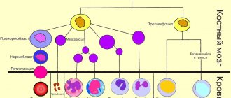
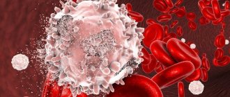
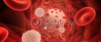
Leukocytes . Their purpose is to recognize foreign substances and microorganisms, fight them, and also remember information about them. There are several types of leukocytes: - lymphocytes: B-lymphocytes get their name from the Latin word “bursa” - bursa (named after the bursa of Fabricius - the organ in which B-lymphocytes mature in birds. In humans, this process occurs in the bone marrow). This type of lymphocyte produces antibodies (immunoglobulins). T lymphocytes control the work of B lymphocytes, that is, the production of antibodies. They got their name from the word “thymus” - the organ in which their maturation occurs. ٭The thymus is the same as the thymus gland. T-helpers are a type of T-lymphocytes that help synthesize antibodies (to help). T-suppressors are a type of T-lymphocytes that suppress the synthesis of antibodies (to suppress, suppress, restrain, prohibit). — natural killer cells (NK) are lymphocyte-like cells that lack the characteristics of T- and B-lymphocytes. They are able to destroy tumor cells and cells infected with viruses; - neutrophilic leukocytes (band-nucleus: the nucleus looks like curved rods; segmented: the nuclei are segmented, have constrictions); - eosinophilic leukocytes; - basophilic leukocytes. Neutrophils, eosinophils and basophils are types of leukocytes, but these cells do not perceive dyes in the same way, which is why they have different names. Eosinophils are involved in the destruction of parasites (they secrete special enzymes that have a damaging effect on them), as well as in allergic reactions (they secrete substances that destroy histamine, preventing the release of enzymes from mast cell granules). Neutrophils are sometimes called "microphagocytes", referring to their ability to phagocytose microorganisms. Macrophages, or phagocytes, “eat” living and dead microbes, antigen-antibody complexes (formed in the process of fighting viruses, bacteria and their toxins), dead cells of the body itself. Without these cells, the activity of lymphocytes is impossible, since they are the ones who “help” the latter recognize antigens and secrete mediators (substances that stimulate or inhibit the activity of other cells of the immune system). The average number of microbes engulfed by one phagocytic cell is called the phagocytic number, and the percentage of phagocytic macrophages is called the phagocytic index. On the surface of cells there are antigens - unique markers (labels) that distinguish some cells from others. These are called clusters of differentiation (CDs). What marks are on the surface of the cell depends on its type (T-lymphocyte, B-lymphocyte, etc.) and its maturity (ability to perform its functions). Labels are numbered in turn, according to when they were opened: the earlier the cluster was opened, the lower its number. In laboratories, clusters of differentiation are detected using monoclonal antibodies. ٭A clone is a collection of cells arising from one common cell. The clone cells are 100% identical. Identical cells synthesize identical antibodies, which are called monoclonal.
The most common types of clusters are: CD2 - cluster of T-lymphocytes, NK-cells; CD3 - T-lymphocyte cluster; CD4 - T-helper cluster; CD8 – T-suppressor cluster; CD16 – cluster of NK cells (natural killer cells); CD20 is a cluster of B cells. Immunoglobulin testing provides information about the state of the humoral immune system. This is used in the diagnosis of primary and secondary immunodeficiencies, autoimmune, infectious, hematological and other diseases. Changes in immunological parameters can be a manifestation of the body’s normal reaction to the influence of physiological or pathological factors (with different patterns of changes at different stages of the disease), reflect excessive activation, depletion of the immune system, or characterize a congenital or acquired defect of individual parts of the immune system.
There are four types of immunoglobulins: IgM - this type of antibody appears first upon contact with an antigen (microbe). An increase in their titer, or content in the blood, indicates an acute inflammatory process. IgG - antibodies of this class appear some time after contact with the antigen. They participate in the fight against microbes: they bind to antigens on the surface of the bacterial cell; then other plasma proteins (the so-called complement) join them, as a result of which the bacterial cell is lysed (its membrane ruptures). In addition, IgG is involved in some allergic reactions. IgA prevents the penetration of microorganisms through mucous membranes. IgE - antibodies of this class interact with receptors located on mast cells (connective tissue cells that secrete physiologically active substances: heparin, histamine, serotonin, etc. They are involved in the processes of inflammation, blood clotting, etc.) and basophils. As a result, histamine and other allergy mediators are released. An allergic reaction itself develops. One of the most important indicators of immune status is complement components C3, C 4. Complement is the set of immune proteins contained in fresh blood serum. They participate in the bactericidal action of blood. C3 is a central component of the complement system, an acute phase protein of inflammation. This is an essential part of the defense system against infections. It is formed in the liver, macrophages, fibroblasts, lymphoid tissue and skin. Therefore, disruption of their normal state significantly affects this component. C4 is a glycoprotein synthesized in the lungs and bone tissue. C4 supports phagocytosis, increases the permeability of the vascular wall, and is involved in the neutralization of viruses. This test is usually prescribed for suspected autoimmune disorders, repeated bacterial infections; during dynamic observation of patients with systemic autoimmune diseases; in the diagnosis of systemic lupus erythematosus, rheumatoid vasculitis and other diseases. Another indicator of immune status is cryoglobulin, an abnormal protein that can be present in the blood in a number of diseases. At low temperatures, cryoglobulins become insoluble, leading to blockage of small blood vessels located in the fingers and toes in cold weather, causing the characteristic rash. The presence of cryoglobulins (cryoglobulinemia) can be a symptom of various diseases, including macroglobulinemia, systemic lupus erythematosus, as well as a number of infectious diseases. Interferons are a special group of proteins that are produced by cells of the human immune system. They are a type of weapon with which the body can resist pathogenic bacteria, parasites and even cancer cells. There are three main classes of interferons: interferon alpha (α), interferon beta (β), interferon gamma (γ), and interferon omega (ω). All of them not only have antiviral and antitumor effects, but also have the property of activating - forcing other protective factors - macrophages and natural killer cells - to act. All types of interferon have a very strong influence on the course of RNA viral infections. In most cases, interferon production is provoked by the penetration of bacteria, viruses or their metabolic products into the body.
Immunoglobulins A, M, G, E
Total immunoglobulins are indicators of the humoral immune response. IgM levels increase during the acute period of the disease and during exacerbation of chronic infection, IgG - during the recovery process, IgA - during the acute period of the disease, persisting longer than IgM, reflecting a protracted course and damage to the mucous membranes. Elevated IgE levels indicate allergic inflammation.
General immunoglobulins do not explain the cause of infection, so it is advisable to look at specific antibodies to specific pathogens. The main indications for determining total antibodies are suspicion of congenital immunodeficiency associated with humoral immunity.
Total immunoglobulin E, immunoglobulin G, immunoglobulin M, immunoglobulin A are examined.
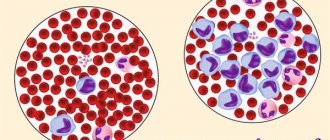
Clinical blood test with leukocyte formula
A clinical blood test with a leukocyte formula can identify many diseases. It reflects the state of hematopoiesis - the maturation and growth of blood cells in the bone marrow. The state of the immune system is indicated by the levels of white blood cells - leukocytes and the leukocyte formula. Monitoring the leukocyte formula in the dynamics of the disease will reveal complications, the spread of infection throughout the body, and the response to treatment.
With bacterial infections, you can see an increase in leukocytes and neutrophils with the appearance of their young forms in the blood.
With viral infections, on the contrary, leukocytes decrease, and lymphocytes predominate in the leukocyte formula.
The predominance of certain populations in the leukocyte formula helps in diagnosing the cause of inflammation. For example, in certain infections (for example, infectious mononucleosis), altered monocytes increase, and in allergic and parasitic diseases, eosinophils increase.
The presence of very young cells (blasts) in the blood is a sign of excessive activation of the bone marrow, and a low level of leukocytes and neutrophils without inflammation is a sign of its suppression.
Lymphocytes, immunophenotyping
There are different populations of lymphocytes that play a key role in directing the immune response - cellular or humoral. Determination of the immune phenotype of a lymphocyte is possible in a special study using flow cytometry. The phenotype of lymphocytes depends on special receptors on their surface - CD differentiation clusters. The presence of one or another receptor indicates that the cell belongs to a certain population of lymphocytes.
Cellular immunity is determined:
- B lymphocytes (CD19+)
- T-lymphocytes (CD3+) common and their subtypes:
- T helper cells (CD3+CD4+)
- T-cytotoxic (CD3+CD8+)
- T-killer cells (CD3+CD16+CD56+)
- Natural killer cells (CD3-CD16+CD56+)
- Lymphocytes with HLA-DR+ marker
- Immunoregulatory index (T-helpers/T-cytotoxic)
Additionally, other lymphocyte CD markers are examined to determine their activity.
Evaluation of the result is possible only in conjunction with data from the clinical picture and immunomodulatory therapy. Immunophenotyping of lymphocytes is usually carried out over time. Thus, in HIV infection that affects T-helper cells, a decrease in the level of CD3+CD4+ cells and the immunoregulatory index (CD3+CD4+/CD3+CD8+) has a poor prognosis for the course of the disease.
Circulating immune complexes (CIC)
CECs are circulating immune complexes, the level of which increases during acute infections and autoimmune diseases. Circulating immune complexes (CICs) are present in many people with systemic lupus erythematosus (SLE) and rheumatoid arthritis (RA), especially those with complications such as vasculitis. There is a positive correlation (* systematic and conditional relationship) between disease activity and the level of CEC in the blood. CEC formation is a physiological defense mechanism resulting in the rapid elimination of either endogenous or exogenous antigens (eg, microorganisms, viruses, parasites, plant antigens, fungal antigens, pollen or food antigens) through the reticuloendothelial system. High levels of CEC in serum and/or other biological fluids are observed in many inflammatory and malignant diseases, which can cause the development of pathology. Determination of serum CEC is an important marker for assessing disease activity, especially in autoimmune diseases.