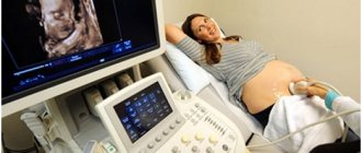
I wonder how a baby feels in the womb? If you take into account only the subjective and emotional feelings of the mother, it is quite difficult to find out the truth. Fortunately, there are special types of examinations that allow you to monitor the development of the fetus, one of them is cardiotocography .
In our clinic you have the opportunity to undergo a full diagnosis and consult with the country's leading specialists, including candidates and doctors of medical sciences, professors. Our expert ultrasound equipment Voluson 10 (only a few clinics can boast of this) is indispensable for diagnosing fetal development, identifying congenital anomalies and malformations (such as heart defects, cleft lip, etc.), early detection of tumors and other serious pathologies.
Modern diagnostics of fetal development includes the following methods:
- Ultrasound examination (ultrasound of the fetus) - helps to see the structure of the child’s organs and body, estimate its approximate weight;
- Doppler ultrasound method - shows the state of fetal blood flow;
- Cardiotocography – allows you to evaluate and record the child’s heartbeat.
1 ultrasound scanner Voluson 10
2 Cardiotocography (CTG)
3 Cardiotocography (CTG)
What is cardiotocography?
This safe and highly effective diagnostic method allows you to assess the condition and development of the fetus by the nature of its heartbeat.
The study takes into account heart rate depending on uterine contractions, fetal activity and exposure to external stimuli. CTG is a mandatory component of comprehensive prenatal screening, along with ultrasound and Doppler sonography of the fetus. The results of the study can seriously affect the management of pregnancy and childbirth itself.
CTG, in addition to heart rate (HR), also records uterine contractions (this is done using a special strain gauge).
Using cardiotocography, you can see signs of oxygen deficiency (hypoxia) of the fetus. Oxygen deficiency is very dangerous because it can lead to delayed fetal development and increases the risk of various disorders during childbirth and the postpartum period.
The study is absolutely safe for a pregnant woman and her child.
What to do if there are deviations from the norm
When using a device to listen to the fetal heartbeat at home, it is necessary to correctly interpret the results obtained in order to react in time and take action if problems arise.
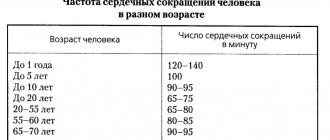
The characteristics of the fetal heart rate include frequency, rhythm, filling and tension. Tachycardia is considered to be an increase in heart rate above 170 beats per minute, bradycardia is a decrease in heart rate below 110. Normally, the baby’s heart sounds are clear and rhythmic.
Changes in these indicators arise due to many reasons: from the banally stuffy room in which a pregnant woman is located, to intrauterine fetal hypoxia. Therefore, if deviations in the child’s pulse occur, be sure to seek help from a doctor for a scheduled consultation. Only he will find out the cause of this condition and help keep the baby healthy. For this, the expectant mother undergoes additional studies (Holter and ECHO-CG (24-hour ECG monitoring)).
Operating principles of cardiotocography
Features of the study:
CTG is best performed at around 32 weeks. In the earlier stages of pregnancy, it is quite difficult to examine the baby’s heartbeat.
For up to 30 weeks, cardiotocography is used if the doctor cannot find the point at which the fetal heart can be heard. In these cases, the device can be more sensitive and accurate than the human ear.
The recording time is from 30 to 90 minutes, which is associated with periods of sleep and wakefulness of the fetus. CTG is done during the active phase. Data is provided in the form of graphs, which are recorded on paper tape.
Before the examination, the woman needs to rest, get enough sleep, and eat lightly.
Immediately before the procedure, a gel is applied to the working sensor, which enhances the conductivity of impulses.
During labor, CHT is used to examine uterine contractions and the fetal heart rate.
Important! An experienced specialist should analyze the results; only he will be able to distinguish normal from pathology.
At the MedicCity clinic, cardiotocography is performed using modern high-precision ultrasound equipment, which allows the gynecologist to get a clear picture of the condition of the fetus and apply the correct tactics for managing pregnancy and childbirth. In some cases, based on cardiotocography data, the doctor may recommend hospitalization to the woman.
1 Cardiotocography in MedicCity
2 Cardiotocography in MedicCity
3 Cardiotocography in MedicCity
Listening to your baby's heartbeat
You can listen to the rhythm and frequency of the embryo’s heartbeat using medical equipment as early as the 3rd week.
- At 6-8 weeks - heart rate varies between 110-130 beats/min.
- From 8 to 11 weeks – indicators reach up to 190 beats/min.
- From 11-12 weeks they remain stably at 140-160 beats/min.
When assessing the baby's heart rate, both the timing of pregnancy and the general condition of the mother are taken into account. After all, all this in its entirety will determine the frequency, intensity of the heartbeat, and the development of the child’s cardiovascular system.
How is CHT performed?
To obtain accurate data during cardiotocography, a woman must be in the correct position: half-sitting or lying on her left side. If the expectant mother lies on her right side, the uterus may press on the inferior vena cava, which will lead to complications.
Before starting the examination, the doctor listens to the pregnant woman’s belly with a stethoscope and finds the point where the baby’s heartbeat can best be heard. It depends on how the baby turned in the womb. It is best to perform CTG within 2-3 hours after the patient has eaten. To avoid obtaining erroneous data, cardiotocography should not be done earlier than an hour after intravenous administration of glucose to the expectant mother.
Control at home
Situations arise when the fetal heartbeat is monitored every day at home. After all, the mother is worried about the baby’s condition; it is important for her to know that his heart is beating and developing.
Listen to the fetal heartbeat at home in several ways. The simplest one is to put your ear to your stomach. With this diagnostic method, the baby’s heartbeat can be heard only after 30 weeks of pregnancy. This method is ineffective because it depends on the woman’s fatty tissue, the thickness of which distorts the conduction of the sound of the beating heart.
A more reliable method is to calculate the fetal heart rate using an obstetric stethoscope, which is applied to the pregnant abdomen with the widest part. However, this requires daily training and a patient assistant. In addition to knowledge of auscultation points, it is necessary to distinguish between extraneous noises (pulse of the expectant mother, intestinal peristalsis). By correctly applying the diaphragm of the device to the woman’s anterior abdominal wall, using a phonendoscope, the typical sound of a fetal heartbeat is heard.
Today, the expectant mother evaluates the baby’s heart function without leaving home, with minimal error. For this purpose, they purchase a device for listening to the fetal heartbeat at home - a fetal doppler. The work is based on the use of ultrasound, the level of which is eight times lower than when performing ultrasound. Therefore, its use is considered safe for children. Such a device for listening to the heartbeat is sold in a pharmacy or medical equipment store.
Advantages of such a device:
- convenience and ease of use;
- the ability to quickly independently assess the fetal heart rate;
- use from 12 weeks.
The disadvantages include:
- limited application time;
- high price.
In what cases is CTG performed?
Cardiotocography is performed to identify pathologies of fetal development at an early stage of pregnancy. The study is carried out in the following cases:
- with unfavorable pregnancies in the past (miscarriages, frozen pregnancies, various chromosomal pathologies in the fetus);
- with gestosis in a pregnant woman (this disease can lead to impaired blood supply to the fetus);
- in case of Rh conflict;
- with high or low water;
- if the expectant mother has chronic diseases (overweight, hypertension, diabetes, heart disease, kidney disease, liver disease);
- changes in the rhythms of fetal behavior (if the child was very active during the day and suddenly calmed down, then perhaps the fetus is experiencing some kind of discomfort);
- in post-term pregnancy;
- if a woman does not stop smoking or drinking alcohol during pregnancy;
- if a pregnant woman falls ill with ARVI, influenza or other acute diseases.
1 ultrasound scanner Voluson 10
2 Cardiotocography (CTG)
3 Cardiotocography (CTG)
Listening to a baby's heartbeat in a medical facility
- Ultrasound is the first of many methods for listening to the frequency and intensity of fetal SB. Allows you to assess not only SB, but also the general condition of the fetus and placenta.
- Auscultation is listening to the heartbeat using a stethoscope. Allows you to listen to the baby’s heartbeat from 28-29 weeks. It is important to remember that in some cases this diagnostic method is not possible. For example, with a large weight of the pregnant woman herself, a small/large amount of amniotic fluid, and so on.
- Cardiotocography, or CTG for short, is an informative, modern method. The method works on the principle of ultrasound equipment, when with the help of special sensors it is possible to accurately determine the frequency and intensity of the fetal heartbeat, uterine contractions and other necessary physiological indicators.
- Echocardiography is used at 18-28 weeks of pregnancy - it is used to diagnose the general condition of the blood flow and the structure of the child’s cardiovascular system. It is prescribed if a woman has previously given birth to children with heart defects, or if she herself has a history of heart muscle defects. It is also indicated in cases where the mother has suffered an infectious disease during pregnancy, and if the woman in labor was 38 years or older at the time of pregnancy and has been diagnosed with diabetes.
All these methods for diagnosing and listening to fetal SB are used in medical institutions. But parents can hear their child’s heartbeat at home, without visiting a medical facility.
What problems are diagnosed using cardiotocography?
Cardiotocography is an additional examination method. Therefore, it is impossible to make an accurate diagnosis based only on its results. However, with the help of CTG it is possible to detect the development of various fetal pathologies, including the following disorders:
- heart rhythm disturbance;
- hypoxia;
- pressing or entwining the umbilical cord.
If CTG reveals abnormalities in the condition of the fetus, additional ultrasound and Doppler sonography are performed.
Cheap methods for determining the sex of a child: what is the reliability?
The content of the article
Not so long ago, the mystery of the sex of the unborn child remained a mystery until the moment of its birth. Our grandmothers used folk methods in solving this issue, from studying the shape of a pregnant woman’s belly to analyzing her taste preferences. Japanese and Chinese conception tables and signs relating to the appearance and behavior of a pregnant woman were used.
But already in those days, gynecologists preferred to trust the laws of anatomy, and knew how to determine the sex of a baby by its heartbeat. This technique is still used today, although it has been established that its reliability does not exceed 70%. Is this not enough? Any other folk methods of determining sex give a probability of 50%, i.e. absolutely useless - guessed right - wrong.
When does the fetal heart begin to beat?
Any expectant mother is interested in this question. We answer: already in the 1st trimester, the heartbeat of the unborn child can be traced and is a significant indicator for determining the baby’s health status.
The fetal heart is formed in the 4th week of pregnancy. At first it looks like a hollow tube. At week 5, the organ begins to function. By about the 9th week, it becomes similar to the heart of an adult, that is, it has the same 2 atria and 2 ventricles (four-chambered heart). But there is also a difference. The baby’s heart works not only as the “motor” of the body. The child in the womb does not breathe air, like you and me, but receives oxygen from the mother, through the blood. To ensure this function, there is an oval window between the right and left atria of the fetus, and the aorta and pulmonary artery are connected by a special vascular trunk (botallian duct). When the baby is born, the window and duct close, and the heart begins to function as in an adult.
Rating of fetal dopplers: which device is better?
If you are in doubt which device is better to choose, we suggest that you familiarize yourself with the rating of fetal dopplers. It is compiled taking into account the functional features of the devices presented in our store.
| Doppler model | Display | US, MHz | Probe | Nutrition |
| Pelvifine MedLar KM-U2 | Eat | 2,5 | Remote | AA, 2pcs |
| Pelvifine MedLar KM-U3 | Eat | 2,5 | Remote | Battery 800ma |
| Sonoline Baby Sound C | Eat | 2,0/3,0 | Remote | AA, 2pcs |
| Sonoline Baby Sound C1 | Eat | 2,0/3,0 | Remote | AA, 2pcs |
| Joylife TK-T806 | Eat | 3,0 | Remote | AA, 2pcs |
| OLIECO AD51A | Eat | 2,5 | Remote | AA, 2pcs |
| Contec Sonoline B | Eat | 3,0 | Remote | AA, 2pcs |
| ELERA tk-t802 | Eat | 2,5 | Remote | AA, 2pcs |
| Cofoe JPD-100A | Eat | 3,0 | Remote | AA, 2pcs |
| Cofoe FD-270G | Eat | 2,0 | Remote | AA, 2pcs |
| Cofoe FD-270B | Eat | 2,0 | Remote | AA, 2pcs |
| Baby Sound B | Eat | 2,0 | Built-in | AA, 2pcs |
| Baby Sound A | No | 2,0 | Built-in | AA, 2pcs |
| JPD-100S (mini) | No | 3,0 | Built-in | "Crown" |
| U-kiss JPD-100S | No | 3,0 | Built-in | "Crown" |
The top lines of the rating are headed by the flagship model Sonoline Baby Sound C1 from the famous manufacturer of medical equipment Contec and its improved version - Sonoline Baby Sound C. If your budget allows, then it is better to choose one of these models. Both devices received such a high rating due to their advanced functionality. The devices are equipped with a highly sensitive remote sensor, a backlit color display, an external speaker and a headphone jack. The kit also includes 30 ml media gel.
Next we should consider the Joylife TK-T806 model. The model is easy to use and clear sound (thanks to the built-in noise reduction program). The external probe makes the process of monitoring the fetal heartbeat comfortable, since the indicators are clearly visible on the screen.
Next on our rating list is the Pelvifine KM-U2, equipped with an external probe, a large backlit screen and a built-in speaker. This model is best to choose if you need a functional device at a low price.
The OLIECO AD51A model occupies the next position in the rating. The device has an external tracking sensor, a large TFT display, and an external speaker with volume control. Dual measurement mode and intelligent dynamic fetal detection are the distinctive features that make it better to choose this device.
Next in the ranking is the popular Contec Sonoline B model. Thanks to the power of the 3 MHz ultrasonic sensor, the device helps detect the fetal heart rate starting from the 8th week of pregnancy. This fetal doppler is equipped with a backlit LCD display, a built-in speaker, and a remote-type probe. The device is worth choosing because of its high level of security.
Portable devices ELERA tk-t802 and HOGNSIGN FD-210 occupy the following positions in our rating. Both models are similar in functionality: they have a waterproof remote probe with a sensor power of 2.5 MHz, an LCD screen, and an external speaker with the ability to listen to the child’s heart rate in real time.
Next on the rating list are the budget models Cofoe JPD-100A Cofoe FD-270B and Cofoe FD-270G. These fetal dopplers have an affordable price with fairly high technical characteristics. The devices are equipped with a large display, a remote tracking sensor and an external speaker with good sound quality. Separately in the rating it is worth noting the function of intelligent noise reduction and dynamic fetal detection.
Model Baby Sound B occupies the next position in the rating. The device is equipped with a built-in ultrasound sensor and a small LCD monitor to record fetal heart rate. The weight of the device is only 80 g, so it is better to use it on any trips.
The next place in the ranking goes to the Baby Sound A device, the simplest and most compact fetal doppler model from Contec. The device does not have a display or built-in speaker; the fetal heartbeat can only be heard through headphones. The high power of the ultrasound sensor allows diagnosis in the early stages of pregnancy (from 8-10 weeks). If you need a reliable fetal doppler at an affordable price, then it is better to choose this particular model from the rating.
Our rating is completed by budget devices AngelSounds JPD-100S (mini) and U-kiss JPD 100S. The devices are equipped with a built-in ultrasonic sensor. A headphone jack is provided to listen to the fetal heartbeat. It is worth choosing one of these models if you need a compact device that is convenient to carry with you.
Methods for assessing the “adequacy” of fetal movements
Counting the number of movements
The easiest way to assess fetal movements is to count the number of movements by the pregnant woman herself. Self-assessment methods are very easy to use, do not require additional equipment or the presence of a doctor, and are easily reproduced by any woman. Their disadvantages are that each woman has different thresholds of susceptibility.
"Count to Ten"
The most common method for assessing fetal movements is called “count to ten” . It can be performed after 28 weeks of pregnancy, when the fetus is mature enough for active movements. Its essence lies in the fact that the expectant mother counts the movements of the fetus over a 12-hour period of time, for example from 9 am to 9 pm. The time when the pregnant woman detects the tenth movement is recorded on a tablet. If the fetus makes less than 10 movements in 12 hours, this is a reason to consult a doctor for further examination.
Sadowski method
In the evening after dinner (approximately from 19 to 23 hours), the woman lies on her left side and counts the movements of the fetus. At the same time, everything is counted, even the smallest movements. If 10 or more movements are noted within an hour, this indicates that the baby is moving quite actively and feels well. If the fetus moved less than 10 times in an hour, then the movements are counted over the next hour. The evening time for this assessment method was not chosen by chance. It is in the evening hours, especially after dinner and the associated increase in glucose, that the greatest activity of the fetus is observed. If the number of fetal movements during this test is less than 10 in two hours, this should be considered as a sign of a violation of its condition and additional research should be carried out.
For an obstetrician-gynecologist, fetal movements are also an important diagnostic criterion for certain deviations in the course of pregnancy from the norm. Too active, violent, painful movement of the fetus or weak, infrequent movements may indicate its unfavorable condition.
Where to determine the sex of a child during pregnancy in St. Petersburg
We invite all pregnant women to screening ultrasounds of all trimesters at the Diana Clinic in St. Petersburg. Here you can also undergo other specific examinations during pregnancy, for example, ultrasound of the symphysis, Doppler ultrasound, fetometry, 4D ultrasound, take tests, treat infections, etc. The examination is carried out using a new expert apparatus. The cost of an ultrasound depends on the stage of pregnancy, starting from 1300 rubles.
If you find an error, please select a piece of text and press Ctrl+Enter
Why do you need to monitor the fetal heartbeat?
Failures in the functioning of the heart can be fraught with serious complications in the future. Even at the stage of embryogenesis, heart rate deviations are associated with problems such as hypoxia, heart disease, and pathologies of the development of fetal membranes and placenta. A slow or excessively fast heartbeat may indicate one of these problems.
Normal heart rate indicators by week are presented in the table.
| A week | Heart rate (beats per minute) |
| 5 | 90-110 |
| 6-7 | 100-130 |
| 8-9 | 130-150 |
| 10-11 | 130-160 |
| 12-13 | 140-170 |
| 14-15 | 140-180 |
| 16-17 | 140-170 |
| 18-19 | 130-170 |
| 20-21 | 140-170 |
| 22-23 | 130-160 |
| 24-40 | 120-160 |
Regular monitoring of fetal heart rate is indicated for various abnormalities during pregnancy. If the expectant mother suffers from anemia or chronic diseases, then the baby’s heartbeat must be constantly monitored. In addition, indications for heart rate control are increased uterine tone, the threat of miscarriage, and the appearance of bleeding.
At what time does fetal movement begin?
The future baby begins to make its first movements early - already at 7-8 weeks of pregnancy . It is at this time that the first muscles and rudiments of the nervous system of the fetus are formed. Naturally, at this time the movements of the embryo are still very primitive - these are muscle contractions in response to nerve impulses.
about 10 weeks of pregnancy, the fetus begins to move more actively in the uterus, and, encountering an obstacle on its way (the walls of the uterus), change the trajectory of movements. However, the baby is still very small and the impacts on the wall of the uterus are very weak; the expectant mother cannot yet feel them. At 11-12 weeks of intrauterine life, the little man already knows how to clench his fists, grimace, wince; by 16 weeks of pregnancy he begins to react to loud, sharp sounds with increased motor activity, at 17 weeks the first facial expressions appear, and at 18 weeks he covers his face with his hands and plays. with the umbilical cord, squeezes and unclenches the fingers.
Gradually, with increasing gestational age, movements become more coordinated and more like conscious ones. As the baby grows up, the pregnant woman begins to feel his movements.
When does fetal movement begin during the first and subsequent pregnancies?
It is generally accepted that during the first pregnancy, the expectant mother feels the first movements of the fetus at 20 weeks of pregnancy, and for repeated pregnancies - at 18 weeks. This is not entirely true. A mother who is expecting her first child, indeed, most often begins to feel fetal movements a little later than a multiparous woman. This is due to the fact that “experienced” mothers know how the baby’s movements feel at first and what they should feel. Some primigravidas perceive the first movements of the fetus as increased intestinal motility, “gas”. Many women describe the first movements of the fetus as a feeling of fluid transfusion in the stomach, “butterflies fluttering” or “fish swimming”.
The first movements are usually rare and irregular. The time of the first sensations of fetal movements naturally depends on the individual sensitivity of the woman. Some expectant mothers feel the first movements already at 15-16 weeks, and some only after 20. Slender women, as a rule, begin to feel movements earlier than plump ones. Women who lead an active lifestyle and work a lot usually feel fetal movements later.
By 20 weeks, due to the formation of the spinal cord and brain, as well as the accumulation of a certain amount of muscle mass in the fetus, movements become more regular and noticeable.
From 24 weeks of pregnancy, the movements of the fetus already resemble the movements of a newborn - the expectant mother feels how the fetus changes position, moves its arms and legs. The motor activity of the fetus increases gradually and its peak occurs in the period from the 24th to the 32nd week of pregnancy. At this time, the activity of the baby’s movements becomes one of the indicators of his normal development. After 24 weeks, the baby begins to “communicate” with his mother through movements, respond to the sounds of voices, music, and the mother’s emotional state. As the gestation period increases beyond 32 weeks, the motor activity of the fetus gradually decreases due to the fact that the baby grows up and simply does not have enough space for active movements. This becomes especially noticeable at the time of childbirth. By the end of the third trimester of pregnancy, the number of fetal movements may decrease slightly, but their intensity and strength remain the same or increase.