Treatment under the compulsory medical insurance policy is possible!
Submit your application
Follow the news, subscribe to our social networks
Details
Paget-Schroetter syndrome is a thrombosis of the subclavian vein, extending to the subclavian and axillary veins, as well as to the veins of the shoulder, which leads to impaired venous outflow in the arm. The syndrome is also called traumatic thrombosis or effort syndrome. Despite its relative rarity, it has become increasingly diagnosed in the last decade. The main complaints are swelling of the tissues of the upper limb and bursting pain.
The first case of the syndrome was described by James Paget in 1875, and in 1894 von Schrötter identified vascular injury as a potential cause of the disease. The term Paget-Schroetter syndrome was first used in 1948.
What is PV and where is it located?
The subclavian vein is located in front of the scalene muscle and receives the veins of the arm and neck. It serves as a continuation of the axillary vein, which receives blood from the arm belt and free upper limb.
The anatomy of the vessel is as follows: it has valves that are located from the lateral edge of the 1st rib to the sternoclavicular joint. The subclavian vein forms a venous angle due to its connection with the internal jugular vein.
The brachiocephalic veins are formed due to the confluence of the subclavian and internal jugular veins. The subclavian vein is separated from the artery of the same name by the anterior scalene muscle.
Clinical anatomy
The subclavian vein collects blood from the upper limb. At the level of the lower edge of the first rib, the axillary vein continues. At this point, it curves around the first rib from above, and then passes along the anterior edge of the scalene muscle behind the clavicle. Placed in the prescalene space. This space is a frontal triangular fissure, which is formed by the vein groove. It is surrounded by the scalene muscle, sternothyroid, sternohyoid muscle and cleidomastoid muscle tissue. The subclavian vein is located at the very bottom of this gap.
It passes through two points, with the lower one located at a distance of 2.5 centimeters inward from the coracoid process of the scapula, and the upper one goes three centimeters below the sternal edge of the end of the clavicle. In children under five years of age and newborns, it passes in the middle of the collarbone. The projection shifts to the middle third of the clavicle with age.
The vein is located slightly obliquely relative to the center line of the body. When moving the arms or neck, the topography of the subclavian vein does not change. This is due to the fact that its walls are very closely connected with the first rib, subclavian muscles, clavipectoral fascia and clavicular periosteum.
Subclavian vein tributary
The venous wall is fused with the fascia of the neck, the periosteum of the 1st rib and the tendon. It is for this reason that the lumen of the vein does not collapse. This factor plays an important role, because when a vein is injured, there is a high probability of developing an air embolism.
The tributaries of the subclavian vein are:
- the dorsal scapular vein corresponds to the basin of the artery of the same name;
- sternal veins, which carry blood from the pectoral muscles.
On the right side of the neck, the Pirogov venous angle is located in front of the subclavian vein, into which the jugular vein flows.
Functions
The main function of the vein is considered to be the outflow of blood saturated with carbon dioxide and metabolic products.
In addition, thanks to it, hormones from the endocrine glands and nutrients that are absorbed in the gastrointestinal tract penetrate into the bloodstream.
The subclavian vein occupies an important place in the regulation of general and local blood circulation, as well as in the spread of various inflammatory processes occurring in the human body.
What diseases are associated with PV?
The circulatory system is considered a complex structure, which plays the role of transferring blood from other channels.
One of the common diseases of the subclavian vein is thrombosis, in which the blood flow along the entire upper limb changes significantly. The reason for the development of this disease lies in blood clotting disorders and excessive physical stress on the body.
Mostly the pathology is diagnosed in young patients and predominantly in men.
With timely diagnosis and effective therapy, the prognosis is quite favorable.
With high physical stress on the body, one arm becomes overstrained, which leads to compression of the veins. The result of this is thrombosis, and this is facilitated by increased blood clotting and too slow blood flow.
A common developmental anomaly of the subclavian arteries is the aberrant right subclavian artery. As a result of compression of the aberrant subclavian artery of the underlying structures, symptoms may appear that directly depend on the patient’s age and anatomical features of development.
Tests and diagnostic methods
To make an accurate diagnosis of vein damage, a specialist carefully studies the patient’s medical history. The doctor clarifies with the patient all the signs that bother the patient. The collected information allows us to clarify the duration of thrombosis.
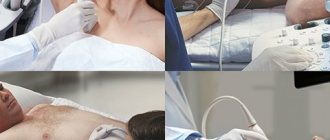
In order to identify venous pathology in acute or chronic form, the following diagnostic methods are usually used:
- MRI and radiography help not only to identify the cause of the disease, but also the location of the thrombus;
- Ultrasound examination of deep veins;
- Dopplerography, which evaluates blood circulation in the damaged vein;
- X-ray using a contrast agent;
- duplex scanning of a vein;
- venography;
- shoulder girdle.
If puncture is necessary, subclavian access is preferably used.
Symptoms of PV pathologies
The main cause of subclavian vein thrombosis is considered to be high physical stress on the body. It is this factor that plays the leading role in the formation of a blood clot. Sometimes it is possible for a blood clot to break off, regardless of the degree of physical stress on the body, but this is extremely rare.
With thrombus formation in the subclavian vein, both increasing and disappearing symptoms may be observed, manifested by certain influxes.
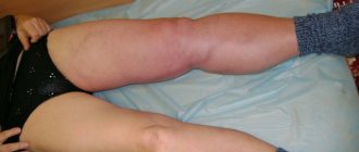
As a result of injury to the subclavian vein, certain symptoms are noted:
- pronounced pain in the arm area;
- too bright pattern of veins, visible through the epidermis;
- severe swelling of the limb and the appearance of a glossy sheen on the skin.
When the subclavian vein is injured, the patient exhibits signs of a neurological disorder. They manifest themselves in the form of numbness of the limbs and their twitching.
Often patients do not notice the appearance of a venous pattern on the arm. The diameter of the veins is determined by the size and increase in hypertension of the thrombus.
Initially, pain occurs during physical exertion on the body. Over time, discomfort can be constant and a feeling of fullness appears.
The pain syndrome is felt throughout the entire surface of the arm, in the shoulder area and collarbone. Sometimes it can radiate to the upper back and chest.
With thrombosis, the entire arm is affected by edema. When pressing on the swollen area, the hole in this area is not preserved. The hand becomes too hard and the unusual heaviness bothers you. With a prolonged inflammatory process, problems with blood circulation are noted, and the manifestations of thrombosis noticeably increase.
The patient may experience twitching of the fingers, a tingling sensation and a slight burning sensation.
During the transition of a patient's thrombosis from an acute to a chronic form, the clinical picture is blurred, and it becomes less pronounced. With this pathology, the patient’s motor activity is limited, muscle atrophy develops, and pain occurs with increased physical activity.
Sometimes, with a disease such as subclavian vein thrombosis, the patient is given a disability.
What doctors treat
A phlebologist treats vein pathologies, including the subclavian vein. It is he who can explain to the patient in detail what the subclavian vein is, its importance for the functioning of the whole body and possible pathologies.
If the blood vessels are not significantly clogged, the problem can be dealt with using local treatment. It is necessary that the hand is at rest.
When in a horizontal position, the upper limb should be positioned slightly above the heart area. While the patient is in an upright position, you need to suspend your arm using a scarf or bandage, bending it slightly at the elbow.
Methods of puncture
Puncture of the subclavian vein can be performed in two ways: supraclavicular access and subclavian. In this case, the puncture can be made from any side. This vein is characterized by good blood flow, which, in turn, reduces the risk of thrombosis. There is more than one access point for catheterization. Experts give the greatest preference to the so-called Abaniak point. It is located on the border of the inner and middle third of the clavicle. The success rate of catheterization at this point reaches 99%.
Therapy methods
Local therapy for damage to the subclavian vein can be carried out using certain groups of medications:
- gel-like products that contain rutoside and troxevasin;
- ointments with heparin;
- alcohol-based compresses;
- non-steroidal anti-inflammatory medications.
In case of acute thrombosis of the subclavian vein and the appearance of painful symptoms, the patient is admitted to the hospital. The course of therapy includes the use of:
- angioprotectors;
- anticoagulants;
- antiplatelet agents;
- fibrinolytic agents.
The main goal of drug therapy for subclavian vein thrombosis is to restore normal blood circulation.
Causes and risk factors
Subclavian vein thrombosis is associated with compression of the thoracic outlet. The subclavian vein originates from the first rib from the axillary vein, and at the level of the sternoclavicular junction with the jugular vein it forms the brachiocephalic vein. Compression of the vein walls in the area between the collarbone and the first rib leads to slower blood flow and thrombosis. It can be considered the venous equivalent of thoracic outlet syndrome.
One of the causes of thrombosis is muscle hypertrophy (increase in the volume or mass of skeletal muscles). As a result, the subclavian vein can become compressed between the ribs (in front of it), the muscle (behind it), and the collarbone (above it).
Another reason is that a person has a congenital small anatomical space between the collarbone and the first rib. In this case, compression of the subclavian vein is possible even without major muscle hypertrophy.
Other causes of thrombosis include:
- incorrect posture,
- bone pathology (in the subclavian region),
- collarbone fractures
- use of subclavian catheters
- incorrect sleeping position
- thoracic outlet syndrome
- Risk factors include excessive physical activity. People who undergo intense physical activity and athletes (wrestlers, weightlifters or bodybuilders) have a higher risk of developing thrombosis due to repeated damage to the subclavian vein from frequent mechanical compression of the vessels between the collarbone, first rib and joint.
The average age of patients with Paget-Schroetter syndrome is 30-40 years, and the male to female ratio is approximately 2:1. It is more common on the right side, probably due to the frequency of right hand dominance, and 60% to 80% of patients have this condition. who performed vigorous exercise involving the upper limbs.
When is surgery required?
If after 2 months the blood clot has not resolved, then surgery is prescribed.
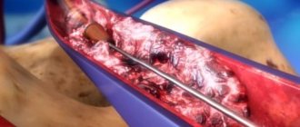
If thrombosis of the subclavian vein is too long, there is a high risk of necrosis of the arm tissue. During the operation, a specialist removes tissue that has died.
If blood circulation through the veins is impaired and there is a chronic form of thrombosis of the subclavian vein, thrombectomy or recanalization may be performed. If it is impossible to carry out this type of operation, they resort to eliminating the damaged section of the main vein, performing plastic surgery and bypass surgery.
In a situation where the pathology is completely untreatable, the arm is amputated.
Puncture and catheterization of the subclavian vein is performed by a surgeon or anesthesiologist. Sometimes these procedures can be performed by a specially trained therapist.
Indications for catheterization are inaccessibility of peripheral veins, too long operations with loss of large amounts of blood and the need for parenteral nutrition.
In case of injuries and bleeding, taking into account the topography of the subclavian vein, it is necessary to ligate it or apply a special bandage from three zones: under, above and behind the collarbone. The patient is placed on his back, a cushion is placed under his shoulders and his head is turned in the direction opposite to where the operation is being performed.
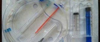
Indications for CPV
The subclavian vein (photo below) has a fairly large diameter, making its catheterization the most convenient.
The procedure for catheterization of this vein is indicated in the following cases:
- Upcoming complex surgery with possible blood loss.
- Need for intensive care.
- Insertion of a cardiac pacemaker.
- The need to measure central pressure in the veins.
- Parenteral nutrition.
- The need for probing the cardiac cavities.
- Open heart surgery.
- The need for X-ray contrast studies.
The role of PV in the diagnosis and treatment of diseases
Thrombosis of the subclavian vein is considered a pathology that almost every person can encounter. It is possible to prevent the development of such a disease by excluding factors that influence thrombus formation.
The risk of damage to the subclavian vein is quite high with an abscess, which must be eliminated as soon as possible. In all other situations, it is almost impossible to avoid the development of pathology.
The role of PV in the treatment and diagnosis of diseases of various nature is high.
This blood vessel is mainly used for infusion therapy for cancer, in intensive care units, and intensive care units.
Possible complications after the procedure
Most often, catheterization of the subclavian vein does not entail serious complications. Any change during catheterization can be determined by bright red pulsating blood. Experts believe that the main reason why complications arise is that the catheter or guidewire was incorrectly positioned in the vein.
Such an error can provoke the development of such unpleasant consequences as:
- Hydrothorax and infusion into fiber.
- Perforation of the venous wall.
- Thrombosis of the subclavian vein.
- Formation of knots and twisting of the catheter.
- Migration of the catheter through the veins.
- Heart rhythm disturbances.
In this case, adjustment of the catheter position is required. After adjusting the port, you need to contact consultants who have extensive experience. If necessary, the catheter is removed completely. In order to avoid deterioration of the patient's condition, it is necessary to immediately respond to symptoms of complications, especially thrombosis.