Damage to the vessels of the neck
Damage to the vessels of the neck (precerebral sections of the arteries of the brain).
The brachiocephalic arteries (BCA) are the main vessels of the body. BCAs include:
- brachiocephalic trunk,
- common carotid artery (divided into internal and external arteries)
- subclavian arteries,
- vertebral arteries.
The anatomy of the vertebral arteries is often individual. The internal carotid and vertebral arteries (also known as the precerebral sections of the arteries of the brain) after penetrating into the skull form the Circle of Willis. This anatomical formation is designed to ensure uniform distribution of blood throughout all parts of the brain.
The problem is that the anatomy of the Circle of Willis is also variable and more than 20% of people in the European population have an open circle. With this anatomical feature, it is enough to stop blood circulation even in one of the arteries for the development of a stroke (death of brain tissue).
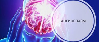
However, the overwhelming number of ischemic strokes are embologenic . This means that the cause of the disease is clogging of the arteries of the brain with material objects.
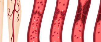
Embolism can be caused by blood clots from the cavities of the heart (if it is pathological), but most often they are fragments of disintegrating atherosclerotic plaques from the carotid arteries and the zone of division of the common carotid artery into internal and external (bifurcation zone). It is the bifurcation zone that is a typical place for the emergence and development of an atherosclerotic plaque.
Plaques, in turn, are divided into different types and classified according to the degree of narrowing of the vessel. The most dangerous are embologenic plaques - that is, those that can collapse at any moment and all their contents will instantly clog many arteries of the brain and cause a stroke.
Precursors of a stroke may be:
- headache;
- dizziness;
- numbness of half the body;
- visual impairment;
- difficulties in formulating thoughts.
- articulation disorder
- disturbances in the fine functions of the hands (for example, when writing)
- difficulty walking, etc.
Very often, a stroke occurs without any warning signs. That is why ultrasound diagnostics of the neck arteries (triplex, duplex scanning) is an absolutely necessary procedure for men and women after 40-50 years.
Angiosurgeons of the St. Petersburg Hospital of the Russian Academy of Sciences use various methods of treating atherosclerosis of the central artery, in the case of the carotid arteries, this is primarily carotid endarterectomy - a radical surgical treatment that allows removing the plaque and restoring the integrity of the vessel wall.
The operation is performed using a technique that ensures the highest possible degree of brain protection and microsurgical restoration of the artery.
Today, carotid endarterectomy gives the most consistent positive results over long-term follow-up periods - from 5 to 20~25 years. That is why in countries with developed medicine, carotid endarterectomy is the main method of treating this pathology.
For other affected areas, the St. Petersburg Hospital of the Russian Academy of Sciences performs arterial transplantations and bypass operations.
You can get expert advice and find out more details through the contact center 323 45 35
Neck veins, swelling
To assess the filling of the external jugular veins, the patient should be placed on his back, with the torso bent at an angle of 45°. Normally, the veins in this position look sunken or fill to a level of no more than 1–2 cm above the manubrium of the sternum, and the filling of the veins during inhalation is less than during exhalation.
Pathomechanism and causes
Vein swelling is a consequence of increased venous pressure. If, in a standing position, the filling of the jugular veins reaches the angle of the mandible, then the venous pressure is ≥25 cmH2O. The causes of swelling of the jugular veins are as follows:
1) bilateral - right ventricular heart failure, a large amount of fluid in the heart sac (including cardiac tamponade), constrictive pericarditis (in this case, swelling increases during inspiration - unusual [paradoxical] venous pulse [symptom] of Kussmaul [sometimes observed with severe right ventricular failure]), impaired patency of the superior vena cava (superior vena cava syndrome (320; causes - lung tumor and enlarged lymph nodes of the upper mediastinum, less often - thrombophlebitis of the superior vena cava, mediastinal fibrosis, thoracic aortic aneurysm, very large goiter), stenosis or tricuspid valve insufficiency (with insufficiency, a positive venous pulse is observed - filling increases during cardiac systole), pulmonary hypertension, pulmonary embolism, tension pneumothorax;
2) unilateral - large goiter; on the left - compression of the left brachiocephalic vein by an aortic aneurysm.
Diagnostics
1. Assess vital signs (respirations, pulse, blood pressure), as there may be an immediate threat to life (especially in the case of cardiac tamponade, tension pneumothorax, or pulmonary embolism).
2. It is necessary to collect anamnesis and conduct an objective examination. Examine the hepatojugular drainage to locate the obstruction that is causing the jugular veins to distend. Place the patient on his back. In this case, his torso should be in such a position that the jugular veins do not fill more than 1–2 cm above the level of the jugular notch of the sternum. For 30–60 s, apply pressure to the area of the right hypochondrium with your hand, and if there is increased sensitivity in this place, to another area of the abdominal cavity; Make sure that the patient is breathing freely and observe the jugular veins. Their bulging above the level of the sternocleidomastoid muscle (positive hepatojugular reflux) is characteristic of congestive heart failure (compression of the liver area increases pressure in the inferior vena cava and right atrium, which is transmitted to the superior vena cava and jugular veins). In healthy individuals or in cases where circulatory impairment is present above the right atrium, compression of the liver does not cause a significant increase in atrial pressure or transmission of increased pressure from the right atrium to the superior vena cava is impossible. Holding your breath during the study of the hepatojugular outflow creates an effect similar to the Valsalva maneuver and swelling of the jugular veins in this case has no diagnostic value.
3. Additional research methods: X-ray of the chest; if heart failure, cardiac tamponade, pericarditis or valvular defects are suspected - echocardiography; for large goiters - ultrasound of the neck plus a study of TSH and thyroid hormones; for superior vena cava syndrome (it is accompanied by swelling of the face and neck, as well as dilatation of the veins of the upper half of the chest) - CT scan of the chest, if lung cancer is suspected - bronchoscopy; if pulmonary embolism is suspected, angio-CT of the chest, possibly ultrasound of the veins of the lower extremities.
Treatment
Pre-hospital assistance
In case of traumatic injuries, the neck must be secured with a cotton-gauze collar. In other cases, it is recommended to provide functional rest to the neck and not self-medicate, since swelling occurs in many diseases that require different approaches to therapy. In case of intense pain, the use of painkillers is allowed. You should not warm your neck - this can intensify inflammatory and other pathological processes.
Conservative therapy
The conservative treatment regimen includes a protective regimen, etiopathogenetic and symptomatic drug therapy, and physical therapy. The following medications are used:
- NSAIDs
. They are used in the form of tablets, injections, and local remedies. They have analgesic and anti-inflammatory effects. - Antibiotics
. Broad-spectrum medications are prescribed, and after culture results are obtained, medications are replaced taking into account the sensitivity of the pathogen. For extensive purulent processes, combinations of two drugs are recommended. - Antithyroid drugs
. Hyperthyroidism is considered as an indication for the use of medications. For hypothyroidism, replacement therapy is carried out. - Antihistamines
. Necessary for neck swelling caused by allergic reactions.
After eliminating acute phenomena, UHF, laser therapy, electrophoresis with novocaine, and anti-inflammatory drugs are used. Patients with cancer undergo chemotherapy and radiation therapy.
Surgery
For acute pain, blockades with local anesthetics are performed. Taking into account the nature of the pathology, the following operations are performed:
- Traumatic injuries
: transoral fixation, application of a Halo-apparatus, PSO of neck wounds, tracheostomy, chordectomy, resection of the larynx, laryngopexy, removal of foreign bodies of the larynx, reconstructive interventions. - Maxillofacial pathologies
: parotidectomy, opening of abscesses and phlegmons of the neck, dilation of the excretory duct of the salivary gland. - Endocrine diseases
: enucleation of the node, subtotal resection of the thyroid gland, hemithyroidectomy. - Otolaryngological diseases
: removal of a median or lateral neck cyst, opening of a retropharyngeal abscess. - Tumors
: excision of lipomas and fibromas of the neck, removal of the thyroid gland, radioiodine therapy for thyroid cancer, laryngectomy, laryngopharyngectomy, hemilaryngectomy, installation of a voice prosthesis, Krail, Vanach operations.
Diagnostics
Therapists are involved in determining the cause of neck swelling. According to indications, patients are referred to otolaryngologists, endocrinologists, maxillofacial surgeons, and other specialists. The examination includes objective, instrumental, and laboratory techniques. The following diagnostic procedures are carried out:
- Questioning, general examination
. A basic study that allows you to establish a preliminary diagnosis and draw up a plan for further examination. Involves studying symptoms, medical history, and assessing external changes. - Sonography
. Ultrasound of the neck is informative when studying the condition of organs and soft tissues, it allows you to identify tumors, areas of inflammation, determine the localization and boundaries of pathological processes. An ultrasound of the thyroid gland is prescribed if an endocrine cause of neck swelling is suspected. Ultrasound of the lymph nodes is recommended for lymphadenitis and cancerous tumors. - Radiography
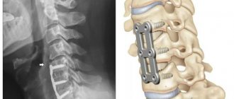
. To exclude subluxations and vertebral fractures, X-rays of C1 or the cervical spine are prescribed. If there are symptoms of damage to the ENT organs, an x-ray of the larynx is performed. - Other visualization techniques
. CT and MRI of the neck are performed to detail the information obtained during sonography or radiography and make it possible to clarify the location, volume, and nature of the pathological process. - Radioisotope scintigraphy.
Effective in studying the thyroid gland, visualizes nodes, confirms the presence of diffuse changes. - Endoscopic examination of the throat.
Direct and indirect laryngoscopy, microlaryngoscopy, fibrolaryngoscopy are prescribed for lesions of the larynx. According to indications, a biopsy is performed during the study. - Lab tests
. Depending on the existing symptoms, general and biochemical blood tests, examination of the level of thyroid hormones and antibodies to them, microbiological analysis of swabs, punctures, blood samples, cytological or histological examination of a biopsy specimen can be performed.
Neck palpation
Venous malformation (venous angioma).
It occurs relatively frequently and is not a true malformation; it is more a variant of the structure of the venous outflow.
The course is usually asymptomatic. Convulsions rarely occur.
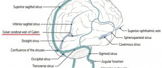
Venous malformation. Scheme. Small dilated venules in the form of an “umbrella”, “jellyfish head” are identified, draining into a large transcortical vein, which, in turn, flows into the superior sagittal sinus.
a) T1 with intravenous contrast. Arrows indicate dilated deep white matter veins draining into the dilated transcortical vein; b) Contrast-enhanced MR venography shows venous dysplasia draining into a dilated internal cerebral vein. Venous malformation.