Infectious causes of aneurysms
Bacterial pathogens cause intoxication and provoke direct tissue damage.
List of infectious diseases that contribute to the formation of aneurysm:
- Meningitis - inflammatory damage to the meninges with transition to the arterial membrane;
- Fungal infections with damage to the cerebral parenchyma;
- Bacterial endocarditis is characterized by blockage of cerebral vessels by blood clots that enter the cerebral arteries from the primary inflammatory focus, which is the heart valves and myocardium.
How is it diagnosed?
Since an arterial aneurysm can occur without significant symptoms, it is necessary to pay special attention to the first signs in women and men. A neurologist must diagnose the disease based on the examination, the diagnostic results obtained and the study of cerebrospinal fluid.
During a neurological examination, focal and meningeal symptoms are detected. Based on the location of complaints, it is possible to determine presumably where the pathological focus is located.
The neurologist obtains accurate information about the patient’s condition using the following studies:
- X-ray of the skull. A cerebral aneurysm is characterized by signs of bone tissue destruction.
- Computed tomography, magnetic resonance imaging of the brain.
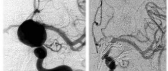
- Angiography. Basic information about the location, shape and size of the brain aneurysm can be obtained.
- Lumbar puncture. The study reveals blood in the cerebrospinal fluid, which indicates the presence of hemorrhage.
Specialists carry out differential diagnosis of cerebral aneurysm. It is important to make an accurate diagnosis in a short time. This will avoid the adverse effects of a brain aneurysm. It is necessary to differentiate an aneurysm from tumor-like processes, cysts and abscesses.
Principles of treatment
Aneurysm treatment is carried out by a neurologist. The operation is performed only by a neurosurgeon. Surgical methods are aimed primarily at preventing aneurysm rupture, which can result in the death of the patient. Conservative therapy for this pathology is used to slow the progression of the disease.
Surgical treatment of a cerebral aneurysm involves two types of main operations: clipping and endovascular occlusion. Surgery prevents the aneurysm from rupturing. A radical method of treatment is the removal of arteriovenous malformation during a microsurgical operation.
Aneurysm clipping
Aneurysm clipping is an open operation during which the neurosurgeon blocks blood flow in a certain area of the vessel and applies a clip. At the stage of preparation for surgery, specialists prescribe a procedure that allows them to stabilize the patient’s condition and minimize the risk of possible complications. Preventive examination makes it possible to identify possible disorders and diseases that can lead to adverse consequences of surgical intervention.
The operation is performed using modern microsurgical techniques through a small trepanation hole. During surgery, the neurosurgeon prevents rupture of the wall of the malformation.
After isolating the neck of the aneurysm, the specialist places a clip on it. Additional control with Doppler ultrasound allows you to assess the state of blood flow and ensure the effectiveness of the operation.
Treatment of an aneurysm using the clipping method is considered quite complex and should only be performed by an experienced neurosurgeon. A qualified specialist will do everything possible to ensure accurate and technically correct application of the clip. Otherwise, dangerous complications and a long rehabilitation period cannot be avoided.
The clip is applied to the neck of the aneurysm vessel. The accuracy of the neurosurgeon’s actions is confirmed by conducting high-quality diagnostics (Dopplerography) during the procedure.
Endovascular occlusion
During endovascular occlusion of an aneurysm, the neurosurgeon blocks the lumen of the dilated vessel with a special implant. The operation is used when clipping is not possible, for example, when the aneurysm is spindle-shaped. During surgery, a specialist inserts a catheter balloon through the femoral artery using angiographic control. It closes the lumen of the aneurysm. You can also use a microspiral to perform thrombosis. The choice remains with the attending physician. The microspiral in the cavity of the affected vascular area forms blood clots, which clog the lumen of the vessel and shut off the aneurysms from the blood circulation.
Aneurysmal dilatation in arterial hypertension
Persistent elevation of blood pressure is a chronic condition. The etiology of the nosology has not been established, but there are provoking factors that need to be eliminated during treatment.
Essential hypertension or essential hypertension is established when there is a persistent increase in blood pressure over 140 mmHg. mercury column.
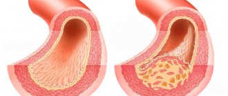
An increase in intravascular pressure against the background of nosology leads to the destruction of the vascular wall, promotes the formation of blood clots, and expansion of the middle and outer lining of the artery.
Other provoking factors are inflammatory vascular processes, connective tissue diseases, hereditary predisposition, atherosclerosis.
Symptoms of cerebral aneurysms
Most forms occur hidden. Mild symptoms appear only in the presence of mild intracerebral hemorrhage (hemorrhagic permeation).
Focal neurological disorders form large intracerebral formations:
- Muscle cramps are formed by large expansions with compression of the hemispheres;
- Vision pathology is associated with damage to the optic nerve;
- Transient ischemic attack (TIA) is caused by a significant decrease in blood flow through the large cerebral arteries;
- Headaches are provoked by compression of the arachnoid and soft membranes, irritation of pain receptors by inflammation, swelling, and neoplasm;
- Pain in the facial area occurs when the cranial nerves that innervate the masticatory and facial muscles are damaged;
- Doubling of objects before the eyes is provoked by compression, damage to the optic chiasm inside the brain;
- Speech disorders.
Weak symptoms of the pathology develop when the wall of the carotid or vertebral artery expands.
Classification of the disease
Experts distinguish between two types of disease: arterial and arteriovenous aneurysm, of which arterial aneurysm is the most dangerous. Aneurysms are classified according to these parameters:
1. By configuration:
- saccular;
- spindle-shaped.
2. By location:
- anterior cerebral artery;
- internal carotid artery;
- vertebrobasilar system;
- middle cerebral artery;
- multiple localizations.
3. By size of education:
- gigantic size;
- large;
- medium size;
- small;
- miliary.