Aortic aneurysm is a pathological expansion of the largest vessel in the human body, which is accompanied by thinning of its wall with the likelihood of rupture and fatal bleeding. Thrombotic masses form in the aneurysmal sac, which can be washed away by the blood flow and cause blockage of the arteries of the lower extremities or internal organs, which can lead to the development of acute arterial insufficiency and gangrene. Complications of aortic aneurysm account for 6% of all deaths in developed countries. However, there is an effective and safe treatment that reduces the risk of such complications tenfold.
What is an aneurysm
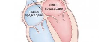
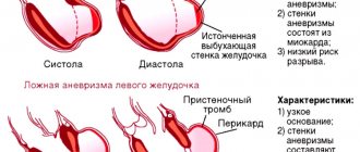
A thoracic aortic aneurysm is an abnormal bulge on the walls of the artery, formed due to intense blood pressure on a weak area of the vessel. An aneurysm can be:
- fusiform - has a convex shape, protrudes from both sides of the aorta;
- saccular - a rounded formation that protrudes from one side of the vessel.
A small aneurysm does not pose a health threat. However, if it is present, you must be observed by a doctor so as not to miss the moment of disease progression.
Diagnostic methods
Thoracic aortic aneurysms are often detected during routine medical examinations: chest x-rays, or ultrasounds of the heart or abdominal organs, which are prescribed for other reasons. If your doctor suspects that you have an aortic aneurysm, this can be confirmed by special tests at the heart center. These include:
- X-ray of the chest organs. Your doctor may first notice that you have a thoracic aortic aneurysm when examining chest X-rays. Your doctor may order a chest x-ray as the first test to detect damage to the upper aorta, or may detect that you have a thoracic aortic aneurysm from x-rays taken for another reason.
- Echocardiography. Thoracic aortic aneurysm can be diagnosed by echocardiography, and this method is often used to evaluate family members of patients with aortic aneurysm. Echocardiography is performed when evaluating for an aneurysm in people with Marfan syndrome. This uses sound waves to produce images of the heart in motion in real time. Echocardiography is an informative way to check the blood vessels of the heart; it shows how the chambers and valves of the heart work. In some cases, to get a better look at the aorta, your doctor may recommend a transesophageal echocardiogram, which uses a device inserted into your esophagus to create sound waves inside your body.
- Computed tomography (CT). This painless test allows your doctor to get a clear image of your aorta. During a CT scan, you will lie on a table inside a barbell-shaped device called a gantry. Detectors inside the gantry measure the radiation that passes through your body and convert it into electrical signals. The only disadvantage of using CT in detecting and monitoring aortic aneurysms is radiation exposure, especially in patients who require a large number of repeat examinations, such as those with Marfan syndrome.
- Magnetic resonance angiography (MRA). MRA is another painless imaging modality. Most MRA devices contain a large magnet that is shaped like a steering wheel or tunnel. For this test, you will be asked to lie on a table that slides into a tunnel. The magnet produces signals that vary depending on the type of tissue being magnetically scanned. Your doctor can use these images to help determine if you have an aneurysm.
Classification of the disease
Pathology is divided into 3 types depending on the extent and location:
- In the ascending aorta with advancement along the arch. Occurs in 50% of the total number of recorded cases of the disease.
- In the ascending aorta. Diagnosed in 35% of cases.
- In the descending part of the aorta with progression down/up along the arch. Occurs in 15% of cases.
Stretching of the vessel occurs quickly - it does not require weeks. The process can be compared to inflating a balloon, which expands several times when filled with air.
Depending on the duration of the process, an aneurysm can be:
- acute – 1-2 days have occurred since the formation of aortic dissection;
- subacute – the endothelial defect appeared 2-4 weeks ago;
- chronic – the aneurysm formed 4 or more weeks ago.
Some patients ignore the symptoms of the disease and do not see a doctor for years. But the fact that the thoracic aortic aneurysm does not rupture is pure chance and luck. This can happen at any time, and in 90% of cases the person will die.
Symptoms
Aortic aneurysms grow slowly and are usually asymptomatic, making them difficult to detect. Some aneurysms are small and do not grow larger over time. Other aortic aneurysms grow slowly, increasing in size at about 1.2 cm per year. Others grow at a faster rate, increasing the risk of rupture. The rate of aneurysm growth is difficult to predict.
As the aneurysm grows, people notice:
- Soreness or pain in the abdomen or chest;
- Backache
Symptoms of pathology
Symptoms of a thoracic aortic aneurysm differ depending on the location of the formation. In some cases, a person does not feel any discomfort at all, which greatly complicates the timely diagnosis of the disease.
Aneurysm of the ascending aorta is accompanied by:
- Acute pain in the heart area. This is due to the pressure of the damaged vessel on nearby organs and tissues;
- Shortness of breath, significantly increasing over time. It is problematic for a person with an aneurysm to walk long distances or climb stairs;
- Constant feeling of heartbeat. This is one of the most common complaints from patients. This phenomenon is associated with blood pressure on weakened vessel walls;
- Dizziness. May be accompanied by a severe headache that is not relieved by pills;
- Swelling of the face, upper half of the body. This phenomenon is due to the fact that the aneurysm compresses the superior vena cava.
Aneurysm of the aortic arch is characterized by the following symptoms:
- Swallowing problems. The swollen section of the vessel presses on the esophagus;
- Hoarse voice, periodic coughing. The main cause of the malaise is the pressure of the aneurysm on the recurrent nerve;
- Increased salivation. Occurs due to pressure from the damaged aorta on the vagus nerve;
- Breathing problems, shortness of breath. It manifests itself in the case of aneurysm pressure on the trachea and bronchi;
- Unilateral pneumonia. An aneurysm can put pressure on the root of the lung, preventing its normal ventilation. When an infection enters the body, in 94% of cases this condition develops into unilateral pneumonia.
An aneurysm of the descending aorta is accompanied by:
- Pain of unknown nature in the left arm, acute pain syndrome in the scapula area;
- Paresis, paralysis. It occurs due to the pressure of the aneurysm on the intercostal arteries, which disrupts the supply of oxygen to the spinal cord;
- Displacement of the vertebrae if the patient’s aneurysm is in a chronic condition;
- Painful sensations in the side, sternum, similar in nature to pain due to radiculitis/neuralgia.
What is the aorta
The aorta is the largest vessel in the human body, which carries blood from the heart to the organs and limbs. The upper section of the aorta runs inside the chest, this section is called the thoracic aorta . The lower part is in the abdominal cavity and is called the abdominal aorta . It delivers blood to the lower part of the body. In the lower abdomen, the abdominal aorta divides into two large vessels - the iliac arteries , which carry blood to the lower extremities.
The aortic wall consists of three layers: internal (intima), middle (media), external (adventitia).
Reasons for development
A disease such as thoracic aortic aneurysm develops as a result of:
- Atherosclerosis. It is this pathology that in 90% of cases is the cause of aneurysm formation;
- Traumatic damage to a vessel. Occurs most often as a result of an accident, a fall from a great height, or a strong blow to the chest.
- Congenital pathologies. When a patient has a systemic connective tissue disease, the walls of blood vessels are in a chronically inflamed state.
- Complications after medical procedures. These include reconstructive operations on the aorta, cardiac catheterization and other interventions in the cardiovascular system.
- Infectious diseases. An aneurysm can develop in patients with tuberculosis, sepsis, syphilis, osteomyelitis, and pericarditis.
Previously, the pathology was diagnosed mainly in patients over 50 years of age. However, every year the disease becomes younger. Already today, young people aged 25-30 years often turn to doctors when an examination reveals a thoracic aneurysm.
The structure of the aorta and features of the localization of the aneurysm
The aorta is usually divided into several sections, in each of which an aneurysm can develop:
- The ascending aorta is the root from the left ventricle of the heart to the first major branch (brachiocephalic trunk). The coronary arteries of the heart (coronary) depart from the ascending aorta. An aneurysm of the ascending aorta leads to stretching of the aortic ring and the development of severe aortic insufficiency, therefore the treatment of aneurysms of the ascending aorta from the heart lies entirely within the competence of cardiac surgeons. Thoracic aortic aneurysm causes complications in the heart, which makes this disease so life-threatening. A thoracic aortic aneurysm causes symptoms and causes valvular heart failure, and with a diameter of about 50 mm, it has a greater tendency to rupture and cause fatal bleeding. Surgeries for aneurysm of the ascending aorta are performed by cardiac surgeons under conditions of hypothermia and artificial circulation.
- The aortic arch is the section from which the arteries that supply blood to the head and arms (carotid and subclavian arteries) arise. Symptoms of an aortic arch aneurysm, in addition to rupture, may manifest signs of cerebral circulatory disorders associated with thrombosis of the aneurysm cavity and emboli in the carotid arteries. Correction of an aortic arch aneurysm is optimally carried out using a hybrid method - installation of an endoprosthesis (stent-graft into the aneurysm cavity) with preliminary inclusion of the carotid and subclavian arteries into the bloodstream, bypassing the pathological area.
- Descending thoracic aorta - from the left subclavian artery to the diaphragm (the muscle separating the chest and abdominal cavity). Aneurysms of the descending aorta also carry a risk of rupture and it is preferable to use the endovascular method (endoprosthetics) for treatment.
- The suprarenal (suprarenal) abdominal aorta is a continuation of the descending aorta - from the diaphragm to the renal arteries. This part of the aorta gives off important arteries to the stomach, liver, small intestine and spleen. The danger is rupture and thrombosis of the arteries of internal organs. Open intervention on such an aneurysm with the inclusion of all branches presents certain difficulties. Complex endovascular operations using a stent graft with additional branches are less dangerous, although they have a fairly high cost of consumables.
- Infrarenal (subrenal) abdominal aorta - from the renal arteries to the division of the aorta into leg arteries (iliac). An artery departs from this section to the large intestine (inferior mesenteric). This is the most common type of aortic aneurysm, causing many complications, but surgery in this section is technically easier to perform than in other locations. It is possible to perform both endovascular surgery (endoprosthetics) and open surgery (aneurysm resection with prosthetics).
Risk factors
The pathology has a number of risk factors, the presence of which significantly increases the likelihood of its development:
- Male gender. According to statistics, thoracic aneurysm is diagnosed 14 times more often in men than in women.
- Smoking. Specialists from the Moscow Regional Research Institute conducted mass screening diagnostics of patients with atherosclerosis and aneurysm. It turned out that 3/4 of the patients had a smoking history of more than 25 years.
- Age over 55 years. During this period, the walls of blood vessels stop producing collagen and elastin, as a result of which they become thinner.
- Hereditary predisposition. 14% of patients with thoracic aortic dissection have a similar pathology in close relatives.
- Insufficient physical activity. A sedentary lifestyle provokes blood stagnation in the extremities and puts a serious strain on the cardiovascular system.
- Obesity. Excess body weight negatively affects the functioning of the heart and vascular patency.
- Increased blood cholesterol levels. Scientists have long proven that the human body does not need cholesterol coming from outside to function properly - the liver produces it in sufficient quantities.
Causes and risk factors for the development of abdominal aortic aneurysm
The causes of the development of abdominal aortic aneurysms are very diverse. The most common cause of aneurysm development is atherosclerosis. Atherosclerotic aneurysms account for 96% of the total number of all aneurysms. In addition, the disease can be either congenital (fibromuscular dysplasia, Erdheim's cystic medianecrosis, Marfan syndrome, etc.) or acquired (inflammatory and non-inflammatory). Inflammation of the aorta occurs when various microorganisms invade (syphilis, tuberculosis, salmonellosis, etc.) or as a result of an allergic-inflammatory process (nonspecific aortoarteritis). Non-inflammatory aneurysms most often develop with atherosclerotic lesions of the aorta. Less commonly, they are the result of injury to its wall.
Risk factors for developing an aneurysm
- Arterial hypertension;
- Smoking;
- The presence of aneurysms in other family members. Which indicates the role of hereditary factors in the development of this disease;
- Gender: men over 60 years of age (abdominal aortic aneurysms occur less frequently in women).
Potential Complications
The most common complication of the disease is rupture of a thoracic aortic aneurysm. In this case, blood flows freely into the chest, abdominal cavity, pericardium, and esophagus. This condition requires emergency hospitalization and surgery. A ruptured aneurysm in the thoracic aorta without medical assistance will invariably lead to the death of the patient.
Other complications of the pathology are:
- Compression, erosion of adjacent structures;
- Thromboembolism;
- Coronary artery occlusion.
These conditions are deadly. The aneurysm itself is a favorable environment for the formation of blood clots, which under pressure from the bloodstream can break off at any time. If a blood clot enters the lungs, heart or brain, the patient has a more than 90% chance of death. That is why it is so important to consult a doctor at the first symptoms of the disease, who will conduct a diagnosis and select the correct treatment tactics.
Complications of aortic aneurysm
- Thromboembolic complications
The diameter of the aorta in the area of the aneurysm is significantly increased, so blood flow in the area of this sac is slowed down. Blood clots can form in the pathological container, which reduce the functioning lumen of the aorta and thereby normalize the speed of blood flow. However, thrombotic masses are a loose and unstable structure. Under certain conditions, individual pieces of these blood clots can break off and be transported with the bloodstream to the lower or higher parts of the vascular bed, leading to blockage of the artery and the development of acute circulatory failure (gangrene, stroke).
In some cases, the lumen of the aneurysm can be completely thrombosed, in which case a picture of acute circulatory failure develops in parts of the body located downstream of the aorta. If this process occurs in the abdominal aorta, then it may be an intestinal infarction (death of the intestine) or death of both legs.
- Aneurysm rupture
The expansion of the aortic lumen develops due to a decrease in wall thickness. The wall of the aneurysm is a stretched thin connective tissue membrane. High blood pressure, minor trauma, and other unidentified factors can lead to sudden rupture of the aneurysmal sac and profuse bleeding. Any aneurysm has a risk of fatal rupture, but the risk depends on the size of the sac - with a diameter greater than 5 cm, the risk of rupture is about 10% per year. The clinical picture is in the nature of shock: severe weakness, decreased blood pressure, pale skin, serious condition, often requiring cardiopulmonary resuscitation. For ruptures, postoperative mortality is at least 50%. Without emergency surgery, all patients die.
Diagnostics
The first thing a patient needs to do is make an appointment with a therapist. He will conduct an examination, collect anamnesis and issue a referral to a specialist.
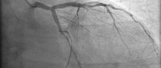
In 50% of cases, pathology is discovered accidentally during an X-ray examination of the lungs. In 70% of cases, the aneurysm is audible with a phonendoscope and manifests itself in the form of characteristic noise. The problem can also be identified during an ultrasound examination of the heart.
Specific methods for diagnosing an aneurysm are:
- Magnetic resonance imaging;
- CT scan;
- angiography.
Based on the data obtained during the examination, the doctor decides on the most appropriate treatment method.
Preparing for a doctor's appointment
If you suspect you have a thoracic aortic aneurysm or are concerned about your risk of developing an aneurysm due to your family history, make an appointment with your doctor at a vascular clinic. If an aneurysm is detected early, treatment may be easier and more effective.
Because most thoracic aortic aneurysms are discovered during a routine medical examination or during an examination for another reason, there is no need to make special preparations for your visit to the doctor. If you are being evaluated for an aortic aneurysm, your doctor will likely ask if anyone in your family has had an aortic aneurysm, so be prepared to answer this question. Be prepared to discuss your diet, physical activity and bad habits. If you are not following a diet or exercise routine, be prepared to talk to your doctor about problems you may encounter when starting treatment. Be sure to tell your doctor if you smoke or have quit smoking.
Surgery
It involves removing the damaged area of the aneurysm and replacing it with an artificial blood vessel - an aortic prosthesis. It is installed forever. The rehabilitation period after surgery ranges from 7 days to 1 month, depending on the patient’s age and the presence of concomitant diseases.
It is important to understand that aortic replacement does not guarantee the recurrence of an aneurysm in another part of the vessel. Therefore, it is necessary to strictly follow the doctor’s recommendations regarding nutrition and lifestyle.
The main indications for surgical treatment of an aneurysm are:
- Large size. The average is more than 5.5 cm in diameter. However, here everything depends on the initial diameter of the aorta. If the patient’s normal size is 3-3.5 cm, then surgery is indicated in case of aneurysm formation larger than 6.5-7 cm;
- Rapid growth - the damaged area of the vessel expands by more than 0.5 cm annually;
- An aneurysm provokes compression of the bronchi, the formation of an aortoesophageal/aortobronchial fistula;
- The vessel was stretched due to traumatic injury.
Types of open surgical operations for aortic aneurysms:
- Bentalla-De Bono operation (replacement of the ascending aorta with a valve-containing conduit with a mechanical prosthetic aortic valve);
- David's operation (replacement of the ascending aorta while preserving the native aortic valve);
- Supracoronary aortic replacement;
- Prosthetics of the ascending aorta and its arch (Borst technique, the use of oblique aggressive anastomosis and other techniques);
- Thoracic aortic replacement;
- Abdominal aortic replacement.
Endovascular intervention
If the shape and location of the aneurysm are not critical, the endoprosthesis replacement technique is used. The operation proceeds as follows:
- a puncture is made on the patient’s thigh, through which a conductor (narrow silicone tube) is inserted into the aorta;
- A vascular prosthesis is inserted through a conductor into the damaged aorta, which is fixed to the normal parts of the vessel above and below the location of the aneurysm.
The intervention is classified as minimally invasive. It does not require abdominal access, making the operation much easier for the patient to tolerate. The rehabilitation period is 2-3 days. During the day after the operation, the patient is under the supervision of a doctor.
Medical observation
For small aneurysms, your doctor may recommend medical monitoring, which includes regular testing to make sure the aneurysm is not getting larger, as well as treatment for underlying conditions that may be contributing to the aneurysm's formation or enlargement.
Your doctor will give you regular tests to determine the size of the aneurysm. Once an aneurysm is diagnosed, you will have an echocardiogram in 6 months, as well as follow-up imaging tests.
If you have high blood pressure or have plaque in your arteries, it is likely that your doctor will prescribe medications to lower your blood pressure and reduce your risk of developing aneurysm complications. Such drugs include:
- Beta blockers. Beta blockers lower blood pressure by slowing your heart rate.
- Angiotensin II receptor blockers. Your doctor may also prescribe these drugs if beta blockers do not lower your blood pressure enough. These medications are recommended for people with Marfan syndrome, even if they do not have high blood pressure.
- Statins. These drugs for the treatment of atherosclerosis of the aorta can lower cholesterol levels, leading to a decrease in plaque deposits in the arteries and a reduced risk of developing aneurysm complications.
If you smoke or chew tobacco, it is important that you quit this bad habit. Tobacco use can worsen the aneurysm.
Forecast
If an aneurysm is detected, the patient needs constant monitoring by doctors. Even after surgery, it is necessary to undergo regular examinations to monitor the condition of the blood vessels. Today, no intervention provides a 100% guarantee that the aneurysm will not reappear in another part of the aorta. Therefore, you should take responsibility for your own health and not neglect regular visits to a specialist.
Statistics on aneurysm operations say that the mortality rate of patients six months after surgery and aortic replacement is 10-15%. Within 10 years from the date of surgery, this figure increases to 40%. This is due to the fact that most patients have concomitant chronic diseases.
In case of endovascular intervention, the prognosis is more favorable. The probability of complications and re-formation of an aneurysm is only 10%, while patient death occurs in only 2% of cases.
Symptoms and signs of abdominal aortic aneurysm
In most patients, abdominal aortic aneurysms occur without any symptoms and are an incidental finding during examinations and operations for other reasons.
When signs of an aneurysm develop, the patient experiences one or more of the following symptoms:
- A feeling of pulsation in the abdomen, similar to a heartbeat, an unpleasant feeling of heaviness or fullness.
- Dull, aching pain in the abdomen, in the navel area, usually on the left.
Indirect signs of abdominal aortic aneurysm are important :
- Abdominal syndrome. Manifested by the appearance of belching, vomiting, unstable stool or constipation, lack of appetite and weight loss;
- Ischioradic syndrome. Manifested by lower back pain, sensory disturbances and movement disorders in the lower extremities;
- Syndrome of chronic ischemia of the lower extremities. Manifests itself in the appearance of pain in the muscles of the lower extremities when walking, sometimes at rest, coldness of the skin of the lower extremities;
- Urological syndrome. It manifests itself as pain and heaviness in the lower back, difficulty urinating, and the appearance of blood in the urine.
Harbingers of rupture may be increased abdominal pain.
When an aneurysm ruptures, the patient suddenly feels an increase or appearance of abdominal pain, sometimes “radiating” to the lower back, groin area and perineum, as well as severe weakness and dizziness. These are symptoms of massive internal bleeding. The development of such a situation is life-threatening! The patient needs emergency medical care!
Prevention
There are no specific measures to prevent such pathology as thoracic aortic aneurysm. Doctors give general recommendations:
- Complete cessation of smoking, including hookah and electronic cigarettes.
- Complete abstinence from alcohol (the maximum allowed is 100g of medium-strength alcohol during the holidays).
- Regular exercise. However, physical activity should be moderate. It is recommended to contact a rehabilitation therapist and exercise therapy specialist, who will select appropriate exercises based on the patient’s diagnosis, the presence of concomitant diseases and the current state of health.
- Control of factors that can provoke a rise in blood pressure. These include stress, kidney pathologies, and prolonged exposure to the open air during the warm season.
- Control of atherosclerosis. This pathology requires treatment, because it can lead to other serious complications in addition to the aneurysm.
- Immediately consult a doctor in case of the slightest suspicion of malfunctions in the cardiovascular system, gastrointestinal tract or lungs. It is necessary to respond to the body's signals to prevent serious complications.
If an aortic aneurysm is already present, prevention consists of preventing the development of complications of the pathology:
- Taking anticoagulants prescribed by a specialist. Such drugs are not cheap, but they are vital. They prevent the formation of blood clots in the lumen of the aneurysm.
- Optimal physical activity. If the patient works in a position that involves heavy lifting or other physical labor, it is necessary to think about changing professions. Excessive force can cause rupture of the aortic wall.
- Control of hypertension. It is necessary to take all medications prescribed by the doctor to avoid increasing blood flow pressure on the thinned wall of the aneurysm. The patient must be aware that it can rupture at any moment.
- Control over psychological state. In medical practice, there have been cases where aortic rupture was caused by a minor stressful situation.
In order to monitor the condition of the cardiovascular system, it is necessary to visit a cardiologist annually. And do this not “for show,” but undergo a comprehensive examination. This way you can detect developing diseases in the early stages, which greatly increases the likelihood of successful treatment without incisions and pain. Take care of your health!
Still have questions about thoracic aortic aneurysm?
Free consultation with AngioClinic specialists
Author
Salmina Daria Vladimirovna
Geneticist. Graduated from the Chelyabinsk State Medical Academy. She completed an internship at the Northwestern State Medical University named after I.I. Mechnikov.
Abdominal aortic aneurysm
Abdominal aortic aneurysm is a chronic degenerative disease with life-threatening complications. An abdominal aortic aneurysm is defined as an increase in its diameter by more than 50% compared to the norm or a local bulging of its wall. Under the pressure of blood flowing through this vessel, the dilatation or bulging of the aorta may progress. The diameter of a normal aorta in the abdominal region is approximately 2 cm. However, at the site of an aneurysm, the aorta can be dilated to 7 cm or more.
Why is an aortic aneurysm dangerous?
An aortic aneurysm poses a major health risk as it can rupture. A ruptured aneurysm can cause massive internal bleeding, which in turn leads to shock or death.
An abdominal aortic aneurysm can cause other serious health problems. Blood clots (thrombi) often form in the aneurysm sac or parts of the aneurysm tear off, which move with the blood flow along the branches of the aorta to the internal organs and limbs. If one of the blood vessels becomes blocked, it can cause severe pain and lead to organ death or loss of a lower limb. Fortunately, if an aortic aneurysm is diagnosed early, treatment can be timely, safe and effective.
How to treat an aortic aneurysm in the absence of indications for surgical treatment
If the size of the aortic aneurysm corresponds to the minimal risk of rupture, then observation is carried out using therapeutic measures to prevent the progression of growth and possible rupture of the aneurysm. Constant monitoring (at least three times a day) of blood pressure is necessary, maintaining its values not exceeding 120/80 mmHg. If you are prone to high blood pressure, you may need to take antihypertensive medications prescribed by your physician or cardiologist.
To prevent thrombus formation in the aneurysm cavity, it is recommended to take blood thinning drugs, such as Cardiomagnyl or ThromboAss. It is important to avoid excessive physical activity, which increases blood pressure, which is a provoking factor for the growth and subsequent rupture of an abdominal aortic aneurysm.
Increased intra-abdominal pressure can also contribute to the growth of aneurysms, which occurs primarily with heavy lifting, bending over, and in patients with chronic cough and chronic constipation.
How to choose a treatment tactic?
All aneurysms should be treated to reduce the risk of complications. Systemic hypertension promotes expansion and rupture. Strict pressure control is carried out in all patients, regardless of the size of the pathology.
For acute aortic aneurysms, first-line treatment for hypertension is with a short-acting beta blocker. The drug reduces the force of contraction, thereby minimizing the shear force exerted on the expansion by decreasing the pressure in the aorta.
If necessary, symptomatic therapy is carried out.
Indications for surgical treatment are based on the size, rate of growth of the pathology, family history of aneurysm, and possible rupture of the thoracic aorta. The operation has no strict contraindications. But there are relative ones, they are individualized based on the patient’s ability to survive the intervention as a whole. Individuals at increased risk of mortality include older people and patients with decompensated diseases of internal organs.
Surgical techniques
| Endovascular surgeries | Through a small incision in the leg, a stent-graft (in the form of a frame) is placed inside the vessel | The advantage is quick and easy recovery for patients. However, the implant is often incompatible with the patient's anatomy, requiring open surgery |
| Open surgery | It takes place under general anesthesia. The incision is usually made along the left side of the chest. The ribs are "opened" and the weakened area of the aorta is replaced with an artificial blood vessel (graft) made of tissue. | Blood flow through the aorta must be temporarily stopped until the graft is sutured. Often the body's circulation is maintained using mechanical pumps while the aorta is clamped. |
Lifestyle and taking medications at home
There are no medications that can prevent the formation of a thoracic aortic aneurysm, but medications that lower blood pressure and cholesterol levels can reduce the risk of developing complications of a thoracic aortic aneurysm.
In order to prevent the formation and growth of a thoracic aortic aneurysm, keep your blood vessels as healthy as possible. The following is required:
- Quit smoking.
- Monitor blood pressure.
- Exercise regularly.
- Reduce cholesterol and fat levels in your diet.
If you have risk factors for developing an aortic aneurysm, talk to your doctor. If necessary, the Doctor will recommend medications to you, including medications that lower blood pressure, thereby reducing the load on weakened arterial vessels. You should also undergo screening examination (EchoCG) for several years.
Disease prognosis
As for the prognosis of the disease, it largely depends on the size of the aneurysm, on the presence of atherosclerosis, on the location and type of defect. If we consider the prognosis as a whole, it remains unfavorable, which is associated with a high risk of death of the patient from aneurysm rupture. If the formation reaches 6 or more centimeters, then the probability that it will rupture within a year is 50%, and with an aneurysm of a smaller diameter, the percentage of its rupture is reduced to 20%.
The earlier an aortic aneurysm is detected, the more favorable the prognosis that the operation will be successful and the lower the mortality rate of patients during its implementation.
Author of the article:
Molchanov Sergey Nikolaevich |
Cardiologist Education: Diploma in Cardiology received from Perm State Medical University named after. I. M. Sechenova (2015). Here I completed my postgraduate studies and received a diploma in Cardiology. Our authors
Recovery after treatment
In the postoperative period, it is necessary to monitor the condition of the nervous system and kidneys, as possible
complications due to blood stagnation or disruption of certain structures during surgery.
In the long term, it is necessary to monitor the state of your health and concomitant cardiovascular diseases: modify your lifestyle, reduce mental and physical stress, quit bad habits (such as smoking), switch to a low-cholesterol diet and undergo annual preventive examinations, periodically monitor your blood pressure parameters and see a cardiologist. Medications are prescribed only if there are special indications.
Such actions will help not only reduce symptoms, but also prevent complications.
Postoperative period
After surgery, the recovery period takes 3-4 weeks. The patient undergoes regular examinations; cardiologists monitor the functioning of the heart and aorta. Depending on the technique of the intervention performed and the size of the aortic aneurysms, clinical recommendations suggest minimal physical activity only under medical supervision.
Physical therapy with the use of special simulators is indicated so that the heart and blood vessels learn to work in new conditions (with a stent or shunt). After a month, if there are no complications after surgery, the patient can return to the pre-operative lifestyle. It is recommended to reduce physical activity and the amount of fatty foods in the diet so as not to provoke the development of atherosclerosis.