This article uses anatomical terminology.
| Abdominal aorta | |
| Aorta segments, with both adrenal and | |
| Abdominal aorta and its branches. | |
| Details | |
| Source | Thoracic aorta |
| branches | Celiac artery, superior mesenteric artery, inferior mesenteric artery, common iliac bone, and 6 more |
| Ven | Inferior vena cava |
| Identifiers | |
| Latin | Abdominal aorta |
| MeSH | D001012 |
| TA98 | A12.2.12.001 |
| TA2 | 4205 |
| F.M.A. | 3789 |
| Anatomical terminology [edit in Wikidata] | |
The abdominal aorta
the largest artery in the abdominal cavity. Within the aorta, it is a direct continuation of the descending aorta (chest).[1]
Structure
The abdominal aorta begins at the level of the diaphragm, crossing through the aortic rupture, technically behind the diaphragm, at the level of the T12 vertebra.[1] It runs along the back wall of the abdomen in front of the spine. Thus, it follows the curvature of the lumbar vertebrae, that is, convex anteriorly. The peak of this convexity is at the level of the third lumbar vertebra (L3) and runs parallel to the vertebra. the inferior vena cava, which is located to the right of the abdominal aorta and becomes smaller in diameter as branches arise from it. This is believed to be due to the large size of its main branches. The 11th rib has a diameter of 122 mm in length and 55 mm in width, and this is due to constant pressure.[2] Clinically, the abdominal aorta is divided into 2 segments:
- The suprarenal abdominal
or
paravisceral
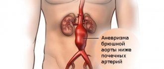
segment is inferior to the diaphragm but above the renal arteries. - The infrarenal
segment is inferior to the renal arteries and leads to the bifurcation of the ilium.
branches
The abdominal aorta supplies blood to most of the abdominal cavity. It begins at T12 and ends at L4 with a bifurcation into the common iliac arteries.[1] and usually has the following branches:
| Artery branch | Vertebra | Type | Paired with? | A/P | Description |
| inferior diaphragmatic | T12 | Parietal | Yes | mail. | Arises above the celiac trunk, below the diaphragm. It passes upward and inward to the adrenal glands and crosses the crus of the diaphragm of the corresponding side. Supplies the diaphragm and gives rise to the superior adrenal arteries. |
| celiac disease | T12 | Visceral | No | ant. | Greater anterior branch |
| superior mesenteric | L1 | Visceral | No | ant. | The large anterior branch arises just below the celiac trunk. |
| middle adrenal gland | L1 | Visceral | Yes | mail. | Crosses the legs of the diaphragm on the sides on each side; supplies the adrenal gland. |
| renal | Between L1 and L2 | Visceral | Yes | mail. | Arises just below the superior mesenteric artery. The right renal artery passes deep into the inferior vena cava, into the right kidney; here it is divided into branches. The left renal artery passes deep into the left renal vein. Divides at the hilum of the kidney. Both arteries give rise to the inferior adrenal arteries and branches of the ureter. |
| gonadal | L2 | Visceral | Yes | ant. | Ovarian artery in females; testicular artery in men |
| lumbar | L1-L4 | Parietal | Yes | mail. | Four on each side, which supply the abdominal wall and spinal cord. The fifth pair is the lumbar branches of the iliopsoas arteries. They pass deep into the calf on the side of the vertebral bodies and pass deep into the psoas and quadratus lumborum muscles to enter the space between the internal oblique and transverse abdominis muscles. Each artery gives off a small dorsal branch, which gives a vertebral branch to the spinal canal and then continues to supply the muscles of the back. |
| inferior mesenteric | L3 | Visceral | No | ant. | Greater anterior branch |
| midsacral | L4 | Parietal | No | mail. | An artery that arises from the middle of the aorta in its lower section. It is a continuation of the primitive dorsal aorta; quite large in animals with a tail, but smaller in humans. |
| common ilium | L4 | Terminal | Yes | mail. | Branches (bifurcations) for blood supply to the lower limbs and pelvis, ending in the abdominal aorta |
Note that the bifurcation (union) of the inferior vena cava is located at the level of L5 and, therefore, below the level of the aortic bifurcation.
Scope of visualization: computed tomography of the vessels of the abdominal cavity and pelvis.
- lower diaphragmatic a.
- celiac disease a. left stomach a.
- splenic a. short gastric arteries (6)
- splenic arteries (6)
- left gastroepiploic a.
- pancreatic arteries
- right stomach a.
- cystic a.
- inferior pancreaticoduodenal a.
- left colic a.
- external ilium a.
communications
The abdominal aorta lies slightly to the left of the midline of the body. It is covered in front by the lesser omentum and stomach, behind which are the branches of the celiac artery and celiac plexus; below them is the selective vein (splenic vein), the pancreas, the left renal vein, the lower part of the duodenum, the mesentery, and the aortic plexus.
Posteriorly, it is separated from the lumbar vertebrae and intervertebral cartilage by the anterior longitudinal ligament and the left lumbar veins.
On the right side it is connected with the above azygos vein, the cisterna chili, the thoracic duct, and the right crus of the diaphragm - the latter, separating it from the upper part of the inferior vena cava, and from the right celiac ganglion; the inferior vena cava is in contact with the underlying aorta.
On the left are the left leg of the diaphragm, the left celiac ganglion, the ascending part of the duodenum and several spirals of the diaphragm. small intestine.
3D illustration of the abdominal aorta at the iliac junction.
Connection with the inferior vena cava
The venous counterpart of the abdominal aorta, the inferior vena cava (IVC) runs parallel to it on the right side.
- Above the level of the umbilicus, the aorta is located somewhat posterior to the IVC, sending the right renal artery following it. The IVC also sends its opposite counterpart, the left renal vein, crossing in front of the aorta.
- Below the level of the navel, the situation is usually the opposite: the aorta directs its right angle. the common iliac artery to cross its opposite side (left common iliac vein) in front.
Collateral circulation
Collateral circulation will be maintained by anastomoses between the internal mammary artery and the inferior epigastric artery; due to free communication between the superior and inferior mesentery, if a ligature was placed between these vessels; or an anastomosis between the inferior mesenteric artery and the internal pudendal artery, when (most often) the ligature point is below the beginning of the inferior mesenteric artery; and, possibly, anastomoses of the lumbar arteries with branches of the internal iliac artery.
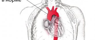
Symptoms of an abdominal aortic aneurysm
Most often, an aneurysm is accidentally diagnosed during an examination for other reasons of concern. The dilation of the vessel usually does not manifest itself in any way, but an aneurysm should be excluded during an examination by a doctor if you experience a dull, aching pain in the chest, under the ribs, or feel pulsation in the abdomen.
If the aneurysm is not detected in a timely manner, there is a risk of its rupture. This condition manifests itself in sharp, severe pain in the abdominal area, a dramatic drop in blood pressure, and fainting. In such a situation, it is necessary to immediately call an ambulance: aortic rupture is life-threatening.
Additional images
- Contrast-enhanced MRA of the abdominal aorta demonstrates normal paired arteries.
- Celiac artery and its branches; the abdomen is raised, the peritoneum is removed.
- Cross section through the middle of the first lumbar vertebra showing the relationship of the pancreas.
- CT scan showing liver and kidney
- Transverse contrast-enhanced CT scan demonstrates an abdominal aortic aneurysm measuring 4.8 by 3.8 cm.
- The standard measurement of the aorta on abdominal ultrasound, such as that used for an abdominal aortic aneurysm, is between the outer edges of the aortic wall.[3]
- Abdominal aorta
- Ultrasound of the abdominal aorta
Treatment
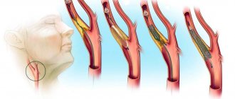
As already noted, the most dangerous complication of an aortic aneurysm is the threat of its rupture and internal bleeding. If the patient did not consult a doctor and the aneurysm was not detected in a timely manner, then in the event of a rupture, the only option would be open surgery: the surgeon will remove the affected area of the aorta and install a prosthesis in its place. This method of emergency treatment of an aneurysm has contraindications; in addition, the operation is a serious intervention with associated risks and a long rehabilitation period (up to three months). Therefore, it is so important to contact a specialist if you are at risk and feel periodic pain and/or throbbing in the abdominal area.
Once an aneurysm is diagnosed, its further expansion is difficult to predict; regular monitoring by an experienced specialist, lifestyle adjustments, and control of risk factors are required. When the attending physician realizes that there is a risk of rupture, an alternative to emergency open surgery will be the planned installation of a stent graft. The installation is minimally invasive, i.e. the stent is inserted through the vessel, advanced to the site of the aneurysm and secured there. The structure of the stent resembles a vessel; it is fixed with one edge above the expansion of the aorta, and with the other below. After the operation, blood will flow through the stent, and the resulting cavity between it and the aortic wall will decrease over time. The stent graft is inserted under local anesthesia and requires only a couple of days of recovery.
Of course, any surgical intervention carries its own risks, which is why it is so important to choose a good medical center with experienced cardiovascular surgeons. Understanding the danger of aortic aneurysm as a disease, not only doctors in the cardiology department, but also in other departments of our center, when conducting routine examinations or examinations due to other diseases, monitor the patient’s condition and analyze the entire volume of data received: if a problem with the aorta is suspected, the patient within one center will be transferred for consultation with a cardiologist. For our patients, we try to organize the most convenient logic for staying in our center and consulting with specialists in order to avoid double examinations and unnecessary manipulations. Comprehensive patient management is one of the main principles of our work. And the quality of surgical and conservative care is maintained thanks to a team of experienced specialists, provided with the necessary diagnostic and treatment facilities.
You can sign up for a consultation using a special form on the website or by phone.
Recommendations
- ^ a b c
Lech, Christy;
Swaminathan, Anand (November 2021). "Emergencies of the abdominal aorta." Urgent Care Clinics of North America
.
35
(4):847–867. doi:10.1016/j.emc.2017.07.003. PMID 28987432. - Jim, Jeffrey; Thompson, Robert W. "Clinics and Diagnosis of Abdominal Aortic Aneurysm." In a timely manner
. - Young, Timothy (28 August 2021). "Bedside ultrasound assessment of abdominal aortic aneurysm - technique." Medscape
.
What function and tasks does it perform?
This vessel is very important because it supplies blood enriched with oxygen and nutrients to the entire abdominal cavity and lower limbs. In fact, such an aorta completely ensures the functioning of the digestive and genitourinary systems of the body, therefore pathologies of the vessel can lead to disruptions in the functioning of the corresponding organs.
In addition, this vessel also plays a significant role in maintaining normal blood pressure due to its elastic properties. At the moment of contraction of the heart, a large volume of blood stretches the wall; during relaxation, it returns to its original position. This mechanism prevents too large a gap between systolic and diastolic blood pressure readings.
The blood flow is greatly influenced by the condition of the aortic walls. Normally, a laminar (or linear) flow of blood should be observed. However, if there are any protrusions (or vice versa, pockets, niches), turbulence appears, which causes a turbulent (chaotic) current. It contains a large friction force, which slows down the speed and leads to disruption of hemodynamics and perfusion (blood supply) of tissues.