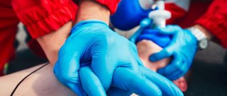
In modern laboratories, dozens of different tests can be performed, which help evaluate many processes in the body and play a huge role in the diagnosis of most diseases. According to statistics, 70% of decisions made by doctors are based on laboratory test data.
Over the past decades, laboratory diagnostics have become very accurate, but, unfortunately, sometimes the test results are erroneous. And it's not always the laboratory's fault. The preanalytical stage and proper preparation of the patient play a huge role. This is where about half of the mistakes occur. The result is distorted results, incorrect conclusions, incorrect treatment and unnecessary additional diagnostic methods.
One possible cause of errors in laboratory diagnostics is lipemia , a condition in which there are high levels of lipids in the blood. It occurs in 1–5 samples out of 200 received by the laboratory, and is much more common in outpatients than in inpatients. The main reason is that patients do not maintain the required interval between the last meal and blood donation. Unfortunately, not everyone knows how to properly take tests on an empty stomach, and doctors do not always explain well and cannot control the situation.
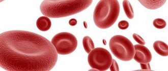
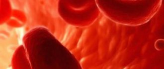
| A brief excursion into biochemistry: what are blood lipids? Fats and fat-like substances (cholesterol, triglycerides, phospholipids) are combined under the term “lipids”. By themselves, they cannot dissolve in water, so they are found in the blood plasma in the form of complexes with proteins - lipoproteins. There are several types of lipoproteins, they differ in composition, size and functions:
Lipemia refers to the cloudiness of a blood sample due to high levels of lipoproteins. |
Why does lipemia occur?
The most common reason is improper patient preparation. After eating a meal, especially a fatty one, the content of chylomicrons in the blood increases for several hours. Often, doctors simply say that you need to show up for tests in the morning on an empty stomach. Everyone can understand this in their own way. For example, a person can have dinner very late, then sleep a little and come to the clinic in the morning without breakfast, but the required interval will not be maintained. In emergency situations, when analysis needs to be done immediately, there is often no time to find out when the patient last ate.
This is why doctors at the Center for Immunology and Reproduction tell patients in such detail how to properly prepare for tests. It is important to strictly follow these recommendations.
Other possible causes of lipemia:
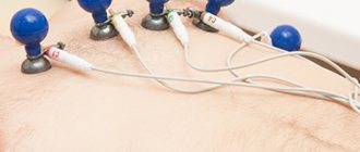
- Recent introduction of fat emulsions for parenteral (intravenous) nutrition. They are usually used in intensive care units and in patients who cannot feed themselves or are unable to feed through a tube. These are drugs such as Intralipid, SMOFlipid, Lipofundin, Lipovenoz.
- Primary lipemia is a hereditary disorder of lipid metabolism.
- Secondary lipemia is a condition that is a symptom of other diseases, such as non-alcoholic fatty liver disease, diabetes mellitus, HIV infection, and renal dysfunction.
- Excessive alcohol consumption, alcoholism.
- Taking certain medications: antiretroviral drugs, glucocorticosteroids, non-selective beta-adrenergic receptor antagonists.
- There is evidence that ketogenic diets increase blood cholesterol and triglyceride levels.
Treatment of reactive pancreatitis
Treatment of reactive pancreatitis includes eliminating inflammation of the pancreas, removing intoxication and restoring normal secretion of pancreatic juice. The treatment process must certainly take place under the supervision of a doctor.
Therapeutic fasting can help relieve inflammation, especially in the acute period. It relieves stress from the inflamed pancreas and the entire digestive tract. After the acute phase has passed, it is allowed to eat in small portions, eating crushed foods. But under no circumstances should you adjust your diet on your own. Only a doctor can prescribe a correct and healthy diet, based on the characteristics of your body.
As a rule, those foods that can provoke re-activation of pancreatic enzymes and intensify the inflammatory reaction in tissues are excluded from the patient’s diet for several months.
For reactive pancreatitis, the following are prohibited:
- alcohol;
- fatty and fried foods;
- legumes;
- sour juices;
- fresh baked goods;
- smoked meats, sausages;
- corn;
- mushrooms;
- sauces, seasonings, spices.
It is recommended to steam or boil the meat. It is advisable to prefer lean poultry, beef or rabbit. In the treatment of pancreatitis, it is very important to strictly follow the diet: regular eating disorders can provoke an exacerbation of the disease.
Drug therapy for pancreatitis includes taking enzyme preparations and antispasmodics that eliminate spasm of smooth muscles and relieve pain. The doctor may also prescribe medications that improve intestinal microflora and increase the content of live bacteria in the body.
To learn more
An important stage of treatment is taking enzyme agents based on pancreatin. An inflamed pancreas does not secrete enzymes in the required quantities to ensure high-quality digestion of food and the correct course of digestive processes. Medicines containing enzymes compensate for the lack of natural digestive elements and prevent the occurrence of fermentation and rotting of food in the intestines.
An example of an enzyme preparation used in the treatment of reactive pancreatitis is Creon®. A modern drug of the latest generation, produced in the form of capsules with active minimicrospheres of pancreatin, enclosed in a gelatin shell. Once in the stomach, the capsule quickly dissolves2, and the minimicrospheres are mixed with food and enter the intestines with it, helping the breakdown process and absorption of nutrients. More information about the drug Creon® can be found here.
How does lipemia interfere with testing?
Lipemia can interfere with blood tests in several ways:
- Increases light absorption. Because of this, the results of spectrophotometric analysis, which are used during biochemical blood tests, are distorted. The accuracy of studies that use light with a short wavelength suffers especially severely. The device determines the concentrations of some substances, for example, ALT, AST, bilirubin, incorrectly.
- Lipoproteins prevent antibodies from binding to antigens. As a result, the results of immunological tests are distorted. Depending on the nature of the reaction, they can turn out to be false positive and false negative.
- Distorts the results of studies in which the method of capillary electrophoresis of whey proteins is used. For example, it was noted that when analyzing blood with lipemia, the alpha-2-globulin fraction changes.
- Leads to heterogeneity of plasma and serum after centrifugation. In a centrifuge, VLDL, due to its low density, accumulates in the upper layer. Accordingly, there will be very few substances that are soluble in water. During analysis, their concentration will be incorrectly determined as low. At the same time, steroid hormones and some drugs accumulate in the upper lipid layer, and their concentration will be low in the lower part of the tube.
- Leads to the effect of volume displacement. Normal blood plasma consists of 92% water and 8% lipids. With lipemia, these figures can be 75% and 25%, respectively. But electrolytes are dissolved only in plasma, they are not in lipids. If the laboratory uses methods that determine the level of electrolytes in the total volume (indirect potentiometry, flame photometry), then their concentration will be lower than it actually is.
- With lipemia, hemolysis occurs more strongly - the destruction of red blood cells and the release of hemoglobin from them. This also interferes with the analysis and affects its accuracy. It is not entirely clear why lipids lead to this effect; it is believed that they act on the wall of red blood cells during the processing and centrifugation of blood as detergents (surfactants).
What determines the effectiveness of cryolipolysis?
The effectiveness of cryolipolysis is assessed by the amount of removed adipose tissue - the difference in the thickness of the fat layer before exposure and after 2 months. after completing the procedure. The more adipocytes are exposed to cold and the more intense it is (which is determined by the temperature and duration of cooling), the more pronounced the result of the procedure. It will also depend on the use of the so-called. effect enhancers - local (massage, lymphatic drainage, shock wave therapy) and general (diet and physical activity). In general, this dependence can be represented as the following formula:
The effectiveness of cryolipolysis (the amount of adipose tissue eliminated) = The amount of cooled adipose tissue × Temperature in the treatment area × Duration of the procedure × Factors that enhance efficiency.
Table 1. Studies on the effectiveness of cryolipolysis*
| Research | Type | Number of participants | Procedure settings | Observation period | results |
| Garibyan, 2014 [29] | P | 11 | CIF 41.6, 60 min | 2 months | The average decrease in the volume of adipose tissue is 56.2 cm3, the average decrease in the thickness of the fat fold is 14.9% |
| Pinto, 2012 [26] | P | 16 | 3.1 °C, 25–35 min, 1–2 sessions | 40 days | After the 1st procedure, the average reduction in the thickness of the fat fold was 19.7%, after the 2nd - 28.5% |
| Boey, 2014 [23] | P | 17 | CIF 42, 60 min + 2 min massage | 4 months | The average reduction in fat layer on the side where the subsequent massage was performed was 68% after 2 months and 44% after 4 months. |
| Dover, 2009 [31] | P | 32 | Manufacturer's CIF | 4 months | The average reduction in fat layer thickness was 22.4% 4 months after the procedure. A decrease in body fat was observed in 100% of patients |
| Sasaki, 2014 [24] | P | 112 | CIF 42, 60 min + 5 min massage | 6 months | The average reduction in the thickness of the fat fold is 21.5%; average reduction in fat layer thickness - 19.6% (abdomen) |
| Klein, 2009 [14] | P | 40 | CIF 42, 30 min | 12 weeks | There were no significant changes in lipid metabolism and liver function during 12 weeks of observation |
| Lee, 2013 [32] | P | 14 | CIF 42, 60 min | 12 weeks | Reduction in hip circumference by 19.55%. Indicators of lipid metabolism and glucose were within normal limits throughout the entire observation period |
| Riopelle, 2009 [15] | P | 10 | Not specified | 12 weeks | Against the background of a decrease in the thickness of the fat layer, no pathological changes in lipid metabolism and liver function were observed in studies 1, 4, 8 and 12 weeks after the procedure |
| Coleman, 2009 [16] | P | 9 | CIF 33, 60 min and CIF 37, 45 min | 6 months | The average reduction in fat layer thickness was 20.4% after 2 months and 25.5% after 6 months. Histological studies showed no long-term changes in peripheral nerves |
| Rosales-Berber, 2009 [33] | P | 42 | Intensity and duration not specified, 1 or 2 sessions | 6 months | 79% of subjects noted the effectiveness of the procedure |
| Dover, 2011 [34] | P | 15 | Not specified | 6 months | 80% of patients are satisfied with the results (6.6 on a scale of 1 to 10) |
| Shek, 2012 [25] | P | 21 | CIF 41.6, 60 min, 1 session | 2 months | Average reduction in fat fold thickness - 14.7% |
| Shek, 2012 [25] | R | 12 | CIF 41.6, 60 min, 2 sessions | 2 months | The average reduction in the thickness of the fat fold after the first procedure is 14% in the abdominal area, 13.4% in the sides; after the second - 7.2% in the abdominal area and 4.3% - in the sides |
| Dierickx, 2013 [35] | R | 518 | Not specified | 1 and 2 months | In 94% of patients, there was a decrease in the thickness of the fat fold by an average of 23% |
| Stevens, 2013 [36] | R | 528 | 60 min | 2 or 3 months | Statistics: increase in the number of procedures performed from 2010 to 2012 by 823% |
| Kaminer 2009 [37] | R | 50 | Not specified | 6 months | In 89% of cases, a difference was recorded between the circumference of the treated area before and after the procedure |
| Brightman, 2011 [38] | KS | 1 | CIF 42, 60 min, 2 sessions | 5 months | Reduction of abdominal circumference by 2.4 cm |
| Jalian, 2014 [20] | KS | 1 | Manufacturer's CIF | 5 months | Gradual, painless growth of adipose tissue in the treatment area (paradoxical fatty hyperplasia), which stabilized after 5 months |
| Bernstein, 2013 [39] | KS | 1 | CIF 42, 60 min | 2 years | 2 years after the procedure, there was a persistent decrease in the circumference of the treated area (comparison of before/after photographs) |
| Bernstein, 2013 [39] | KS | 1 | CIF 34, 60 min | 5 years | 5 years after the procedure, there was a persistent decrease in the circumference of the treated area (comparison of before/after photographs) |
* Measurement of the thickness of the fat fold in the studies was carried out using a caliper, the thickness of the fat layer - using ultrasound scanning. P—prospective study. R—retrospective study. CS is a clinical case. CIF - Cooling Intensity Factor, introduced by Zeltiq as a measure of the rate of heat removal from fabric, high CIF determines a higher rate of heat removal or a colder cycle: CIF 33 corresponds to heat removal with an intensity of 64 mW/cm2; CIF 37 - 68.3 mW/cm2; CIF 42 - 72.9 mW/cm2
How do laboratories detect lipemia?
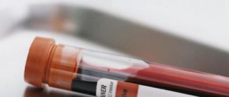
Lipemia can be seen with the naked eye. The blood of such patients appears cloudy. This method is not reliable. Lipemia can be noticed only if the concentration of triglycerides is very high: in the supernatant - more than 3.4 mmol/L, in whole blood - more than 11.3 mmol/L. This is very subjective; not every laboratory technician can examine the changes that are recorded using special tests.
Many laboratories test for triglyceride concentrations. This method makes it possible to indirectly judge the presence of lipemia and, in combination with determining the L-index (more about it below), judge its cause. But this study has two shortcomings:
- The content of triglycerides in different types of lipoproteins varies: for example, in VLDL it is about 50%, in chylomicrons – up to 85–90%.
- In most of these assays, triglyceride concentration is inferred from the oxidation of glycerol to dihydroxyacetone phosphate. The more triglycerides, the faster glycerol oxidizes. If there is a lot of glycerol in the sample, a false positive result will be obtained. Such cases are known in patients with certain genetic disorders, among lovers of beer that contains glycerin.
Currently, automatic determination of the L-index is most often used. The essence of the method is that a blood sample is diluted in saline or a special buffer solution and the absorption spectrum of light of a certain wavelength (300–700 nm) is measured. The more lipids in a sample, the more it absorbs light.
Automatic determination of the L-index is a fast, inexpensive and fairly accurate method for detecting lipemia. But it also has some disadvantages. You can get a false positive result when the blood plasma is cloudy due to the content of other substances, for example, paraprotein (an “incorrect” immunoglobulin protein that appears in the blood in some diseases), contrast dyes that are used during some medical procedures.
Test systems for automatically determining the L-index are produced by different manufacturers; there is no single standard, and this makes it difficult to interpret the analysis results. Thus, errors are also possible when using this method, although they are rare.
History of cryolipolysis
The first scientific report on the specific effect of low temperatures on adipose tissue appeared more than a century ago - the Austrian pediatrician Carl Hochsinger published an article in 1902 about cases of cold panniculitis (inflammation of subcutaneous fat tissue). He described children aged 4 to 10 years who, after exposure to unusually cold weather, experienced the appearance of dense, slightly reddened nodules in the submental area, rich in subcutaneous fat, which spontaneously resolved within 2–3 weeks [1]. In 1941, Danish dermatologist Holger Haxthausen reported similar cases in 4 young children and one teenage girl after exposure to extreme cold in winter (Fig. 1) [2]. He called such changes adiponecrosis e frigore - “freezing adiponecrosis.” Over the following years, reports of cold panniculitis following severe cooling in both children and adults appeared in the scientific literature [3].
Rice. 1. Cold panniculitis - diponecrosis e frigore - in a boy
In 1970, American doctors Ervin H. Epstein and Mark E. Oren encountered a case of buccal fat necrosis in an infant who ate ice cream, and coined the new term “popsicle panniculitis” [4 ]. At the same time, there was an assumption about increased sensitivity to cold effects of tissues with a high lipid content. However, scientists began testing it much later. In 2008, a group of researchers from Harvard Medical School led by Dr. Dieter Manstein conducted the first studies in pigs and coined the term “cryolipolysis” to refer to the destruction of fat tissue under the influence of cold [5]. A year later, in 2009, the first cryolipolysis device was created - Coolsculpting (Zeltiq® Aesthetics, Inc., USA).