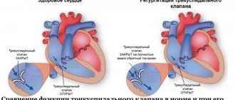
Tricuspid regurgitation is the reversal of blood flow from the right ventricle back into the atrium, but is not a diagnosis in itself. This is not even a disease, but a consequence of a malfunction of the tricuspid valve, which closes the passage from the right atrium to the corresponding ventricle.
The condition can be primary or secondary, depending on the origin of the pathological process. Recovery is carried out surgically.
The prospects for a complete cure are good, but only in the early stages, when there are no anatomical defects in the heart or distant systems yet.
Fortunately, the duration of the initial phase is sufficient for a thorough diagnosis. The intervention is planned, except for exceptional cases.
The approximate time frame from the moment the deviation occurs to the formation of a clear clinic is 3-6 years.
Development mechanism
{banner_banstat0}
The essence of the pathological process is the disruption of hemodynamics at the local level and the formation of a persistent anatomical defect.
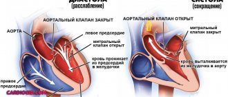
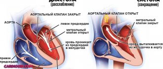
In the normal state of affairs, blood in the cardiac structures moves strictly in one direction, ending the cycle in the left ventricle and being transported to the aorta, and from there to its branches in a large circle.
The heart is represented by a group of chambers, each separated from the other by valves, which does not allow fluid connective tissue to move in the opposite direction.
The tricuspid structure closes the gap between the right atrium and ventricle. In case of weakness, insufficiency, or defects of the connective tissue, a reverse flow of blood or regurgitation occurs, which is called according to the name of the valve that causes the condition.
The result of the deviation is, firstly, a disruption in the transport of blood in the small circle, and secondly, an insufficient amount of it, which is released into the aorta.
This leads to generalized hemodynamic deviations, tissue hypoxia, and multiple organ failure in the future.
1.What is mitral valve regurgitation?
Mitral valve regurgitation
characterized by an unnatural flow of blood from the left ventricle into the left atrium during
systole
- contraction of the heart muscle.
When the heart valve is working properly, blood moves from the atrium to the ventricle. Against the background of rheumatic fever, dilation of the mitral valve ring, ischemic dysfunction of the papillary muscles and other unfavorable factors, the direction of blood flow changes in the opposite direction.
According to statistics, about 70% of the world's population is affected by mitral regurgitation. Minor manifestations of this pathological process can occur even in absolutely healthy people.
A must read! Help with treatment and hospitalization!
Forms of violation
{banner_banstat1}
Typing of the pathological process is carried out on two grounds.
Based on the origin of the anatomical defect, they talk about:
- Primary form. It develops spontaneously, against the background of cardiac problems themselves. Including aortic insufficiency, previous inflammatory, infectious conditions and others.
It is characterized by greater complexity from the point of view of cure and prospects for recovery, since correction requires not only the symptomatic component, but also the acquired defect.
This group also includes congenital factors caused by genetic defects and spontaneous deformations of the tricuspid valve.
- Secondary variety. Against the background of current pathologies of distant organs and systems.
Degrees of regurgitation
Another basis for classification is the degree of deviation from the norm. Also called stages of the pathological process.
Accordingly, they distinguish:
- Weakly expressed type. 1st degree. The amount of blood returning is not known exactly. The volumes of the jet do not exceed 1 cm in diameter. The intensity of manifestations with minimal tricuspid regurgitation is insignificant or completely absent, which makes early diagnosis a matter of luck. This is the best time to start therapy under the supervision of cardiac surgeons.
- Moderate type. 2nd degree. Characterized by disruption of normal blood flow in a volume of 2 cm, no more. Recovery is carried out surgically. The clinical picture is minimal, characterized by chest pain, shortness of breath during intense physical activity. There is a chance for a complete cure, the likelihood of the formation of persistent cardiac and extracardiac defects is present, but it is not yet great. Even if these occur, the likelihood of a high-quality, long life is maximum.
- Expressed type. 3rd degree. A stream of blood more than 2 cm in diameter. Chronic heart failure of congestive type develops. There are prospects for recovery, but they are not complete, and long-term, lifelong maintenance therapy is required.
- Terminal phase. 4th degree. Surgical assistance does not make much sense, since the heart, kidneys, liver, and brain are significantly changed. Recovery is impossible; palliative care is required to ensure an acceptable quality of life for the short remaining period of life. Death occurs from acute heart failure.
Classifications are used to accurately assess the patient’s condition, prospects for treatment, and determine diagnostic and therapeutic tactics.
How dangerous is the disease?
{banner_banstat2}
Complications arise starting from the third, less often the second stage of the pathological process. Tricuspid valve regurgitation determines the following consequences for health and life:
- Acute heart failure. Disruption of the normal functioning of cardiac structures. It is characterized by a triad of signs: a decrease in blood output, a drop in local and generalized hemodynamics, and arrhythmic processes. It has a short period of development in an acute case; in a latent course, the duration of formation of a full-fledged picture is 2-4 weeks; death occurs as a result of stopping the work of a muscular organ.
- Cardiogenic shock. The condition is lethal in almost 100% of cases. There is no prospect of cure. Even with partial recovery, there is a guarantee of a repeat episode.
- Heart attack. Myocardial nutritional disturbances, acute tissue necrosis and, as a result, decreased functional activity. Heart failure develops with all its consequences.
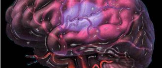
- Stroke. Cerebral ischemia.
- Dangerous forms of arrhythmia leading to cardiac arrest.
Minor regurgitation provokes fatal complications in 0.3-2% of cases, often the result of a random coincidence.
Hemodynamically significant forms determine the risk of death in a wide range: from 10 to 70% and higher.
The main cause of death is not regurgitation, but organic defects of the heart and systems developing against its background.
Trouble is not a problem: tricuspid regurgitation in the fetus
In the ultrasound screening program of the 1st trimester of pregnancy, it is required (but not by all, not always, and not always performed as expected) to study the blood flow on the tricuspid valve of the fetus.
The fact of detection of tricuspid regurgitation in the fetus (i.e., reverse natural blood flow, and tricuspid valve insufficiency) is considered by the recommendations of domestic and foreign luminaries of prenatal and ultrasound diagnostics to be a marker of the risk of fetal developmental anomalies and its chromosomal aberrations.
Indeed, with various structural anomalies of the heart, such as atrioventricular septal defect (AVS), tricuspid valve dysplasia, Ebstein's disease, as well as stenosis or atresia of the pulmonary valve, stenosis and premature closure of the ductus arteriosus, hypoplastic left heart syndrome, and fetal arrhythmias , we encounter tricuspid valve insufficiency - tricuspid regurgitation. And, of course, it occurs in fetal cardiomyopathies and myocarditis, fetal heart failure, and various vascular malformations of the fetus (a classic example is the vein of Galen aneurysm).
But, excuse me, all of these listed diseases are obvious, and have more important, main manifestations, and tricuspid insufficiency with them is just one of many other symptoms, and not an independent finding!
At the same time, we (as cardiologists) know that almost everyone has physiological tricuspid regurgitation - both in a newly born child and in elderly patients. It is distinguished from pathological tricuspid valve insufficiency by the low degree and relatively low flow rate of regurgitation, and, of course, by the absence of other serious congenital or acquired pathology of the heart and blood circulation in general.
But the legislators of prenatal diagnostics stubbornly replicate completely alarming figures: only 5% of fetuses with tricuspid regurgitation have no pathologies, the remaining 95% are carriers of chromosomal pathology, the risk of trisomy 21 chromosomes is especially high, i.e. Down's disease!
Internet forums are shouting with thousands of voices: “Help, my unborn child has been diagnosed with tricuspid regurgitation! What to do! Nobody can explain anything!” A non-professional will not explain; he has no idea what this regurgitation is caused by.
It’s good if you don’t initially confuse the flows of mitral and tricuspid regurgitation, or (even worse), the normal blood flow in the main artery of the heart with this very tricuspid regurgitation (and, believe me, there are heaps of such “finds” - half of those shouting in these very forums, and I refer specialists who doubt this statement to the published ultrasound images in the same online resource).
The easiest way for this specialist is to disown you, sending you to the hellish circles of additional endless research, additional examinations and consultations.
In our practice, when tricuspid regurgitation is detected in the fetus, we carry out a number of additional diagnostic measures that allow us to assess in much more detail both the degree and significance of tricuspid regurgitation, as well as the fetal heart and its circulation as a whole, and, if possible, exclude congenital malformations of the fetal heart , fetal heart failure, etc.
If an intact heart is detected, as well as in the absence of other ultrasound markers of the risk of fetopathies and chromosomal diseases of the fetus, the diagnostic significance of the detected tricuspid regurgitation in the fetus is negligible.
I think that a high-quality repeated ultrasound examination of the fetal heart when tricuspid regurgitation is detected in the first ultrasound screening of pregnancy will not only not hurt, but is actually necessary. It is carried out (fetal echocardiography) at 20-23 weeks of pregnancy, and it is during this period that an experienced specialist is able to meticulously examine literally every millimeter of the heart of your unborn child.
It's a matter of the patients' discipline and diligence, and, alas, she is not always at her best either! And the choice of a research specialist is yours, dear future mothers and fathers! Choosing “nearby and quickly” is not wise even when buying a loaf of bread!
Pregnancy 16 weeks. Fetal echocardiography. PWD – dopplerography. There is a physiological orientation of transtricuspid blood flow filling the right ventricle at a speed of 0.60 m/s, and a regurgitant flow with a maximum speed of 1.30 m/s.
Pregnancy 11 weeks. TVI. Fetal echocardiography. Color Dopplerography of the heart. The blood flow on the mitral and tricuspid heart valves, symmetrical in spectrum and speed, is clearly visualized (the filling flows of the ventricles of the heart are displayed in red). No regurgitation is noted! We draw your attention to the diagnostic capabilities of the equipment: the total size of the heart is less than 5 mm, the ventricles of the heart are 2 mm in diameter, the main arteries are 1 mm in diameter, but how informative the study is!
Pregnancy 31 weeks. Fetal echocardiography. Color Dopplerography of the heart. The regurgitation flow on the tricuspid valve is clearly visualized (red flow). Physiological filling blood flow of the right ventricle through the tricuspid valve is mapped in blue in this study.
anomalies of cardiac development fetal arrhythmias dopplerography of the fetus blood flow on the tricuspid valve fetal heart failure pulmonary aortic stenosis tricuspid regurgitation of the fetus ultrasound of pregnancy Pyatigorsk ultrasound Pyatigorsk 4D echocardiography of the fetus
Causes
{banner_banstat3}
Formation factors are divided into primary and secondary, according to the main forms of the pathological process.
Primary factors
{banner_banstat4}
- Burdened heredity. Leads to the development of tricuspid valve insufficiency. Problems arise during the prenatal period. In this case, there is a genetic predisposition. The exact mechanism, however, is not known.
One thing has been proven: in the presence of a sick parent, children are born with the defect in question and regurgitation in 12-15% of cases. Spontaneous defects of the perinatal period are possible, caused by internal and external factors.
- Spikes in the heart. These are small fibrin strands that disrupt the normal anatomical structure of the organ. They develop as a result of inflammatory processes of any type, especially infectious. This is a kind of protective mechanism, as well as further deposition of calcium salts to isolate the affected area.
- Previous heart attack. It ends with the replacement of functionally active tissues with weak, scarred ones, incapable of contraction, signal transmission, or spontaneous excitation.
If the process affects the tricuspid valve, the following options are possible: its complete fusion, stenosis, or functional failure, immediately leading to severe regurgitation. Recovery is urgent, surgical.
- Inflammatory pathologies of the heart (myocarditis and others). Accompanied by rapid destruction of tissue of cardiac structures. Treatment is urgent, in a hospital, with the use of antibiotics and NSAIDs, as well as steroids and diuretics.
- Rheumatism. Inflammatory pathology of a chronic nature, with frequent relapses and short periods of remission. Therapy is lifelong, using supportive tactics. If necessary, surgical correction of the consequences is performed.
Secondary factors
{banner_banstat5}
The secondary pathological process is caused by cardiac problems and extracardiac issues:
- Pulmonary hypertension and the development of specific abnormalities in the anatomical development of the heart. Requires urgent treatment in the early stages, since in the later stages there is no longer any sense. The main risks are smokers, alcoholics, asthmatics and patients with long-term COPD.
- Cardiomyopathy.
- Endocrine pathologies: hyperthyroidism, excess of adrenal hormones, their deficiency, diabetes mellitus and others.
Risk factors
{banner_banstat6}
They do not directly cause tricuspid regurgitation, but lead to the onset of the pathological process:
- Long-term smoking.
- Consuming alcohol in immoderate quantities.
- A long period of immobilization, without the possibility of vigorous activity. Development takes a long time, from six months or more.
- Drug addict.
- Excessive use of “dangerous” drugs: glycosides, antiarrhythmics, progestin agents, also hormonal medications, broad-spectrum antibiotics.
- Harmful working conditions affect chemical, hot production, and mines.
The reasons are considered as a whole; a system of development factors is possible.
Statistics and prognosis for the treatment of tricuspid valve insufficiency in children in Israel
Defective valve function detected in the first years of life is most often a congenital anomaly (organic) and, as a rule, not isolated.
The acquired form (relative or functional, not associated with deformation of the valve leaflets) is caused by diseases that result in the development of rheumatic pathologies. Such a lesion is recognized in 15–30% of cases.
A child with severe tricuspid valve insufficiency, especially in combination with other severe myocardial pathologies, develops serious complications that require immediate treatment, which is successfully carried out in the best clinic in Israel, Hadassah . In addition, corrective or therapeutic measures are required for all types of congenital heart disease.
| Otherwise, once diagnosed, the five-year survival threshold is rarely surpassed. |
The prognosis of therapy depends on the severity of reverse blood flow (there are 4 degrees), damage to the myocardium itself, and the severity of associated complications. The rich experience and professional skills of the doctors at our medical center allow us to adequately assess the condition of a small patient and, if appropriate, select effective drug treatment. But the prospects of the technique are considered short-lived, since the medicinal effect can stabilize well-being, without excluding the progression of the disease. The mortality rate remains high.
Without timely assistance from cardiac surgeons, not only stagnation in the body, but also irreversible processes can develop.
Open heart surgery carries an increased risk but has a better prognosis. Mortality after surgery is 1–3%, and for plastic surgery this figure is lower than for prosthetics (about 14% of the total).
Characteristic symptoms
Manifestations depend on the stage of the pathological process. A hemodynamically insignificant variety has no signs at all.
Typical signs in other situations include:
- Liver lesions. They make themselves known in the later stages. They are determined by pain in the right hypochondrium, an increase in the size of the organ, and yellowness of the skin due to excess bilirubin. A gradual formation of insufficiency is possible.
- Abdominal pain of unknown localization. Wandering, radiating to the iliac regions. Acute discomfort is not typical, therefore it is impossible to confuse it with the clinical picture of appendicitis.
- Shortness of breath for no apparent reason. It develops first against the background of intense physical activity, then occurs in a state of complete rest. Significantly reduces quality of life.
- Polyuria. As a result of developing renal failure. At later stages (3-4), with primary damage to the excretory system, it is replaced by a reverse process. Daily diuresis is 500 ml or less.
- Tachycardia. The heart rate reaches 120-150 beats. They are full-fledged, regular. Type - sinus. Less often paroxysmal.
- Weakness, lack of ability to work.
- Feeling of constant cold. The patient freezes because the intensity of peripheral circulation decreases.
- Increased pressure in the veins. Objectively, the symptom is manifested by swelling of the cervical vessels, their intense pulsation, and visible tension. Not only the doctor, but also the patient himself or the people around him can determine the sign. However, blood pressure drops in most cases. Not significant, however, clinical significance is present.
- Swelling of the lower extremities. As a logical continuation of increasing renal failure.
- Breathing problems.
As a result, the patient has a whole range of symptoms from both distant organs and systems, and the cardiac structures themselves. The reason for all the sensations lies in the disruption of blood circulation, both in the large and in the small circle.
4. Diagnosis and treatment of mitral valve regurgitation
Diagnosis of mitral valve regurgitation may include:
- medical examination;
- echocardiogram - a procedure based on the use of sound waves to determine the shape, size and structure of heart tissue;
- chest x-ray;
- electrocardiogram of the heart, which allows to detect irregularities in the heart rhythm;
- cardiac catheterization - examination of the cavities of the heart, as well as nearby blood vessels, using a hollow flexible tube.
These tests not only detect mitral regurgitation, but also determine the degree of mitral regurgitation. The information obtained during the study is the basis on which further treatment is based.
The choice of treatment for mitral valve regurgitation depends primarily on the form of the disease, as well as the degree of its progression.
For example, in chronic cases, doctors most often prescribe constant monitoring of the patient’s heart condition and the use of special medications to eliminate the symptoms of the disease. These drugs include:
- vasodilators – a group of vasodilating drugs;
- diuretics and diuretics;
- anticoagulants - drugs that prevent the formation of blood clots.
If necessary, your doctor may recommend surgery to repair or replace the mitral valve. Patients with mitral regurgitation need to radically change their lifestyle in order to reduce the load on the heart. Doctors recommend avoiding intense physical activity and emotional stress, leading a healthy lifestyle and eating right.
Diagnostics
{banner_banstat7}
The examination is carried out under the guidance of a cardiologist, and if the process is proven, the specialized surgeon continues to work. He is also responsible for prescribing treatment.
Scheme of events in the correct order:
- Oral questioning of the patient regarding complaints, their duration, as well as collecting anamnesis. This way the doctor understands the direction of further examination.
- Blood pressure measurement. Usually it is slightly reduced. Heart rate is higher than normal. The rhythm is correct; as it progresses, spontaneous premature beats (extrasystoles) occur.
- Listening to sound (auscultation). A sinus noise of reverse blood flow is detected. Tones can be either normal or dull.
- Daily monitoring. To record cardiac performance indicators over 24 hours in dynamics. It is most often used as the first method, after a routine examination. Provides comprehensive information about the movement of blood pressure and heart rate during the day.
- Electrocardiography. Assessment of the functional state of the heart.
- Echocardiography. Method of visualization of cardiac structures. It is carried out as a matter of priority, since it allows one to detect organic abnormalities on the part of the tricuspid valve.
- MRI or CR (much less often). It is carried out to detail the image of the heart and surrounding tissues.
- Pulmonary artery pressure measurement.
- Load tests. At an early stage, later not used due to significant danger.
The methods are aimed both at establishing the fact of an anatomical defect and at verifying the alleged diagnosis.
Treatment methods
{banner_banstat8}
Therapy is carried out under the full supervision of a cardiac surgeon. Methods of exposure depend on the stage of the pathological process.
Grade 1 tricuspid regurgitation is the best time to start therapy. But there are no symptoms yet, identification is incidental (random), and does not pose any difficulties during a targeted search.
At this stage, dynamic observation for 3-5 years is indicated. In the absence of progression, with stagnation of the process, there is no need for treatment. Sometimes patients can live a quality life without knowing about their condition, without major restrictions.
Tricuspid regurgitation of grade 2 and higher is corrected strictly surgically. There are several intervention options.
But before the treatment stage, it is necessary to stabilize the patient’s condition, if there is time for that (planned operations).
Drugs used:
- Antiarrhythmics in the minimum dosage to restore an acceptable heart rate (Amiodarone, Hindin).
- Beta blockers (Metoprolol).
- Glycosides. In order to normalize myocardial contractility.
- Cardioprotectors.
- Anticoagulants. To prevent the formation of blood clots, which cause frequent premature death of patients.
- Diuretics in the treatment of early manifestations of renal conditions.
The duration of the preparatory period varies from 2 to 4 months, possibly more.
By the time of surgery, the rhythm should be stable, correct, blood pressure within the reference value or close to it.
Depending on the stage of the pathological process and the nature of the changes, plasty or prosthetics of the tricuspid valve is indicated. Both methods are generally equivalent.
Correction of pathologies and defects of distant organs is carried out under the supervision of specialized specialists. The list of techniques is wide and is determined based on the severity of the process.
Attention:
The use of folk remedies is impossible. Since the effect of them in case of organic deviation on the part of the cardiac structures is zero.
Changing your lifestyle will also not play a key role. It makes sense to give up smoking, alcohol and drugs. When carrying out severe therapy for third-party pathologies, correction by a treating specialist is recommended.
Modern methods of treating tricuspid valve insufficiency in children in Israel
The pediatric cardiology department at Hadassah is headed by Dr. Azariah Rein. After receiving higher medical education at the Hebrew University, he trained at the Shaare Zedek Clinic. After receiving his degree, he became a university professor in Jerusalem and Harvard. Permanent member of the Academic Council of Israel, engaged in scientific research.
Cardiac surgery is under the direction of Professor Eldad Erez, holder of two higher qualifications, specializing in the treatment of congenital defects in children. The future doctor of medicine's internship took place in Haifa and Beilinson, his residency in Atlanta (USA), where the doctor mastered the technology of heart transplantation. Erez teaches medical students in Tel Aviv and Jerusalem, and is the author of scientific works.
The first day after hospitalization in the hospital is reserved for examination, medical history, and documentation in Hebrew. Throughout the entire stay in the hospital, the little patient and his parents are accompanied by a Russian-speaking consultant.
Treatment for tricuspid valve insufficiency in children is selected strictly on an individual basis and depends on the degree of regurgitation of blood flow (1, 2, 3 or 4):
- 1 - subtle throw or touch of the tricuspid valve;
- 2 — reverse ejection is determined at a distance of 2 cm from the atrioventricular opening;
- 3 — return of blood from the right ventricle to a distance of more than 2 cm;
- 4 - regurgitation in the right atrium over a significant extent.
The first stage may be limited to medical observation or conservative therapy. Starting from the second, the nature of the intervention will depend on the intensity of casting, complications caused by the defect, and the presence of combined anomalies.
Elimination of the defect and the consequences of improper functioning of the heart is carried out through medication or surgery.
Drug therapy
The conservative approach includes drugs that ease the work of the heart and reduce pulmonary hypertension. This list includes diuretics, anticoagulants, b-blockers, glycosides, drugs that increase metabolism, and ACE inhibitors.
Surgical intervention
The main types of surgical techniques are repair of the patient's own valve or prosthetics using a bioprosthesis or a mechanical analogue.
Indications for prosthetics are considered to be severe changes in the valves. The material for the manufacture of a biological prosthesis is the aorta of an animal (pig). Unlike a mechanical one, a bioprosthesis significantly reduces the development of thromboembolism, but lasts only 10 years, requiring repeated replacement.
Preference is given to plastic correction, but only if no pronounced changes in the structure of the valves are detected. The obvious advantages of this procedure are minimal postoperative complications and a reduction in the rehabilitation period. In addition, restoration using one’s own, normally functioning tissues increases resistance to infections and eliminates the need for constant use of special medications.
Forecast
{banner_banstat9}
Depends on the stage and nature of therapy.
- At the first stage, the survival rate is 100%, especially if there is no progression of the condition.
- The second is associated with a probability of 85%.
- Third - 45%.
- The fourth or terminal puts an end to the patient, giving no chance. The median is 1-2 years, often even less.
When carrying out complex therapy, it is possible to stabilize the conditions of even the most severe patients, prolonging life for several years.
Favorable prognostic factors:
- Period of youth.
- Absence of somatic pathologies, bad habits, complications after the operation.
- Good family history.
- Response to treatment.
- Reduction of symptoms.
Determining the possible outcome falls on the shoulders of the cardiologist. In order to say anything concrete, you need to at least carry out a full diagnosis.