© Author: Sazykina Oksana Yuryevna, cardiologist, especially for SosudInfo.ru (about the authors)
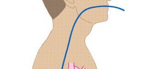
Transesophageal electrophysiological study (TEPE) is a technique for studying cardiac contractility and heart rhythm by applying physiological doses of electric current to the area of the heart most closely adjacent to the esophagus.
For the first time, the PPE technique began to be widely introduced into practice by arrhythmologists in the 70-80s of the last century. The study is otherwise called a non-invasive electrophysiological study, as opposed to an invasive one, in which electrodes are usually inserted through large arteries directly into the heart cavity from the inside. A little later, TEE began to be actively used in pediatrics, significantly expanding diagnostic capabilities in arrhythmology.
The essence of the method is based on the anatomical proximity of the esophagus and the heart. Often, in many patients, heart rhythm disturbances (arrhythmias) cannot be detected using a standard ECG or even with 24-hour blood pressure and ECG monitoring. Then stimulation of the heart muscle is applied, and if the patient has an arrhythmia, this will manifest itself during TEE. In other words, in some cases the myocardium must be “provoked” in order to understand whether electrical impulses are carried out correctly or whether there is a rhythm disturbance.
Using an electrode inserted through the esophagus, impulses are sent to the heart muscle, and electrodes placed on the chest record the rhythm during the study.
In addition, the rapid heart rate that occurs due to electrical stimulation can cause episodes of myocardial ischemia in patients suffering from coronary heart disease that is not detected on a standard ECG.
In addition to transesophageal EPI, there are invasive methods:
insertion of electrodes into the heart during invasive catheter EPI
- With endocardial EPI, a catheter with an electrode is delivered through the femoral vein into the ventricle or atrium, and the diagnostic procedure is often combined with subsequent RFA of pathological areas of the myocardium.
- EPI is also performed through open access (during cardiac surgery). Stimulation occurs epicardially - from the “outer” surface of the heart.
The essence of invasive procedures is generally similar to transesophageal procedures. Naturally, such interventions are correspondingly traumatic, but can be more effective.
Advantages and disadvantages of PPEFI
The undoubted advantage of this technique is a more accurate diagnosis of rhythm disturbances, especially short runs of supraventricular tachycardia . What is meant here is local diagnostics, that is, with the help of TEE, in 90-100% of cases (according to various studies), it is possible to establish the exact place in the conduction system of the heart in which conditions exist for the occurrence of tachycardia or, conversely, for blocking the propagation of an impulse through the heart . Both rhythm disturbances can be life-threatening (depending on the type of arrhythmia), so identifying them using TEE can save the patient’s life and health.
The method has no disadvantages (besides the patient’s sensations), and other advantages include low trauma, non-invasiveness, the possibility of repeating the study, and the accessibility of the method to the general population.
Indications for electrophysiological examination of the heart
The main indications are various rhythm disturbances. Thus, performing TEE is justified in diagnostically unclear cases when the doctor suspects the patient:
- Paroxysmal tachycardia - atrial or supraventricular,
- Paroxysmal atrial fibrillation,
- Bradyarrhythmia, accompanied by a heart rate of less than 50 per minute, for example, with sick sinus syndrome, with atrioventricular block, etc.
- Tachycardia with SVC syndrome (Wolf-Parkinson-White syndrome) or KLK syndrome (Clerk-Levi-Christesco) - paroxysmal types of tachycardia when the atria contract at a high frequency (more than 100 per minute), and due to the presence of additional conduction pathways between the atria and ventricles (Kent and James bundles), tachycardia can spread to the ventricles, which poses a threat to the patient’s life.
In some cases, the study is performed when diagnosing coronary heart disease, for example, in patients in whom it is not possible to register episodes of myocardial ischemia using an ECG or 24-hour monitoring. In addition, TEE helps in diagnosing ischemia in the presence of contraindications for testing with physical activity, for example, in the presence of severe deforming osteoarthritis or other osteoarticular pathology.
In addition to direct diagnosis of diseases, repeat TEE is performed to monitor drug treatment of arrhythmias or after surgery, for example, after radiofrequency ablation (RFA). TEE is also performed before installing an artificial pacemaker.
What symptoms may indicate the need for TEE?
Rhythm disturbances requiring transesophageal pacing can be suspected based on the presence of the following signs in the patient:
- Attacks of sudden rapid heartbeat with rapid pulse (more than 100-120 per minute), also suddenly stopping spontaneously or stopping only after the administration of antiarrhythmic drugs,
- Feelings of irregular heartbeat with a rare pulse (less than 50 per minute),
- Attacks of loss of consciousness that are not associated with neurological problems or with situationally occurring conditions (stuffiness in the room, stress, etc.), but are caused by rhythm disturbances and are called Morgagni-Edams-Stokes attacks (MES episodes).
If a doctor, while examining a patient, hears about the presence of the symptoms described above, then he should think about a more accurate diagnosis of heart rhythm disturbances. And if the ECG and 24-hour monitor do not reveal any types of arrhythmias, and the patient’s complaints persist, it is necessary to perform TEE. If it was possible to register an arrhythmia on the ECG, for example, a delta wave, characteristic of latent SVC syndrome, the patient should be further examined.
In any case, the need for this technique is determined only by a doctor during an in-person examination of the patient.
Contraindications
Regarding electrical stimulation of the heart, contraindications include the presence of obvious disturbances in heart rhythm and conduction detected on the electrocardiogram. Thus, the impact of impulses on the heart muscle is contraindicated in the case of atrioventricular block of 2 and 3 degrees, as well as in the presence of existing atrial fibrillation-flutter.
In addition, an absolute contraindication is the presence of a blood clot in the atrial cavity, identified by the results of an ultrasound of the heart, since rapid heartbeat at the time of the study can lead to the separation and spread of the blood clot through the bloodstream.
Regarding the insertion of a probe into the lumen of the esophagus, contraindications are the presence of tumors in the patient, strictures (adhesions) of the esophagus, scar narrowing of the esophagus and an inflammatory process - esophagitis.
Transesophageal electrophysiological examination is also contraindicated in the presence of acute infectious processes in the body, fever and severe mental disorders in the patient.
Specifics of the EPI method and its varieties
The essence of the method is to stimulate cardiac activity in order to detect a pathological rhythm, which was not detected during a conventional single electrocardiological study and 24-hour monitoring. Using the device, electrical impulses are sent to the body, which lead to physiological tachycardia. With the work of the heart obtained in this way, it is quite easy to detect the source of the disturbance.
There are two EPI methods:
- non-invasive – transesophageal EPI;
- invasive – endocardial and epicardial EPI.
Invasive method
Let's take a closer look at what it is:
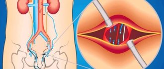
- Transesophageal electrical stimulation (TEES) involves inserting a special electrode into the lumen of the esophagus. Stimulation of cardiac activity is achieved due to the close proximity of the anterior wall of the esophagus to the left atrium. This makes it possible to record atrial depolarization, sometimes indistinguishable in conventional leads. This method is characterized by low trauma and low cost. However, its diagnostic capabilities are inferior to invasive methods.
Transesophageal electrical stimulation
- Endocardial invasive EPI involves inserting an electrode through the femoral and vena cava directly into the cavity of the ventricle or atrium. Epicardial pacing can only be performed through the outer wall of the heart during abdominal surgery.
In this way, an increase in rhythm is achieved. These methods require modern equipment in the clinic, sufficient financial support and are risky due to the frequent development of complications. However, these diagnostic measures enable the doctor to stimulate not only the left atrium, but also the ventricles, thus controlling ventricular arrhythmias.
See also:
What is Holter ECG monitoring and its interpretation?
How to prepare for the procedure?
The patient must prepare for the study as follows:
- Stop taking prescribed antiarrhythmic drugs no later than a week before the test, but only in agreement with the doctor who referred you for TPE,
- Of the prescribed drugs, it is permissible to use only short-acting nitroglycerin under the tongue for angina attacks,
- A few weeks or days before the examination, perform an ECG, ultrasound of the heart and 24-hour monitoring of blood pressure and ECG,
- It is advisable not to smoke or drink alcohol during the week before the procedure.
- On the eve of the study, a light dinner is allowed, it is advisable to exclude coffee,
- On the day of the study, the patient must appear for the procedure on a completely empty stomach, even drinking water is excluded.
Cost and reviews
The procedure is considered safe, patients leave positive reviews about it. Cardiac EPI can only be performed in specialized medical centers equipped with modern equipment. Quotas from the Ministry of Health are allocated for the procedure, but the queues are quite long and the wait will be long.
In private clinics, prices for TEE can vary between 1000-4000 rubles. The cost of EndoEFI is over 20,000 rubles.
Did you like the article? Save it!
Still have questions? Ask them in the comments! Cardiologist Mariam Harutyunyan will answer them.
Ivan Grekhov
Graduated from the Ural State Medical University with a degree in General Medicine. General practitioner
How is the procedure performed?
TPEFI can be performed in a clinic or in a hospital, depending on the personnel and technical equipment of the institution. Typically, such techniques are carried out in the department of functional diagnostics.
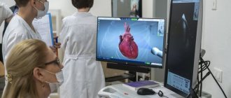
equipment for CPEFI (photo: doktora.by)
At the appointed time, after pre-registration, the patient visits this department. In the TPE room, he is placed on a couch, and after preliminary measurement of blood pressure and recording of a standard ECG, the procedure begins. To do this, the doctor treats the root of the tongue with a solution of a local anesthetic - lidocaine or dicaine to reduce discomfort when inserting the probe.
The probe used is thin and sterile, and is most often inserted through the nasopharynx, in some cases through the mouth. This procedure at this stage is no more unpleasant than examining the stomach during fibrogastroscopy. The probe contains an electrode as a tip that will send signals to the heart muscle. Electrodes that read the electrical activity of the heart are placed on the chest wall, similar to a regular ECG.
The probe is placed approximately 30-35 cm from the nasopharynx, and signals begin to be sent to the heart. During this time, the patient may experience a rapid heartbeat or a burning sensation in the chest, and such sensations are normal. At the same time, an ECG is recorded using electrodes on the chest.
probe and TEE
After the end of stimulation, the doctor interprets the results and draws up his conclusion in the form of a protocol, which the patient must provide to the treating arrhythmologist or cardiologist. The entire procedure takes no more than 40 minutes and does not cause significant pain in the patient.
Detailed examination - TEE of the heart
The proximity of the esophagus and the heart is used to study the contractile function and rhythm of the myocardium. To do this, small doses of electric current are applied to the nearest area of the muscle layer.
In patients with latent forms of arrhythmia, pathological contractions occur under the influence of impulses supplied through the electrode. They can be detected by sensors attached to the chest.
A transesophageal electrophysiological study (TEPE) is used if a conventional ECG or Holter monitoring does not reveal any abnormalities, and the patient has symptoms of irregular heartbeat.
Advantages and disadvantages of the method
The advantages of this diagnostic method are the following:
- examination of rare and short forms of tachycardia of supraventricular origin;
- studying the exact place where it is possible to generate frequent signals or block the passage of impulses;
- can be used as an alternative to stress tests if the patient is unable to complete tasks for such studies;
- minor trauma;
- availability;
- no penetration into the vascular bed is required;
- There are no contraindications for repeated use.
Disadvantages include discomfort during insertion of the electrode.
We recommend reading the article about how phonocardiography is performed. From it you will learn about why auscultation is not enough, indications for phonocardiography, options for its implementation, contraindications and interpretation of the results.
And here is more information about Doppler echocardiography.
Indications for cardiac PPE
The examination is indicated in complex diagnostic cases when the doctor must confirm or remove the following diagnoses:
Using TEE, you can monitor the success of treating arrhythmias, performing heart surgeries, and cauterizing the myocardium with radio waves; it is recommended to perform this before installing an artificial pacemaker.
Suspicions of a significant rhythm disturbance arise if the following symptoms are present:
- an attack of rapid heartbeat with a sudden onset and end, relieved by antiarrhythmic drugs;
- rare pulse with sensations of interruptions in the heart;
- syncope after excluding neurological or other causes.
Additional examination is prescribed for patients with complaints of arrhythmia, which could not be detected during a long-term ECG examination, or signs that require detailed detail have been identified.
Watch the video about when chest pain is cardiac and when it is anginal:
Preparation for the event
The procedure has specific features; preparation for it begins 7-10 days in advance :
- discontinue medications for the treatment of arrhythmia or reduce their dose in consultation with the cardiologist;
- during angina attacks, only Nitroglycerin is left for sublingual use;
- before TEE they undergo ECG, EchoCG, Holter monitoring and measure blood pressure several times a day;
- give up smoking and alcohol at least a week in advance;
- Dinner should be very light, and on the morning of the examination you should neither eat nor drink.
How is a transesophageal electrophysiological study performed?
TEE can be carried out in an outpatient or inpatient department of functional diagnostics. After measuring blood pressure and pulse rate, a baseline electrocardiogram is taken. Then the throat is treated with an anesthetic solution to alleviate discomfort when the probe is inserted.
TPE probe
A sterile tube with a tip that is equipped with an electrode for generating signals is passed through the nose or mouth. Sensors for a standard ECG are fixed on the chest. The depth of insertion of the device is 35 - 40 cm. If all stages were successful, then the supply of electrical impulses begins. During this time, there may be frequent heart contractions or a burning sensation in the chest.
The procedure itself lasts about half an hour. At this time, the doctor applies an electric current to the myocardium and takes readings of its activity with a cardiograph. During TPE, there is discomfort, but there is no severe pain.
Research results
A test is considered normal if, when exposed to impulses supplied through the esophageal electrode, no rhythm disturbances are detected. If complaints of rapid heartbeat or attacks of loss of consciousness remain, and TPEFI is negative, then it is either repeated after 1 - 2 weeks, or an electrophysiological study is performed with an electrode inserted through the vascular network.
When rhythm disturbances are detected, its properties are recorded:
- type of arrhythmia,
- duration,
- appearance time,
- characteristics of electrical signals.
Example of PPEFI results
Price
To carry out diagnostics, special equipment is required, its cost can be within the following limits (depending on the choice of clinic):
- in Russia – from 700 to 2000 rubles;
- in Ukraine – from 300 to 700 hryvnia.
We recommend reading the article about how bicycle ergometry is performed. From it you will learn about what this examination method is, indications and contraindications for it, methodology and preparation, as well as the results.
And here is more information about stress echocardiography.
Cardiac examination using transesophageal electrical stimulation is used to identify hidden forms of disorders of myocardial contractile function.
Prescribed for diagnosing arrhythmia and ischemia that could not be detected using a conventional ECG or Holter monitoring.
A week before the procedure, special preparation is carried out, as well as an examination to identify contraindications. TEE is relatively safe and less traumatic for the patient.
Source: https://CardioBook.ru/chpefi-serdca/
Interpretation of results
The doctor who performed cardiac EPI and the attending doctor should tell the patient what was revealed during the examination. If the patient has not achieved any type of arrhythmia using all cardiac stimulation protocols, the electrogram is considered normal. However, if the patient, despite normal EPI results, continues to complain about heart failure in the future, it is necessary to be observed by the attending physician, and, if necessary, undergo a repeat transesophageal electrophysiological study or endocardial (invasive) EPI.
If, as a result of the procedure, rhythm disturbances were identified and registered, the functionalist doctor gives a complete description of the arrhythmia - the time of occurrence, type, duration of the rhythm disturbance and the stimulation parameters that provoked this arrhythmia.
PPEFI result. Rapid heartbeat (tachycardia) is a normal, transient condition during electrical stimulation of the heart.
What is EFI?
Many heart diseases accompanied by rhythm disturbances are quite difficult to identify. It is rarely possible to detect such deviations using a conventional electrocardiograph, so the prescribed treatment may not always be adequate.
Electrophysiological research methods began to be introduced into medicine gradually. When performing a standard cardiogram and even when monitoring throughout the day, isolated heart rhythm disturbances cannot always be detected. Therefore, patients should know about cardiac EPI: what is it, how and why is it performed?
Electrophysiological stimulation of the heart helps provoke an arrhythmia so that it can be recorded on an ECG. This is achieved through impulse action, causing a physiological increase in heart rate, which in most cases becomes the cause of disruptions in cardiac activity.
EPI can be either invasive or non-invasive. The latter involves influencing the heart through the esophageal tube using special equipment. Invasive techniques are used for abdominal surgery or when an electrode is inserted into the heart chambers through a vein in the thigh.