© Author: Sazykina Oksana Yuryevna, cardiologist, especially for SosudInfo.ru (about the authors)
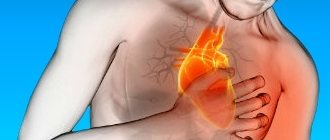
Sudden cardiac death in recent years is not as rare as it used to be, and has become more common even in young patients. This condition can occur anywhere - on the street, in public transport, at sporting events, etc. In this regard, any person, and not only a medical worker, should know how to correctly and timely provide emergency assistance to the victim. This is especially true for such techniques as precordial stroke. Of course, when carrying out such a strike, there are rules, which will be discussed below.
So, a precordial stroke is a method of physical impact on the chest of a patient who has suffered cardiac arrest. Such an effect can translate physical vibrations of the anterior chest wall and the wall of the heart into electrical excitation of the fibers of the heart muscle, since the heart tissue has the property of electrical excitability, as a result of which its mechanical irritation can provide an electrical impulse response. In other words, the mechanical effect on the heart area is a kind of mechanical pacemaker, thanks to which the normal cardiac cycle can start again. However, a number of authors are inclined to believe that such an effect is not enough to produce a full-fledged electrical systole, capable of ensuring adequate ejection of blood into the aorta, and, consequently, ensuring blood flow to the brain. There has been a lot of debate about this effect on the heart in the medical literature, and yet at the present time:
A precordial shock is considered an effective resuscitation aid, but only if the patient actually has a cardiac arrest, and the shock was carried out in the first 30-40 seconds after that.
When is it necessary to perform a precordial stroke?
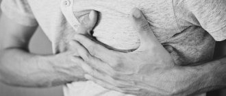
The indication for carrying out this resuscitation aid when providing first aid is the absence of an independent heartbeat in the patient, which is caused by ventricular fibrillation and/or asystole (cardiac arrest) due to other rhythm disturbances. Clinically, asystole, which caused clinical death, is accompanied by such signs as:
- Loss of consciousness
- Absence of pulse in the carotid and femoral arteries,
- Dilated pupils with lack of reaction to light,
- Lack of independent breathing movements,
- The presence of a blue tint on the skin of the face, neck, and hands.
If the doctor has the ability to conduct an ECG or cardioscopy using a monitor on a defibrillator, ventricular fibrillation, electromechanical dissociation of the heart and asystole can be reliably detected.
The algorithm for diagnosing cardiac arrest is as follows:
- If a person falls and loses consciousness, you should call out to him and shake him by the shoulder. It is unacceptable to hit a person on the cheeks; you can sprinkle water on your face.
- If there is no reaction, feel the pulse fluctuations of the carotid artery (at the angle of the lower jaw), assess the presence of independent respiratory movements - see if there is an excursion of the chest, listen to the sound of exhaled air with your ear or feel the exhaled air with your cheek (the “look, listen, feel” algorithm ).
- In the absence of pulse and respiratory movements, immediately begin performing a precordial stroke with further indirect cardiac massage and artificial ventilation using artificial respiration.
Possible complications
This method is quite traumatic. When performing a series of precordial impulses, complications are possible in the form of the following reactions:
fracture of the ribs and fracture of the xiphoid process of the sternum;- damage to the pleura and lung tissue by rib fragments;
- internal bleeding due to rupture of the lung tissue and pleura, which can cause massive hemothorax and respiratory arrest due to imbibition of the lung tissue with blood;
- in childhood - damage to internal organs (anatomically close location), which leads to disruption of the functioning of all systems.
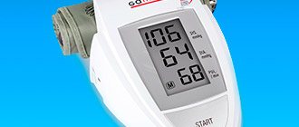
Upon arrival of the ambulance, the victim’s condition is monitored with a heart rate monitor (a finger sensor or a pad with a gel conductor on the heart area) and an ECG machine to avoid negative consequences.
When should you not apply a precordial blow?
This resuscitation aid is strictly not applied in the presence of a pulse in the carotid artery and in the presence of independent respiratory movements. This is fraught with cardiac arrest in a person who has simply lost consciousness or is in a coma, as well as in a patient with convulsive syndrome. That is, the absence of consciousness in a patient with a normal heart rhythm can be mistakenly regarded as clinical death, as a result of which a precordial blow can cause irreparable harm to the patient.
If the victim has open injuries to the chest (open gaping wounds with massive bleeding, prolapse of chest organs into the lumen of the wound), and it is also possible to visually determine rib fractures (deformation of the ribs, protruding parts of the ribs), performing a precordial blow is pointless. In this case, you should wait for doctors or rescuers to arrive.
Thus, the only contraindication for performing a precordial blow with an intact chest frame without visible damage is the presence of a pulse in the carotid or femoral arteries, as well as the presence of an independent heart rhythm on the cardiogram or on the cardioscope of the defibrillator.
Regarding pediatric patients, it should be noted that performing a precordial stroke is strictly contraindicated in children under 7 years of age due to the high likelihood of damage to internal organs. This category of victims immediately begins performing indirect cardiac massage.
What is the essence of the method?
The mechanism of precordial shock is chest compression. With sharp compression, the contractile activity of the ventricles of the heart is provoked, their blood filling and depolarization - an artificial start of the rhythm.
The method is effective at the first symptoms of cardiac arrest for up to 40-60 seconds, then compression strokes do not have the desired effect due to the progression of fibrillation, low blood discharge, and cardiac arrest.
When performing the procedure, a mechanically generated impulse occurs in the electrically excitable heart muscle from a sharp compression of the skeletal muscles. The heart muscles are anatomically built in such a way that they react with automatic excitation to an external trigger - a compression push of the chest.
An electrical impulse response most often occurs when mechanical and electrical effects are combined - performing a sequential complex of precordial shock and defibrillation electrodes on the patient’s body.
Technique for performing a precordial stroke
So, the precordial blow is applied correctly in a certain way. After a person has fallen and lost consciousness, the person providing assistance (hereinafter referred to as the resuscitator) must perform a series of sequential actions within 30-60 seconds:
- Turn the patient onto his back , placing him on a hard, flat surface (floor, ground). In no case should resuscitation be performed on a soft bed, since the chest will be “pressed” into the soft surface, and the desired effect from mechanical shaking of the chest wall will not be achieved.
- Check the pulse in the carotid artery (at the angle of the lower jaw) or in the femoral artery (in the groin).
- If there is no pulse, you should give a voice command to nearby people in order to urgently call an ambulance or rescuers. It is unacceptable to waste precious time searching for a phone yourself, as seconds count. If there are no other people nearby, you should loudly call passers-by for help.
- Simultaneously with the call for help, it is necessary to expose the victim's chest . It is unacceptable to perform a precordial blow on a person wearing a shirt, since the resuscitator may not notice small metal objects in the sternum area (pectoral cross, metal jewelry, coins, etc.). In the latter case, traumatic injury to the chest is more likely.
- After exposing the chest, the resuscitator places the index and middle fingers of the non-working hand (left for right-handed people, right for left-handed people) on the lowest part of the sternum directly above the epigastric region, that is, where the ribs meet the sternum. It is here that the most vulnerable part of the sternum to traumatic injury is located - the xiphoid process . Thus, the resuscitator covers it with two fingers at the moment of impact.
- Now you need to raise your working hand, clenched into a fist, to a height of 20-30 cm above the victim’s sternum. The correct position of the resuscitator in this case is that the resuscitator stands to the side of the victim, facing him. The working arm is raised above the patient’s chest, with the forearm directed parallel to the victim’s sternum (the resuscitator’s elbow is directed towards the victim’s stomach). When the forearm is positioned transversely (the elbow is directed towards the resuscitator), damage to the sternum is inevitable. With considerable force, one or two short blows should be made twice to the area of the uncovered part of the sternum.
You should immediately check the pulse in the major arteries. In any case, both in the presence of a pulse and in its absence, indirect cardiac massage immediately begins at a rhythm of 60-100 compressions per minute.- Precordial shock is no longer performed, but cardiac massage is performed until rescuers arrive or no less than 30 minutes from the start of resuscitation.
Figure: Performing a precordial beat
If cardiac arrest occurs in a medical facility, there is no point in wasting time looking for a defibrillator, since it is necessary to immediately begin delivering a precordial shock. If a defibrillator is at hand, for example, when cardiac arrest occurred in the intensive care unit, the resuscitator should immediately determine the type of asystole using a cardioscope and begin defibrillation using electrical pulse therapy.
Main indications
Resuscitation is performed in case of sudden cardiac arrest. This is a state of cessation of effective cardiac activity in the following cases:
- ventricular fibrillation;
ECG picture of ventricular fibrillation - electromechanical dissociation of the heart (EMD);
- asystole.
Ventricular fibrillation is chaotic, ineffective contractions of the heart muscle that occur as complications of diseases of the cardiovascular system, as well as due to electrical trauma, wounds to the heart, drug and drug poisoning. On an ECG, this condition is recorded as a sinusoidal curve. In 80% of cases, sudden cardiac arrest is caused by this.
Cardiac EMD is a condition in which the myocardium suddenly stops contracting, but the electrical activity of the heart remains. EMD occurs in cases of severe heart disease (heart attack, tumor, traumatic or necrotic rupture of myocardial walls), drug poisoning, pulmonary embolism, and repeated electrical defibrillation. An ECG sign of electromechanical dissociation is single QRS complexes. Accounts for about 5% of cases.
Asystole is the cessation of electrical activity of the heart. If during fibrillation and EMD electrical impulses are recorded on the ECG, then asystole on the cardiogram looks like a straight isoline. Its causes are heart disease with damage to its conduction system, electrolyte disturbances, drug overdose, and severe hypothermia. Asystole accounts for no more than 20% of cases.
Precordial shock is more effective for ventricular fibrillation. In this case, electrical stimulation, converted from mechanical stimulation, can help restore normal heart rhythm. With EMD and asystole, the effectiveness of the precordial stroke is less than 1%, because there are deeper damage to the myocardium and conduction system that require medication.
Mechanism of action
The precordial beat acts as a mechanical pacemaker. The kinetic energy obtained during its conduction is converted into an electrical impulse, causing the ventricles to contract. The less time has passed since cardiac arrest, the higher its effectiveness (optimally no more than 40 seconds).
In any case, this is only a temporary measure to restore heart rhythm. The resulting ventricular depolarization gradually weakens and cardiac output decreases again. Therefore, after the stroke, drug support for cardiac activity is necessary.