Portal hypertension is a complication of liver cirrhosis. This is the phenomenon of increased blood pressure in the portal vein caused by an obstruction to the blood flow from the vein.
Highly qualified doctors at the Yusupov Hospital use classical and innovative techniques, taking into account the individual characteristics of each patient’s body. Modern medical equipment allows for accurate diagnosis and effective treatment of pathology. Doctors saved the lives of a huge number of patients.
Portal hypertension syndrome: causes
Each group of hypertension occurs for different reasons.
Suprahepatic blockade occurs due to:
- pressure of a tumor or scar on the lumen of the vena cava inf.;
- Budd-Chiari syndrome;
- sticking of the leaves of the heart sac during inflammation, which impedes liver flow and increases pressure.
The hepatic form of hypertension develops for the following reasons:
- tumor development;
- taking cytostatics that destroy hepatocytes; poisoning with toxic substances. In this case, blood pressure resistance increases;
- formation of adhesions in the liver;
- chronic inflammatory processes;
- cirrhosis.
Chronic diseases and systematic intoxication of the body can be a consequence of fibrosis, which excludes the organ from the bloodstream. This scenario is possible in the absence of the necessary treatment, even taking into account the fact that the liver is an organ that can quickly regenerate in large areas of damage.
Liver cysts with heterogeneous echostructure on ultrasound
What is hidden behind a cystic formation with a heterogeneous echostructure:
- hemorrhagic or infected cyst;
- hematoma;
- abscess;
- biloma (isolated accumulation of bile);
- seroma;
- large biliary cystadenoma;
- metastases of malignant tumors with cystic or necrotic degeneration.
| Photo. Liver abscess on ultrasound. A - A cystic formation with a heterogeneous echostructure contains a liquid fraction and sediment - this is a liver abscess. B — Isoechogenic liver damage with anechoic zones. This is an abscess partially filled with pus that mimics liver tissue. The boundaries of an abscess are easy to miss. It is necessary to pay attention to the absence of vessels and the slight edge effect from the curved surface of the abscess. B — After contrast administration, the boundaries of the abscess are visible. | |||
| Photo. A - Liver hematoma. B — Focal liver lesion with a hyperechoic inclusion with a shadow behind (large arrow) is an abscess with gas. Hyperechoic gas bubbles in the bile ducts of the left lobe of the liver (small arrows). Clostridium perfringens detected. B — A 9-year-old girl, after two courses of drug treatment, has an inactive 2.5 cm echinococcal cyst in the liver - a hyperechoic arch (calcification) with a shadow behind, which is why the boundaries of the cyst are poorly visible. | ||
A cystic formation with a heterogeneous echostructure may be a benign or malignant formation, as well as a metastasis. There are no specific signs that will allow you to make a diagnosis without additional research methods.
| Photo. A — Liver metastases with a focus of necrosis in the center in a patient with bronchopulmonary carcinoma. B — A hemorrhagic cyst of the right lobe of the liver was mistakenly mistaken for an adrenal tumor. B - Echinococcal liver cyst. | ||
Portal hypertension: symptoms
The primary cause of portal hypertension is cirrhosis. With this pathology, the pressure in the portal vessel of the liver increases.
As the disease progresses, the following signs of portal hypertension appear:
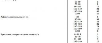
- indicators of laboratory tests change - the norms for the content of platelets, leukocytes and erythrocytes are violated;
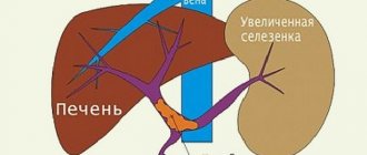
- the spleen enlarges;
- blood clotting worsens;
- accumulation of fluid in the abdominal area (ascites) is diagnosed;
- varicose veins of the digestive tract develop;
- in many cases, patients experience bleeding and anemia.
In the early stages, signs of portal hypertension in liver cirrhosis manifest themselves in the form of deterioration in general health, bloating, and heaviness under the right rib. Next, the patient develops pain in the area under the right rib, the liver and spleen increase in size, and the normal functioning of the digestive tract is disrupted.
Portal hypertension of the prehepatic form appears in childhood, the portal vein is replaced by a cavernoma, thus forming vessels of different sizes. The pathology in this case is manifested by a violation of blood clotting, clogging of the lumen of the portal vessel. Bleeding in the esophagus is quite common.
Portal prehepatic hypertension occurs with mild symptoms and has a very positive prognosis.
The internal phase of portal hypertension is characteristic of liver cirrhosis and is characterized by yellowing of the mucous membranes and skin, bleeding, ascites, and the development of liver failure.
Suprahepatic block is characteristic of Chiari disease. In this case, the symptoms have a pronounced form and manifest themselves in the form of increased body temperature, intense enlargement of the liver, severe pain under the right rib and in the abdominal area.
Fibrous-fatty degeneration of the liver on ultrasound
In fibrofatty degeneration, normal liver tissue is replaced by fatty and fibrous tissue. Fibrofatty liver degeneration on ultrasound:
- the liver is hyperechoic,
- the walls of the portal veins are not visible,
- dorsal attenuation of the signal - part of the diaphragm is not visible (severe fatty degeneration),
- The echostructure of the liver is homogeneous with diffuse fatty infiltration or heterogeneous with focal infiltration.
| Photo. Diffuse fibrofatty infiltration of the liver with a homogeneous echostructure. A, B — Against the background of hyperechoic parenchyma, the walls of the portal veins are not visible (arrow). B - Pronounced fibrofatty infiltration of the liver: hyperechoic parenchyma, the walls of the portal veins are not visible, signal attenuation - part of the diaphragm is not visible. | ||
With focal fatty infiltration, areas of preserved liver against the background of fatty infiltration can be mistaken for a mass. Preserved parenchyma is found subcapsularly, around the large trunks of the portal veins, as well as around the right and left longitudinal grooves of the liver. In order not to make a mistake, you need to evaluate the internal structure of the liver and make sure that there is NO displacement of the vascular structures .
| Photo. Focal fatty infiltration of the liver with a heterogeneous echostructure. A - A hypoechoic focus is a small area of preserved parenchyma (the walls of the vessels inside are determined) against the background of fatty degeneration. B - The echostructure of the liver is heterogeneous - normal parenchyma alternates with hyperechoic zones of fatty infiltration. B - Two hypoechoic zones near the gallbladder are areas of intact parenchyma against the background of fatty infiltration of the liver. |
Portal hypertension: degrees
In total, there are 4 degrees of pathology:
- 1st degree – functional (initial);
- 2nd degree – moderate. Accompanied by moderate dilation of the esophageal veins, enlarged spleen and ascites;
- Portal hypertension of the 3rd degree is a severe form of pathology. At this stage, pronounced hemorrhagic and ascitic syndromes are observed;
- 4th degree (complicated). The patient develops bleeding in the esophagus and stomach, gastropathy and spontaneous bacterial peritonitis occur.
Hyperechoic liver formations
When identifying a hyperechoic formation in the liver, there are no specific ultrasound signs that will allow an accurate diagnosis to be made without additional research. Hyperechoic can be:
- hemangioma;
- abscess;
- hemorrhagic cyst;
- local fat;
- adenoma;
- focal nodular hyperplasia;
- malignant tumor;
- metastasis;
- hepatocellular carcinoma;
- lymphoma.
Homogeneous hyperechoic liver formations very often turn out to be hemangiomas. If a patient without complaints and a history of malignant formations has one homogeneous hyperechoic focus less than 3 cm with clear boundaries, then the diagnosis of hemangioma can be made by ultrasound without additional research methods.
| Photo. Hemangioma is a homogeneous hyperechoic focus less than 3 cm with clear boundaries. | ||
| Photo. Atypical liver hemangiomas on ultrasound. A - Heterogeneous (heterogeneous) hemangioma of the liver. B — Not completely hyperechoic hemangioma of the liver. B — Hemangioma with a cystic cavity. D — Hypoechoic hemangioma of the liver. |
Important!!! Some lesions may look like hemangioma. If the patient is over 40 years old and has painful symptoms, additional examinations must be carried out to clarify the diagnosis.
| Photo. A - A hyperechoic formation with a hypoechoic rim (a sign of malignancy) turned out to be a metastasis of colon cancer in the liver. B — Metastasis of ovarian cancer. B - metastases of pancreatic cancer. |
Portal hypertension: diagnosis
The types of diagnostics at the Yusupov Hospital are as follows:
- Ultrasound: allows you to determine the size of the splenic, portal and superior mesenteric veins. If the diameter of the portal vein is more than 15 mm and the splenic vein is more than 7-10 mm, one can accurately state the presence of portal hypertension. Also, ultrasound examination can reveal an enlargement of the liver and spleen;
- Doppler ultrasound: allows you to examine the structure of blood vessels, as well as measure the speed of blood flow through them;
- FGDS (fibrogastroduodenoscopy): allows you to identify varicose veins of the cardial part of the stomach and esophagus, which cause bleeding in the gastrointestinal tract.
Fulminant (fulminant) hepatitis on ultrasound
Fulminant (fulminant) hepatitis is a rare but serious disease that is easy to miss. Fulminant hepatitis on ultrasound:
- the liver is heterogeneous,
- zones of reduced echogenicity with bright walls of the portal veins (a “starry sky” picture) alternate with hyperechoic areas.
A starry sky pattern indicates acute edema or necrosis, and hyperechoic areas are normal liver tissue.
| Photo. A, B — A woman developed liver failure while taking sulfonamides. On ultrasound, the liver is heterogeneous; zones of low echogenicity with bright walls of the portal veins alternate with hyperechoic areas. I remember another patient with similar changes on ultrasound. A 33-year-old woman with complaints of slight jaundice died 24 hours after admission to the hospital. An autopsy revealed lupus myocarditis and lupus hepatitis. B — A 39-year-old woman was admitted with complaints of jaundice. Ultrasound shows the liver is heterogeneous, with multiple hyperechoic foci in both lobes of the liver. Liver biopsy revealed multiple necrosis. The diagnosis is autoimmune hepatitis. During treatment with prednisone, the patient's condition noticeably improved. Delay could lead to death. | ||
| Photo. A 22-year-old man was admitted with jaundice. On ultrasound, the liver parenchyma is heterogeneous - hypoechoic zones with bright walls of the portal veins alternate with areas of normal echogenicity. Anabolic bodybuilding hormones caused acute hepatitis and liver failure, requiring a liver transplant. |
Portal hypertension: treatment
Treatment of portal hypertension in liver cirrhosis is aimed at preventing bleeding. For this purpose, the patient is prescribed somatostatin, propranolol and terlipressin. Taking medications in combination with sclerotherapy or clamping of varicose veins reduces the likelihood of recurrent bleeding by half.
The effectiveness of sclerotherapy is about 80%. The procedure involves injecting somatostatin into the damaged veins using an endoscope. Thus, the lumen of the veins becomes blocked and their walls “glue together.” This treatment method is considered classic.
As a treatment, a method such as esophageal tamponade is also used. The essence of the procedure is to inflate a special balloon inside the esophagus to stop bleeding from a varicose vein. This method of therapy can be used for no more than a day, since there is a high probability of developing peritonitis.
Surgery for portal hypertension is performed in cases where the patient’s condition is stabilized and liver function is normalized. Also, the surgical method is indicated when other treatment methods are ineffective.
The last resort treatment is liver transplantation. It is indicated in cases of liver cirrhosis with two previous bleeding events.
The prognosis depends on the degree of development of the disease, the characteristics of the underlying pathology that caused portal hypertension, the degree of liver dysfunction, as well as the frequency and intensity of bleeding.
The treatment tactics chosen by the doctor play an important role in the prognosis. A large number of patients with this diagnosis are admitted to the Yusupov Hospital every day. However, in many cases, patients consult a doctor at advanced stages of the disease, which complicates the treatment plan.
Simple cysts in the liver on ultrasound
A simple liver cyst, like any other cyst in our body, is anechoic, has smooth edges and excellent transmission of the ultrasound signal - enhancement behind the cyst (arrows).
| Photo. Simple liver cysts. | ||
An old hematoma, hydatid abscess, biloma (limited collection of bile) and seroma (limited collection of serous fluid) may look like a simple cyst.
| Photo. A — A 9-year-old girl living in Turkey was admitted with complaints of abdominal pain. Ultrasound of the liver revealed three hydatid cysts at different stages of development. Around each larva of echinococcus, connective tissue grows and a hydatid cyst is formed. B - simple thick-walled cyst with a small detachment of the inner layer. B - A thick-walled cyst curls into a “snake” sign. D — Simple cyst without endothelial detachment. Keep in mind that an hydatid cyst grows 1-5 cm per year. | |||
| Photo. A - Multiple liver cysts in adults are often associated with autosomal dominant polycystic kidney disease. B - Daughter cysts inside the main hydatid cyst in the liver. B - Echinococcal cyst with daughter cysts in the kidney. | ||
Cysts in the liver may be dilated bile ducts
There are 5 types of bile duct cysts. In types 4 and 5, ultrasound reveals multiple cysts.
| Photo. Five types of bile duct cysts. Type 1 - isolated dilatation of the common bile duct. Type 2 is a small common bile duct diverticulum. Type 3 is a small protrusion of the common bile duct into the duodenum. Type 4 - dilated bile ducts throughout the liver and pouch-like dilatation of the common bile duct. Type 5 (Caroly's disease) - dilatation of the intrahepatic bile ducts, without dilatation of the common bile duct. | ||||
| Photo. A, B — Common bile duct cyst type I (CC). GB—gallbladder, PH—pancreas, VP—portal vein. B - Liver cysts are connected to each other in a convoluted tubular system. These are bile duct cysts. The common bile duct may be normal (type 5) or dilated (type 4). | ||
| Photo. Caroli disease is a dilatation of the intrahepatic bile ducts associated with a defect in the development of the wall. Normally, each portal vein is surrounded by bile ducts. In Caroli disease, a very important sign on ultrasound is the central point, which is surrounded by dilated bile ducts. A - We see points surrounded by dilated bile ducts. B - With CDK, blood flow is determined at central points - these are the portal veins. B - The central points are clearly visible on the CT scan (arrow). In Caroli disease, dilatation of the bile ducts is often accompanied by polycystic kidney disease. A kidney exam can sometimes help make the diagnosis. | ||
Important!!! If the cystic formation is not very round, it may be a vascular structure - an aneurysm, arterio-portal or portal-hepatic fistula.
Portal hypertension: prevention
Measures to prevent the development of the disease include:
- maintaining a proper diet and nutrition regimen;
- playing sports;
- vaccinations against viral hepatitis;
- refusal to abuse alcoholic beverages;
- avoiding exposure to harmful production factors in the form of poisoning with toxic substances.
Preventative measures for liver diseases are:
- a complete examination to make a diagnosis in the early stages of liver disease and initiate treatment;
- strict compliance with all doctor’s recommendations;
- complex therapy in a hospital setting under the strict supervision of doctors.
Measures to prevent the development of bleeding include:
- control of blood clotting function;
- sigmoidoscopy - that is, examination of the sigmoid and rectum, annually;
- fibrogastroduodenoscopy twice a year.
Types of liver echostructure
Types of liver echostructure: centrilobular, normal, fibrofatty.
| Photo. Centrilobular liver. B - Normal liver. B - Fatty liver. | ||
| The centrilobular liver is edematous, therefore the echogenicity of the parenchyma is reduced. It seems that there are a lot of small portal veins, and their walls shine brightly - a symptom of the “starry sky”. In fact, their number has not increased, the image is simply more contrasting. The aperture is clearly visible - a very bright line. | In fibrofatty degeneration, normal liver tissue is replaced by fatty and fibrous tissue. The liver becomes hyperechoic. The contrast between the parenchyma and the walls of the small portal veins disappears - they are poorly visible or not visible at all. The liver is dense, so the diaphragm is difficult to see. |
|
|
Important!!! The centrilobular picture of the liver can also be seen in healthy people. They are usually thin young adults and teenagers. If biochemical blood parameters (ALT, AST, GGT, bilirubin) are normal, then we can say with confidence that the patient is healthy.
Where to go for portal hypertension?
Oncologists at the Yusupov Hospital have extensive experience in the treatment of portal hypertension. Modern medical equipment allows for accurate diagnosis and effective treatment of pathology. The clinic’s doctors saved the lives of a huge number of patients.
Experienced doctors use classical and innovative techniques during treatment, taking into account the individual characteristics of each patient’s body. The problem is examined from all angles, any risks are weighed, and only after that the doctor makes a decision regarding the choice of the optimal and most effective treatment method.
Your health is in your hands. Trust it only to high-level professionals.
Call the Yusupov Hospital by phone or make an appointment using the appointment form on the website. The coordinating doctor will answer all your questions.
Hypoechoic liver formations on ultrasound
There are no specific ultrasound signs for the differential diagnosis of hypoechoic liver formations. In the liver, hypoechoic can be:
- abscess;
- adenoma;
- focal nodular hyperplasia;
- typical or atypical hemangioma;
- local fatty degeneration;
- microabscesses;
- malignant formation;
- metastases;
- lymphoma.
| Photo. A - Hypoechoic formation in the liver is benign focal nodular hyperplasia. The literature describes that it is characterized by a central scar (white arrow). However, this may not be a diagnostic criterion in 100% of cases. Pictures B and C show two liver masses with a central scar. These are hepatocellular carcinoma and giant hemangioma, respectively. |
| Photo. A - Hypoechoic formation (red arrow) in the liver is a fungal abscess. B — Diffuse, homogeneous, hypoechoic foci (microabscesses) throughout the liver are liver candidiasis in a seven-month-old boy with immunodeficiency. B - Numerous hypoechoic formations against the background of hyperechoic liver parenchyma are liver lymphoma. | ||
Forecast
The average lifespan of patients with this pathology is 10-15 years after surgery to apply artificial anastomoses. This is a fairly high figure considering the scale of the damage. This period can be extended by adequate prevention of complications, supportive therapy, and regular medical examinations.
Clinical recommendations include normalizing the daily routine and diet. It is necessary to limit the consumption of salt and other foods that increase blood pressure. It is also important to monitor the volume of fluid consumed and not exceed the recommended volume (set by the doctor on an individual basis). Heavy physical activity is strictly prohibited.
The fatal factor is most often internal bleeding; this complication accounts for more than 50% of deaths due to this pathology.