It branches like a tree, first into large branches (trunks), then into smaller branches and twigs, and is conventionally divided into several parts or sections:
- 1. The ascending aorta is the area from the aortic valve to the brachiocephalic trunk.
- 2. The aortic arch is a short section from which all the vessels supplying the arms and head (brachiocephalic arteries) depart. They anatomically form an arch connecting the ascending and descending aorta.
- 3. The descending (thoracic) aorta begins from the mouth of the left subclavian artery and continues to the diaphragm.
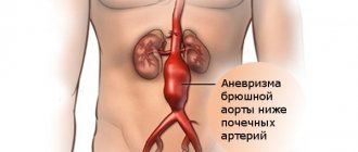
- 4. Below the diaphragm and before the bifurcation of the aorta (bifurcation) is the abdominal aorta.
Dividing the aorta into sections is very important for assessing risk and choosing optimal treatment tactics in patients with aortic aneurysms.
An aortic aneurysm is an area of local expansion.
Causes of aortic enlargement
Congenital systemic connective tissue diseases: Marfan syndrome, Ehlers-Danlos syndrome, caused by genetic changes in which the aortic wall has an abnormal structure, can cause the development of an aneurysm.
Acquired diseases that cause aneurysmal changes in the aortic wall: most often this is atherosclerosis. About 80% of all complicated aortic aneurysms are aneurysms caused by an atherosclerotic process, which leads to weakening of the vessel wall and the inability to withstand normal blood pressure, and as a result, to its expansion.
Less commonly, an aortic aneurysm develops in inflammatory diseases caused by external agents (syphilis, fungal infection, tuberculosis) or in autoimmune diseases (nonspecific aortoarteritis).
Symptoms of aortic aneurysm
Unfortunately, the diagnosis of aortic aneurysm cannot always be established during the “cold period” (before complications develop), since this disease is usually asymptomatic. Most often, it is discovered accidentally during fluorography, ultrasound or tomography studies performed in connection with other diseases. Treatment of an aneurysm of the ascending aorta before complications develop is much safer for the patient, therefore, in the early diagnosis of an aortic aneurysm, routine medical examination is important.
It is worth noting that every hundredth patient who died suddenly dies from aortic dissection.
Complaints usually appear when the aneurysm begins to stratify or, enlarging, compresses surrounding organs and tissues. Pain or dysfunction of those organs located in the area of the aneurysm appears. At first, this is not of a bright nature and, therefore, does not alarm either the patient or the doctor.
However, the pain intensifies as these deadly complications of an aortic aneurysm develop—it is some of the most severe pain a person can experience. It is localized in the chest if the aneurysm is located in the ascending, descending sections or in its arch, or in the abdomen if it formed in the abdominal section. Characterized by severe weakness, pallor, and often the person loses consciousness.
Impaired blood supply to organs located in the area of aneurysm rupture or aortic dissection (brain or spinal cord, kidneys, intestines, upper or lower extremities) leads to loss of function of these organs, and a large volume of blood loss during aortic rupture represents the most serious danger. To save a life, minutes count. If early surgical treatment is not available, the first-day mortality rate for aortic dissection is 1% per hour (one person in a hundred dies every hour). Within the first 24 hours, 33% of patients die from aortic dissection, 50% of patients within 48 hours and 75% within two weeks. Only early surgical intervention makes it possible to save a significant proportion of patients.
Treatment
Timely treatment creates opportunities to improve the quality of life and prevent complications. Usually, first of all, the doctor recommends adjusting the patient’s lifestyle.
For the treatment of pathology it is very important:
- Stick to proper nutrition. The patient should avoid salty, spicy and fried foods. It is important to reduce the amount of animal fats. The diet should be based on vegetables and fruits, legumes, low-fat dairy products, and whole grains.
- Devote time to physical activity. They must be regular. Physical activity is not only good for the body, but also allows you to normalize your weight.
- Get proper rest and saturate your body with nutrients. You should sleep at least 7-8 hours, spend time in nature, take vitamins and minerals.
The pathology is directly treated with medication.
Typically patients are prescribed:
- Statins. These substances help reduce the concentration of cholesterol in the blood. Statins reduce the synthesis of aggressive substances.
- Nicotinic acid and derivatives. These substances increase the content of high-density lipoproteins, reduce the level of cholesterol and triglycerides.
- Bile acid sequestrants. These substances remove cholesterol.
- Fibrates. These substances suppress the synthesis of triglycerides in the liver and promote their rapid removal.
- Beta blockers. These substances help eliminate chest discomfort and pain, as well as reduce pressure.
If drug treatment of the pathology is ineffective, operations are performed.
Today, specialists carry out the following interventions:
- Shunting. The operation consists of applying a shunt, which normalizes the impaired blood flow.
- Angioplasty. This operation involves reconstruction of the vessel. As a result of the intervention, its lumen is restored.
If an aneurysm is found, it is excised. The removed area is replaced with a prosthesis. If the aortic valve annulus becomes enlarged, it is excised and replaced with an artificial one.
Important! A ruptured aneurysm can only be treated with surgery. The operation is performed urgently.
Our clinic provides complex therapy for pathologies. Specialists not only eliminate the manifestations of the disease, but also its causes. This increases the patient's standard of living.
Therapy prices depend on numerous factors.
Among them:
- complexity of therapy;
- chosen technique;
- patient's condition;
- presence of concomitant diseases, etc.
The price for complex therapy can be announced in advance. This will allow you to plan the costs of our clinic’s services.
Important! Doctors approach therapy individually. They must take into account all factors that may affect its success.
Diagnosis of aortic aneurysm
In the diagnosis of aortic aneurysms, the so-called imaging techniques (ultrasound, MRI, CT, AG) are of greatest importance. In the ascending aorta, its arch and in the abdominal section, an aneurysm can be detected using ultrasound methods (ultrasound). To diagnose an aneurysm of the descending (thoracic) aorta, X-ray methods (x-ray, computed tomography) are required. To establish a final diagnosis and select a treatment method, contrast research methods are performed. Currently, the optimal diagnostic method, which provides the most complete information about the location, extent, diameter of the aneurysm and its relationship to nearby organs, is multislice computed tomography - aortography.
Instrumental examination
X-ray examination. For aneurysms of the thoracic aorta, radiography is performed in three projections with mandatory contrasting of the lumen of the esophagus. The expansion of the shadow of the vascular bundle is characteristic. Aneurysms of the descending aorta bulge into the left pulmonary field. Most patients experience displacement of the contrast-enhanced esophagus. Sometimes calcification (calcification) of the aneurysmal sac is determined. In case of aneurysms of the abdominal aorta, plain radiography of the abdominal cavity in two projections allows us to identify calcification of the aortic wall and urination of the lumbar vertebral bodies.
Ultrasound examination (ultrasound) of the aorta and heart. Ultrasound allows us to identify the presence and size in diameter and along the length of aneurysms of the ascending, descending aorta, aortic arch, abdominal aorta, the condition of the vessels extending from the aorta, as well as the presence of aortic valve defect, the nature of changes in the aortic wall.
Computed tomography (CT). When the lumen of the aorta is more than 4 cm, its expansion is considered to be aneurysmal. When performing computed tomography, it is possible to determine the involvement of large arteries in the process and identify signs of dissection (dissection) of the walls (in case of dissecting aortic aneurysm). Angiographic examination (aortography). It is used, as a rule, before surgery when planning its nature and volume.
Treatment methods for aortic aneurysm
The main method of treating an aneurysm of any part of the aorta is surgical. The point of the method is to replace the dilated section of the aorta in order to prevent its further stretching and rupture. To replace the aorta, two methods are used - the endovascular (intravascular) method using a special intravascular prosthesis (stent graft), and open surgery - aortic replacement.
Each method has its own indications, and each of them has its own advantages and disadvantages.
The advantages of the surgical method lie in its versatility, that is, the ability to correct all disorders associated with an aortic aneurysm, regardless of the department and nature of the lesion. For example, in case of an aneurysm of the ascending aorta and damage to the aortic valve, aortic and aortic valve replacement is performed in combination with coronary bypass surgery.
To perform surgery on the ascending aorta and its arch, it is necessary to use artificial circulation, systemic hypothermia, and often complete circulatory arrest.
Indications for surgical treatment
- transverse size of the aneurysm,
- aneurysm growth rate;
- the formation of complications of this disease.
For each section of the aorta, there is a cutoff limit for the transverse size of the aorta, after which the risk of aortic rupture statistically significantly increases. Thus, for the ascending and abdominal aorta, the transverse diameter of the aneurysm is 5 cm dangerous in terms of rupture, for the thoracic aorta - 6 cm. If the diameter of the aneurysm increases by more than 6 mm in 6 months, then this is also an indication for surgery. Also threatening in terms of rupture and dissection of the aorta are the saccular form of the aneurysm and expansion of the aorta, which is smaller than the diameter that is the indication for surgery, but accompanied by pain at the site of expansion and dysfunction of the presenting organs. Dissections and completed ruptures of aneurysms are absolute indications for emergency surgery.
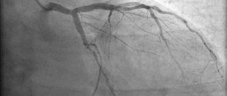
Aortography of the aorta with intravenous enhancement
What is the aorta? This is one of the largest vessels of the human body. The walls of the aorta are subject to enormous stress with each systolic impulse of blood. Due to its large diameter, the aorta experiences significantly greater stress compared to other vessels, as a result of which it is susceptible to a large number of diseases and pathological processes.
Diagnosis of various changes in the aorta is difficult. One of the modern methods that allows you to verify the diagnosis is MR aortography with intravenous enhancement. What does MR aortography reveal?
You can find out the cost of an MR aortography study by calling the phone number located on the contacts tab
Let's consider several examples of pathological changes in the aorta detected during MRI examination:
Aortic aneurysm
A true aneurysm is characterized by expansion of all 3 layers of the vascular wall. Why does it occur? Age, male gender and smoking are the most important risk factors for the development of this pathology. Both limited and generalized expansion of the aorta is possible. A combination of aortic aneurysm and abdominal arteries is often found.
What are the signs of an aortic aneurysm? Clinically, an asymptomatic course of the disease is possible. It happens that patients complain of pain in the abdomen and back. Aneurysms larger than 5 cm in diameter are palpable in most cases.
Read the material on the topic: Why do you need contrast when performing magnetic resonance imaging?
How can MRI help with an aortic aneurysm? MR angiography allows you to determine the location of the aneurysm and its relationship to the branches of the aorta; inflammatory and infiltrative changes in the surrounding tissue, blurred contours of the aneurysm and the threat of perforation when visualizing a contrast agent in the extravascular space.
Aneurysm of the infrarenal aorta, stenosis of the mouths of both iliac arteries
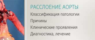
Aortic dissection
What it is? Dissection of the aotra is its dissection due to rupture of one of the layers of the wall. When the inner layer is damaged, blood penetrates between the layers, followed by delamination of the vessel wall. This can be caused by high blood pressure, diseases of the connective tissue of the vessel wall, and, less commonly, atherosclerosis. The most common procedure is dissection of the thoracic aorta.
Clinically, aortic dissection manifests as unexpected severe chest pain. In this case, such a condition can spontaneously disappear.
Read material on the topic: Chest pain - what is it: heart or nerves?
Aortic dissection
The picture shows MIP reconstruction, MR aortography with intravenous contrast.
a) oblique-sagittal MIP image. The detached intima (head of the black arrow) and the true (1) and false (2) lumens of the aorta are shown;
b) T2-axial MIP image at the level of the pulmonary trunk. A detached intima is visualized (black arrow), an oval-shaped true lumen (1) and a crescent-shaped false lumen (2)
As a result of the accumulation of blood between the layers, a pocket may form, leading to a limitation of the patency of the aorta itself. In addition, the dissection may spread to affect the arteries leaving the aorta. Such situations are also well differentiated by MR aortography with IV contrast.
True and false lumen of the aorta
In the figure - 1 - the true lumen of the aorta; 2 - false lumen of the aorta; SMA-superior mesenteric artery; SA-celiac trunk; LRA-left renal artery; RRA-right renal artery.
The mouth of the left renal artery opens into the false lumen of the vessel, the blood flow through it is impaired. The mouth of the superior mesenteric artery opens into both lumens. The right renal artery receives blood from the true lumen of the aorta
Chronic occlusive lesion of the aorta
The most common signs of occlusions and stenoses of atherosclerotic origin. Isolated damage to the aorta is typical, but there may be a combination of damage to the aorta and the iliac arteries.
Stenosis or occlusion of the aorta is usually detected in its distal section, near the bifurcation (posteriorly). As a rule, the severity of collateral circulation is noted.
MRA allows you to visualize the location, extent and degree of stenotic lesions and features of collateral blood flow.
Chronic occlusive lesion of the aorta
In the figure, occlusion of the infrarenal aorta, left renal and inferior mesenteric arteries is determined. Against this background, a fusiform aneurysm of the right renal artery is visualized and the vessel supplying blood to the left parts of the colon (arrow heads) is identified.
Leriche syndrome
What it is? This is complete obliteration of the aortic bifurcation (aortoiliac occlusion). A rare case of occlusive lesions of peripheral arteries.
The occlusive process begins from the infrarenal aorta, spreading to the iliac arteries. Maintaining peripheral perfusion usually occurs due to a well-developed system of collaterals (arteries of the chest and abdominal walls).
What are the symptoms of Leriche syndrome? Clinical picture: symptoms of occlusive lesions of peripheral arteries, impotence. Acute bilateral ischemia of the lower extremities may develop.
They don't like to talk about this problem. She tends to get scared. She can “drive you into depression.” But is everything really so tragic? Read about the causes of erectile dysfunction here
When performing MR aortography with IV contrast in patients with Leriche syndrome, the distal blood flow and collaterals are well visualized; length of the occlusion area; state of collateral blood supply and the presence of occlusive lesions of the branches of the aorta (mesenteric, renal) and peripheral arteries.
Leriche syndrome
In the figure, the occlusion of the infrarenal aorta, the level of the bifurcation and both common iliac arteries is determined. In the femoral arteries, blood flow is visualized due to well-developed collaterals. The arrow heads show the collateral from the right VI intercostal artery entering the right femoral artery. The arrow indicates concomitant stenosis of the right renal artery.
Other materials on topics:
MR angiography in the study of cerebral arteries
How will MRI of cerebral vessels help a patient?
Contraindications for MRI
Types of open surgical operations for aortic aneurysms:
- Bentalla-De Bono operation (replacement of the ascending aorta with a valve-containing conduit with a mechanical prosthetic aortic valve);
- David's operation (replacement of the ascending aorta while preserving the native aortic valve);
- Supracoronary aortic replacement;
- Prosthetics of the ascending aorta and its arch (Borst technique, the use of oblique aggressive anastomosis and other techniques);
- Thoracic aortic replacement;
- Abdominal aortic replacement.
Endovascular interventions
They allow you to dramatically reduce the amount of surgical trauma, shorten hospitalization periods and reduce the inevitable suffering of the patient associated with surgical approaches. One of the main disadvantages of the method is the need for repeated interventions.
Types of endovascular operations for aortic aneurysm:
- implantation of a stent graft into the abdominal aorta,
- implantation of a stent graft into the ascending (thoracic) aorta.
The most modern method of treating aortic aneurysm is a hybrid method, which allows achieving optimal treatment results with the least surgical trauma.
Hybrid surgeries combine the advantages of open and endovascular interventions.
To prevent the development of aortic aneurysms, the most important thing is the need to control risk factors, namely arterial hypertension. In addition to arterial hypertension, the most significant risk factors are age (over 55 years), male gender, smoking, the presence of aneurysms in direct relatives, and high cholesterol levels.
Prevention of aortic atherosclerosis
Prevention of aortic atherosclerosis helps prevent the development of the disease. This is easier than treating pathology for a long time.
To eliminate reversible risk factors, it is enough to:
- stop smoking and drinking alcohol;
- avoid overeating and stressful situations;
- get rid of excess weight.
If there is a hereditary predisposition to aortic atherosclerosis, you should regularly donate blood to assess cholesterol levels. This will allow you to quickly identify any disturbances in lipid metabolism and correct it.
It is especially important to prevent pathology for people with:
- obesity;
- hypertension;
- diabetes;
- angina pectoris.
All people over the age of 40 should visit a cardiologist regularly. It is important to pay close attention to health for those who experience angina attacks or have suffered a stroke.