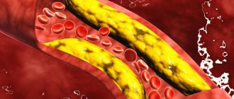
The term "hypoplasia" means "underdevelopment". Such defects of intrauterine development are also found in the arteries of the brain. In the arteries of the carotid artery basin, these anomalies exist in approximately 4% of people, and in the arteries of the vertebrobasilar basin - in every tenth person.
Despite the fact that the brain is the most energy-intensive organ and requires massive blood supply, hypoplasia of the arteries that supply it can go unnoticed for a long time. This is due to the characteristics of the cerebral circulatory system.
Most of the hemispheres receive blood from the system of internal carotid arteries, which form the anterior and middle cerebral arteries, as well as the anterior communicating artery. The posterior part of the hemispheres, the brainstem and the cerebellum are fed from the basin of two vertebral arteries, which merge in the cranial cavity into the main artery, from which the posterior cerebral and posterior communicating arteries are formed.
Due to the anterior and posterior communicating arteries, a connection is formed between the arterial basins of the brain, forming a circle. It is called Willis, and within its limits a compensatory flow of blood into areas with reduced arterial blood flow is possible, including due to hypoplasia of any segment of the artery. It is this feature of the brain’s blood circulation that ensures the long-term asymptomatic course of most hypoplasias, which often become an accidental discovery.
Clinical manifestations of hypoplasia of the cerebral arteries occur against the background of another pathology that worsens the conditions for compensation of cerebral circulation. Most often this is atherosclerosis of cerebral vessels or diseases of the cervical spine. As a result, transient ischemic attacks and ischemic strokes may occur.
Symptoms
Symptoms of chronic cerebral circulatory disorders depend on the vascular pool in which ischemia of brain tissue develops. All patients may be concerned about:
- headache;
- dizziness;
- noise in the head;
- memory impairment;
- sleep disorders.
Hypoplasia of the carotid arteries is also characterized by weakness and/or numbness in the limbs and speech impairment. For ischemia in the vertebral artery basin - impaired coordination, gait instability.
To diagnose hypoplasia of cerebral arteries and determine treatment tactics, the following are carried out:
- cerebral angiography - X-ray examination of the arteries of the brain with a contrast agent injected into them;
- CT angiography;
- MR angiography;
- ultrasound examination (duplex scanning) of the vessels of the neck and head;
- PAT;
- single photon emission computed tomography.
Conservative therapy with drugs that improve blood supply and metabolism of the brain is carried out if the examination data suggests that it can prevent further worsening of cerebral ischemia and ischemic stroke.
Hypoplasia of the vertebral artery
If you complain to a neurologist about dizziness and instability, then most likely you will be advised to undergo an ultrasound examination of the cerebral vessels. And now you are reading a medical report, where it is written in black and white “small diameter vertebral artery” or “vertebral artery hypoplasia.” It sounds alarming, and the attending physician is in no hurry to allay the concern. What actually lies behind the incomprehensible diagnosis and what to do about it now?
What it is
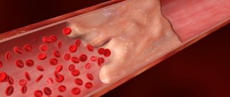
Hypoplasia of the vertebral artery (small diameter artery) is a congenital narrowing of the vertebral artery, most often the right one. A person normally has two vertebral arteries (sometimes there are more), which flow into the basilar artery, which is part of the circle of Willis.
The circle of Willis is, in fact, the connection of the branches of all large vessels of the brain. Normally, this circle is closed, which ensures blood supply and brain function when any of the arteries is “switched off” (for example, due to a blood clot). Therefore, if one of the vertebral arteries is narrowed and the blood flow through it is changed, then the second takes on part of the load.
Symptoms of hypoplasia
Vertebral artery hypoplasia usually has no symptoms if other vessels are functioning normally. In this case, blood circulation is compensated by the second vertebral artery and larger carotid arteries (remember the circle of Willis?). If, nevertheless, compensation does not occur, then patients complain of blurred vision, unsteady gait, and impaired coordination of movements.
Causes
The following reasons give rise to it:
- Hereditary factors. For example, one of the parents may carry a recessive gene, which, due to consanguineous marriages, manifests itself in the child. Characteristic of closed communities where incest is permitted. A striking example is cerebellar hypoplasia due to disruption of the VLDLR gene, which manifests itself in cases of consanguineous blood mixing.
- Teratogenic factors: physical, biological and chemical effects on the body of the mother and child. For example, neuroinfections, living in areas with high levels of radiation, drugs that have not passed the teratogenicity test.
- Injuries during pregnancy.
- Maternal toxicosis.
- Smoking, alcoholism and drug addiction of parents.
- Pathologically reduced amount of amniotic fluid.
Main causes and consequences of pathology
Hypoplasia of the structures of the circulatory system of the brain is usually a congenital pathology, the debut of which usually occurs in the first years of a child’s life. In this case, the provoking factor for the disease is intoxication with harmful substances during pregnancy.
This can lead to:
- Smoking;
- Drinking alcoholic beverages;
- Poisoning of the body with waste products of pathogenic organisms (rubella, influenza);
- Uncontrolled use of medications, intoxication with them.
Improper formation of the vertebral arteries can be a consequence of stress and depression in a pregnant woman.
Mild hypoplasia of the brain may not appear for quite a long time, but during the period of exacerbation of chronic diseases of the cardiovascular system it will seriously complicate the situation.
Acquired pathology most often appears due to mechanical damage to the corresponding structures of the circulatory system or their pinching between the vertebral discs. Such changes occur due to a violation of the integrity of the cervical vertebrae or prolonged wearing of a cervical brace.
A severe form of vascular hypoplasia is always associated with serious consequences. These include:
- The risk of developing aneurysms and, as a consequence of their rupture, stroke;
- Instability of blood pressure, hypertension;
- Deterioration in general health, fatigue, drowsiness;
- Dizziness, fainting, and severe vomiting.
Underdevelopment of cerebral vessels in the fetus is fraught with serious consequences, including the development of defects in the structures of the central nervous system. Subsequently, they can lead to dysfunction of the corpus callosum, underdevelopment of the cerebellum, etc.
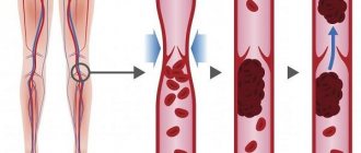
Difficulties in diagnosing thrombosis of the cerebral veins and venous sinuses
Thrombosis of the cerebral veins and venous sinuses is more common in young women than in men, but is generally rare (up to 1% of all cases of cerebral infarction) [1]. According to ISCVT (International Study on Cerebral Vein and Dural Sinus Thrombosis, 2004), the incidence annually is 3–4 cases per 1 million in adults and up to 7 cases per 1 million in children. The mortality rate for this disease ranges from 5 to 30%; during more than 2 years of observation, a corresponding figure of 8.3% was recorded. At the same time, more than 90% of patients had a favorable prognosis. The main risk factors for the development of thrombosis of the cerebral veins and venous sinuses in the population are infectious inflammatory processes (otitis, mastoiditis, sinusitis, septic conditions) and non-infectious causes. Non-infectious causes of thrombosis of the cerebral veins and venous sinuses can be localized or general. The most commonly cited are traumatic brain injury, tumors, head and neck surgery, and pacemaker implantation or central venous catheter placement. Common conditions that contribute to thrombosis of the cerebral veins and venous sinuses include conditions such as hemodynamic compromise (eg, congestive heart failure, dehydration), blood disorders (polycythemia, sickle cell disease, thrombocytopenia), and coagulopathy (disseminated intravascular coagulation syndrome). , deficiency of antithrombin, protein C and protein S), as well as thrombophilic conditions associated with pregnancy, childbirth and the use of oral contraceptives, antiphospholipid syndrome, systemic vasculitis. However, in 15% of cases the cause of sinus thrombosis remains unknown [5]. Difficulties in diagnosing thrombosis of the cerebral veins and venous sinuses are due to the polymorphism of its clinical picture and the variability of the structure of the cerebral venous system. Anatomical features of the structure of
the cerebral venous system
In the development of thrombosis of the cerebral veins and sinuses, an important role is played by the anatomical features of the structure of the cerebral venous system (Fig. 1).
Unlike arteries and peripheral veins, cerebral veins lack a muscular wall and valve apparatus. The venous system of the brain is characterized by “branching”, a large number of anastomoses and the fact that one vein can receive blood from the basins of several arteries. Cerebral veins are divided into superficial and deep. Superficial veins - the superior cerebral vein, the superficial middle cerebral vein (Labbe vein), the inferior anastomatic vein (Trolar vein), the inferior cerebral veins - lie in the subarachnoid space and, anastomosing among themselves, form a network on the surfaces of the cerebral hemispheres. The main mass of venous blood from the cortex and white matter flows into the superficial veins, and then into the nearby sinus of the dura mater. Blood enters the deep cerebral veins from the veins of the choroid plexus of the lateral ventricles, basal ganglia, thalamus, midbrain, pons, medulla oblongata and cerebellum. The superficial and deep veins drain into the sinuses of the dura mater. The superficial veins drain mainly into the superior sagittal sinus. The main collectors of the deep veins are the great cerebral vein (vein of Galen) and the straight sinus. Blood from the superior sagittal and direct sinuses enters the transverse and sigmoid sinuses, which collect blood from the cranial cavity and drain it into the internal jugular vein. In the development of thrombosis of the cerebral veins and venous sinuses, 2 mechanisms are involved that determine the symptoms of the disease. The first is occlusion of the cerebral veins, causing cerebral edema and impaired venous circulation. The second link in the pathogenesis of thrombosis of the cerebral veins and venous sinuses is the development of intracranial hypertension due to occlusion of large venous sinuses. Normally, cerebrospinal fluid is transported from the cerebral ventricles through the subarachnoid space of the lower and superolateral surfaces of the cerebral hemispheres, adsorbed in the arachnoid plexuses and flows into the superior sagittal sinus. With thrombosis of the venous sinuses, venous pressure increases, as a result of which the absorption of cerebrospinal fluid is impaired and intracranial hypertension develops [5]. Both of these mechanisms determine the clinical symptoms of sinus thrombosis. Clinic and diagnosis
Clinical manifestations of thrombosis of the cerebral veins and venous sinuses depend on the location of thrombosis, the speed of its development and the nature of the underlying disease.
Severe disorders of the venous circulation are characterized by headache, vomiting, swelling of the optic discs, focal and generalized convulsions, and progressive depression of consciousness. However, with early recognition of the process, the clinical picture may be less pronounced. Focal neurological disorders can occur with isolated thrombosis of deep or superficial veins or with the spread of thrombosis from the sinuses to the veins. Meningeal syndrome is considered a rare manifestation of uncomplicated sinus thrombosis. Cerebrospinal fluid pressure, according to most authors, is normal or moderately elevated. The composition of the cerebrospinal fluid can be either unchanged or with a slightly increased protein content and pleocytosis of no more than 200/3 [1, 11–13]. Septic thrombosis of the transverse and sigmoid sinuses is characterized by a pronounced range of body temperature, leukocytosis, and accelerated ESR, but the use of antibiotics can smooth out these manifestations. Swelling of the mastoid area, pain on palpation, and less filling of the internal jugular vein on the affected side are very common. Sometimes thrombosis of the sigmoid sinus spreads to the internal jugular vein, which is accompanied by local inflammatory changes along the vein [1]. In contrast to arterial thrombosis and thromboembolism, neurological symptoms in thrombosis of the cerebral veins and venous sinuses more often develop subacutely - within a period of several days to 1 month. (50–80% of cases), although an acute onset may also occur (20–30% of cases) [6]. The most common symptom of thrombosis of the cerebral veins and venous sinuses is intense headache (92% of patients), which is a reflection of the development of intracranial hypertension. It resembles the pain of subarachnoid hemorrhage and is not relieved by analgesics. In addition, according to ISCVT et al. [1, 7–13], the following symptoms are identified: – motor disorders – 42%; – convulsive syndrome – 37% (including status epilepticus – 13%); – psychomotor agitation – 25%; – aphasia – 18%; – visual impairment – 13%; – depression of consciousness (stunning, stupor, coma) – 13%; – disorders of the innervation of cranial nerves – 12%; – sensitivity disorders – 11%; – meningeal syndrome – 5%; – vestibulo-cerebellar disorders – 1%. In the long-term period, the most common symptoms are headache (14%) and convulsions (11%). The cavernous and superior sagittal sinuses are relatively rare sites of infection. More often, the intracranial process is the result of the spread of infection from the middle ear, paranasal sinuses, skin near the upper lip, nose and eyes. With thrombophlebitis of the marginal sinus, which usually occurs against the background of inflammation of the middle ear or mastoiditis, pain in the ear and pain when pressing on the mastoid process appear. After a few days or weeks, fever, headache, nausea and vomiting appear due to increased intracranial pressure. Swelling in the mastoid area, dilation of the veins and pain along the internal jugular vein in the neck occur. When the internal jugular vein is involved in the pathological process, pain in the neck and limitation of its movements are observed. Drowsiness and coma often develop. In 50% of patients, swelling of the optic discs is detected (in some patients it is unilateral). Seizures occur, but focal neurological symptoms are rare. The spread of the pathological process to the inferior petrosal sinus is accompanied by dysfunction of the abducens nerve and trigeminal nerve (Gradenigo syndrome). Thrombophlebitis of the cavernous sinus is secondary to oculonasal infections. The clinical syndrome is manifested by swelling of the orbit and signs of dysfunction of the oculomotor and trochlear nerves, the orbital branch of the trigeminal nerve and the abducens nerve. Subsequent spread of infection to the opposite cavernous sinus is accompanied by the occurrence of bilateral symptoms. The disease can begin acutely, with the appearance of fever, headache, nausea and vomiting. Patients complain of pain in the eyeball area, pain in the orbital area when pressed. Chemosis, edema and cyanosis of the upper half of the face are noted. Consciousness can remain clear. Ophthalmoplegia, sensory disturbances in the area of innervation of the ophthalmic branch of the trigeminal nerve, retinal hemorrhages and papilledema may occur. Infection of the superior sagittal sinus can occur when the infection is transferred from the marginal and cavernous sinuses or spreads from the nasal cavity, the focus of osteomyelitis, the epidural and subdural areas. The disease is manifested by fever, headache, swelling of the optic discs. Characteristic is the development of convulsive attacks and hemiplegia, first on one side and then on the other due to the spread of the pathological process to the cerebral veins. Movement disorders may manifest as monoplegia or predominant involvement of the lower extremities. All types of thrombophlebitis, especially those caused by infection of the ear and paranasal sinuses, can be complicated by other forms of intracranial purulent processes, including bacterial meningitis and brain abscess. Due to the absence of pathognomonic clinical symptoms of the disease, instrumental and laboratory research methods are of utmost importance in diagnosing thrombosis of the cerebral veins and venous sinuses. In recent years, the improvement of neuroimaging technologies has opened up new opportunities for the diagnosis of sinus thrombosis (MRI, MR, CT venosinusography). For example, when performing MRI in standard modes, it is now possible to determine signs of venous thrombosis, expressed in increased signal intensity in the T1 and T2 modes, as well as T2-FLAIR from the altered sinus (Fig. 2). When performing MR venosinusography, a decrease in the signal from blood flow in the right transverse sinus is detected, as well as a compensatory increase in the signal from blood flow in the left transverse sinus (Fig. 3). If after an MRI or CT examination the diagnosis remains unclear, it is possible to perform contrast digital subtraction angiography, which can detect not only sinus thrombosis, but also the rare isolated thrombosis of the cerebral veins. Also, during this study, it is possible to identify dilated and tortuous veins, which is an indirect sign of thrombosis of the cerebral venous sinuses [14]. At the same time, a careful assessment of neuroimaging data is necessary to exclude errors in diagnosis, which may include, for example, hypoplasia or congenital absence of the sinus [6, 15]. Treatment
As already noted, the development of symptoms in thrombosis of the cerebral veins and venous sinuses is based on occlusion of the cerebral veins and sinuses, changes in brain tissue and the development of intracranial hypertension. This combination is potentially dangerous and may be associated with a poor prognosis in patients with thrombosis of the cerebral veins and venous sinuses. Therefore, it is necessary to carry out complex therapy, including pathogenetic therapy (recanalization of sinuses) and symptomatic therapy (fighting intracranial hypertension, infection) [1, 4, 10]. The main goal of therapy for thrombosis of the cerebral veins and venous sinuses is to restore their patency. Currently, the drugs of choice in this situation are anticoagulants, in particular low molecular weight heparins (LMWH). According to research, the use of direct anticoagulants in the acute period of thrombosis of the cerebral veins and venous sinuses improves the prognosis and reduces the risk of death and disability [16]. In addition, the ISCVT study obtained the following data for 80 patients with cerebral vein and venous sinus thrombosis treated with LMWH: 79% of them recovered, 8% remained mildly symptomatic, 5% had significant neurological impairment, 8% of patients died [1]. These data indicate the effectiveness and safety of the use of LMWH in the acute period of thrombosis of the cerebral veins and venous sinuses. In case of development of septic sinus thrombosis, antibacterial therapy should be carried out using high doses of broad-spectrum antibiotics, such as cephalosporins (ceftriaxone, 2 g/day IV), meropenem, ceftazidine (6 g/day IV), vancomycin ( 2 g/day i.v.). At the same time, there is no consensus on the feasibility and safety of anticoagulant therapy, although most authors adhere to the tactics of managing such patients with LMWH [18]. At the end of the acute period of thrombosis of the cerebral veins and venous sinuses, it is recommended to prescribe indirect oral anticoagulants (warfarin, acenocoumarol) at a dose at which the international normalized ratio (INR) is 2–3. Direct anticoagulants are administered to the patient until the INR reaches target values. In case of thrombosis of the cerebral veins and venous sinuses during pregnancy, the administration of indirect anticoagulants should be avoided due to their teratogenic potential and the possibility of penetration through the placental barrier. In such cases, continued treatment with indirect anticoagulants is recommended [18].
Currently, there are no studies clearly regulating the duration of use of oral anticoagulants. According to the recommendations of EFNS (2006), indirect anticoagulants should be used for 3 months. for secondary thrombosis of the cerebral veins and venous sinuses, which developed in the presence of a so-called transient risk factor, from 6 to 12 months. – in patients with idiopathic thrombosis and in the presence of “minor” thrombophilic conditions, such as deficiency of proteins C and S, heterozygous mutation of Leiden factor or mutations in the prothrombin gene (G20210A). Lifelong anticoagulant therapy is recommended for patients with recurrent venous sinus thrombosis, as well as for the diagnosis of congenital thrombophilic conditions (homozygous Leiden factor mutation, antithrombin deficiency) [17]. In addition to basic therapy, measures should be taken to prevent complications such as seizures and intracranial hypertension. These conditions require management according to general rules (prescription of anticonvulsants, elevated head of the bed, mechanical ventilation in hyperventilation mode with positive expiratory pressure, administration of osmotic diuretics). The effectiveness of glucocorticosteroids for cerebral edema resulting from thrombosis of the cerebral veins and venous sinuses has not been proven [19]. In some cases, in severe forms of thrombosis complicated by dislocation of brain structures, the issue of performing decompression hemicraniotomy, which is a life-saving operation, may be considered [20, 21]. This article presents the case histories of 3 patients who were at different times in the 2nd neurological department of the National Center of Neurology with a diagnosis of cerebral sinus thrombosis. The goal is to demonstrate modern capabilities for diagnosing and treating venous circulatory disorders. Patient K., 31 years old,
was admitted to the National Center of Neurology with complaints of intense headache, nausea, and vomiting.
History of illness:
within 2 weeks.
received treatment for herniated intervertebral discs using large doses of glucocorticosteroids and diuretics. On February 8, 2010, an intense headache that was not relieved by taking analgesics, nausea, vomiting, and photophobia suddenly appeared. The condition was regarded as increased intracranial pressure, and therefore diuretics were prescribed on an outpatient basis. On February 16, 2010, a generalized tonic epileptic seizure suddenly developed. The ambulance team hospitalized the patient to the clinic with a diagnosis of “Subarachnoid hemorrhage. Brain contusion,” which was subsequently removed. After treatment (venotonics, glucocorticosteroids, nootropics), the headache regressed. However, on March 7, 2010, headache, nausea, and vomiting suddenly reappeared. On March 19, 2010, the patient was hospitalized at the NCN. On examination:
pronounced expansion of the saphenous veins on the face.
The neurological status shows mild stiffness of the neck muscles. There are no focal symptoms. Laboratory research methods.
Lupus anticoagulant – 1.10%, negative result.
Antibodies to cardiolipins IgG – 15.8 U/ml, the result is weakly positive (normal is up to 10 U/ml). Homocysteine – 16 µmol/l (normal – up to 15 µmol/l). Antigen to von Willebrand factor – 273% (normal – up to 117%). Blood coagulation factors – without deviation from normal values. A blood test for thrombophilic mutations is negative. An examination by an ophthalmologist
revealed signs of intracranial hypertension: hyperemia and swelling of the optic discs, dilation and congestion of the veins in the fundus.
Instrumental research methods:
when performing MRI in T2 mode, an increase in the intensity of the MR signal from the superior sagittal and left sigmoid sinuses was noted (Fig. 4). When performing MR venosinusography, there is no blood flow in both transverse, superior sagittal and left sigmoid sinuses. Noteworthy is the increase in blood flow through the superficial cerebral and facial veins (Fig. 5). Diagnosis: thrombosis of both transverse, left sigmoid and superior sagittal sinuses. Treatment was carried out: nadroparin 0.6 ml 2 times/day subcutaneously for 10 days with a transition to warfarin (INR level 2–3), venotonics (diosmin orally, aminophylline intravenously), carbamazepine (convulsive syndrome). 10 days after the start of therapy, an improvement in well-being was noted - the headache decreased. During MR venosinusography, positive dynamics were noted - blood flow was restored in both transverse sinuses. After 4 months after treatment, the appearance of blood flow through the superior sagittal sinus was noted (Fig. 5). Anticoagulants are continued.
Patient M., 36 years old.
She was admitted to the National Center of Neurology with complaints of intense headache and pulsating noise on the right.
Life history:
migraine attacks without aura have been recurring since adolescence.
She took estrogen-containing contraceptives for a long time. Medical history:
On August 11, 2009, an intense headache suddenly developed, different in nature from ordinary migraine pain, which was not relieved by analgesics.
Later, a pulsating noise in the right ear, a feeling of “heaviness” in the head, nausea, staggering when walking, and weakness appeared. On August 21, 2009, she was hospitalized at the National Center of Neurology. Upon examination:
no cerebral, meningeal or focal symptoms were detected.
Laboratory research.
Lupus anticoagulant – 1.15%, negative result.
Antibodies to cardiolipins IgG – 30 U/ml (normal – up to 10 U/ml). After 3 months upon repeated examination at the Rheumatology Center - 10 U/ml, within normal limits. Homocysteine – 11 µmol/l (normal – up to 15 µmol/l). Coagulogram – without features. Blood coagulation factors – without pathology. A blood test for thrombophilic mutations is negative. Instrumental research methods.
Standard CT and MRI revealed no pathology. Contrast CT angiography revealed the absence of a signal from blood flow along the right sigmoid sinus (Fig. 6). Thrombosis of the right sigmoid sinus was diagnosed.
Treatment carried out:
nadroparin 0.6 ml 2 times/day subcutaneously with a transition to warfarin to achieve INR figures of 2–3 (6 months), aminophylline, rutoside. Due to repeated migraine attacks and prolonged pain syndrome, propranolol and antidepressants from the selective serotonin reuptake inhibition group were prescribed. During treatment, the headache disappeared. After 6 months During a control study (MR venosinusography), restoration of blood flow through the right sigmoid sinus was noted (Fig. 7). Taking into account the absence of signs of coagulopathy and a verified cause of sinus thrombosis, anticoagulant therapy continued for 6 months.
Patient K., 56 years old,
received on August 13, 2010. She made no complaints due to decreased criticism of her condition.
Life history:
arterial hypertension, deep vein thrombosis of the legs.
History of the disease:
On August 13, 2010, the color perception of surrounding objects suddenly became impaired (the color of the house changed).
Relatives noticed inappropriate behavior - the patient began to “talk.” Weakness developed in the left arm and leg, walking was impaired, and convulsive twitching appeared in the left arm and leg. The ambulance team transported her to the National Center. On examination:
partially disoriented in place and time.
Adynamic. Sleepy. Reduced criticism of one's condition. Speech is not impaired. Meningeal symptoms are negative. There are no oculomotor disorders. The face is symmetrical, the tongue is in the midline. There are no bulbar disorders. Mild left-sided hemiparesis with increased muscle tone of the plastic type. Tendon and periosteal reflexes are alive, S>D. Babinski reflex on the left. In the left limbs there are periodic clonic convulsive twitches of varying amplitude, lasting up to 1 minute. There are no sensory disorders. Laboratory tests:
homocysteine – 39 µmol/l (normal – up to 15 µmol/l), von Willebrand factor antigen – 231% (normal – up to 117%).
Lupus anticoagulant – 1.1%, negative result. Antibodies to cardiolipins IgG – 24 U/ml, the result is weakly positive (normal is up to 10 U/ml). Blood coagulation factors – without pathology. An examination by an ophthalmologist revealed signs of obstructed venous outflow. Instrumental studies:
MRI of the brain revealed an infarction with a hemorrhagic component, complicated by subarachnoid hemorrhage in the right hemisphere of the brain, as well as thrombosis of the right transverse sinus (Fig. 8).
Diagnosis
: infarction with a hemorrhagic component in the right hemisphere of the brain due to thrombosis of the right transverse and sigmoid sinuses, complicated by subarachnoid hemorrhage.
Treatment carried out:
enoxaparin 0.4 ml 2 times/day subcutaneously with switching to warfarin (continuous use) under the control of INR 2–3; venotonics (rutoside, aminophylline). Due to the detected hyperhomocysteinemia, folic acid and vitamin B12 were also prescribed. In addition, antihypertensive therapy was carried out. After 10 days from the start of therapy, the disappearance of clonic twitching in the left arm and leg, an increase in strength and range of motion were noted, the patient became more adequate, oriented in place and time. Upon repeated examination, the appearance of a signal from the blood flow along the right transverse sinus was noted (Fig. 9). Taking into account thrombosis of the right transverse sinus, a history of deep vein thrombosis of the legs, and an increase in homocysteine levels, the patient was recommended for continuous therapy with anticoagulants and folic acid.
Conclusion
In the 3 demonstrated cases, among the possible causes, the increased thrombogenic potential of the blood (increased level of antigen to von Willebrand factor, hormonal therapy, hyperhomocysteinemia) should apparently be put in first place. The thrombophilic state could serve as a trigger for the development of venous sinus thrombosis. Thus, in this situation, the main direction of pathogenetic therapy is the prescription of direct anticoagulants with a transition to indirect anticoagulants and maintaining INR values at 2–3. In addition, the importance of a thorough history taking has been demonstrated, in particular, close attention to the presence of an infectious process, traumatic brain injury, venous thrombosis, and taking medications that can provoke the development of a hypercoagulable state. The importance of a physical examination is also emphasized, which may reveal indirect signs of impaired venous outflow through the cerebral veins and venous sinuses (dilation of the facial veins in the first patient). In 2 patients, examination of the fundus revealed signs of impaired venous outflow and intracranial hypertension: congestive, swollen, hyperemic optic discs, dilated, congested veins in the fundus, absence of spontaneous venous pulse. All these signs, along with an indication of a new-onset, intense, difficult-to-treat headache, should give the clinician a reason to exclude disorders of cerebral venous circulation, which in turn is the key to successful treatment of patients and secondary prevention of this type of pathology.
Treatment methods
The treatment tactics for the patient are determined based on the characteristics of the pathology and the degree of its influence on the blood supply to the brain. This can be conservative treatment or surgical intervention.
As a supplement for vascular hypoplasia, experts recommend combining the main treatment with traditional methods. However, self-medication is dangerous, since medications may be incompatible with medicinal plants.
Treatment with medications
Conservative treatment consists of taking medications, the action of which is aimed at improving blood counts and tissue metabolism. In some cases, hormonal agents may be used.
It is important to remember that even if you follow all the specialist’s recommendations, the problem will not disappear, but it will protect the brain from more serious diseases.
Surgery
In an acute condition, when blood flow cannot be restored with drugs, the patient is prescribed surgery.
This is usually an operation using an endovascular technique: a special dilator is inserted into the affected vessel using an endoscope, which will subsequently hold its walls and prevent their spasm.
Hypoplasia/aplasia of the transverse/sigmoid sinus
Hypoplasia and aplasia of the transverse/sigmoid sinuses on the right or left are common findings, in which 2D MR venograms indicate a decrease in the flow signal or its complete absence in case of aplasia. These types of venous sinus structures are normal variants of the anatomical structure.
In most cases, these options should not cause difficulties in the absence of clinical data, the absence of changes in the brain substance of the corresponding area and normal characteristics of the MR signal in the area of the corresponding sinus, in addition, it should be taken into account that on the left these “anomalies” are quite common.
However, sometimes there are situations that confuse not only beginners, but also specialists with a trained eye. For example, if there is a history of ipsilateral headaches or if there is a history of trauma, testing may reveal cortical hemorrhage, which may represent either contusional changes or venous infarction.
Key points
- frequency and location: hypoplasia and aplasia are more common on the left than on the right (Figure 1).
- sinus dimensions: look at the sinus in cross section; with hypoplasia/aplasia, the transverse and sigmoid sinuses will be smaller (Figure 2)
- MR signal: no pathological increase in signal in the sinus area on T2 and FLAIR. A thrombosed sinus will be "bloated" and have abnormally high signal levels on T2 images, instead of normal flow loss (Figure 3)
- sagittal T1 images: can also be used to assess whether an increase in the T1 signal in the sinus region due to methemoglobin in the acute stage of thrombosis excludes hypoplasia and aplasia (Fig. 4).
- sinus territory: in case of venous infarction, it should be on the side and territory of the corresponding sinus or adjacent veins.
- post-contrast study: firstly, you can see normal enhancement of the meninges in the sinus area, secondly, in the case of hypoplasia or aplasia there will be no increase in the MR signal in the sinus area; in the case of sinus thrombosis, the thrombus will show marked enhancement.
- jugular foramen: compare the diameter of the jugular foramina. The jugular foramen on the side of the hypoplastic sinus will be smaller in diameter compared to the opposite side. CT images in the bone window are better suited for this purpose.
Differential diagnosis
- Chronic sinus thrombosis... This is a real trap for the radiologist! In this situation, only analysis of previous MRI studies can help. Always insist on providing previous studies if you are in doubt.
Imaging for chronic sinus thrombosis
- Chronic sinus thrombosis produces a high signal on FLAIR and an abnormally elevated heterogeneous signal on T2, instead of the usual “void flow.”
- The sinus is usually smaller on cross-sectional imaging.
- eccentric pinpoint zones of T2 flow void in the sinus region indicate recanalization and give a weak signal on MR venograms.
- in some cases, contrast enhancement may be necessary to demonstrate enhancing thrombus on cross-sectional images. In the case of the transverse sinus on sagittal sections, and in the case of the superior sagittal sinus on coronal and axial images.