Septic shock is a systemic pathological response to severe infection. It is characterized by fever, tachycardia, tachypnea, and leukocytosis when identifying the source of primary infection. In this case, microbiological blood testing often reveals bacteremia. In some patients with sepsis syndrome, bacteremia is not detected. When arterial hypotension and multiple systemic failure become components of the sepsis syndrome, the development of septic shock is stated.
What is total peripheral resistance?
Total peripheral resistance (TPR) is the resistance to blood flow present in the body's vascular system.
It can be understood as the amount of force opposing the heart as it pumps blood into the vascular system. Although total peripheral resistance plays a critical role in determining blood pressure, it is solely an indicator of cardiovascular health and should not be confused with the pressure exerted on arterial walls, which is an indicator of blood pressure.
Components of the vascular system
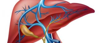
The vascular system, which is responsible for the flow of blood from and to the heart, can be divided into two components: the systemic circulation (systemic circulation) and the pulmonary vascular system (pulmonary circulation).
The pulmonary vascular system delivers blood to and from the lungs, where it is oxygenated, and the systemic circulation is responsible for transporting this blood to the body's cells through the arteries, and returning the blood back to the heart after being supplied.
What is opps in cardiology
Total peripheral resistance influences the functioning of this system and can ultimately significantly affect the blood supply to organs.
The total peripheral resistance is described by the partial equation:
OPS = change in pressure/cardiac output
The change in pressure is the difference between mean arterial pressure and venous pressure.
Mean arterial pressure equals diastolic pressure plus one-third of the difference between systolic and diastolic pressure. Venous blood pressure can be measured using an invasive procedure using special instruments that physically detects the pressure inside the vein.
Cardiac output is the amount of blood pumped by the heart in one minute.
Factors influencing the components of the OPS equation
There are a number of factors that can significantly influence the components of the OPS equation, thereby changing the values of the total peripheral resistance itself.
These factors include vessel diameter and the dynamics of blood properties. The diameter of blood vessels is inversely proportional to blood pressure, so smaller blood vessels increase resistance, thus increasing OPS. Conversely, larger blood vessels correspond to a less concentrated volume of blood particles exerting pressure on the vessel walls, meaning lower pressure.
Hydrodynamics of blood
Blood hydrodynamics can also significantly contribute to an increase or decrease in total peripheral resistance.
Behind this is a change in the levels of coagulation factors and blood components that can change its viscosity. As one might expect, more viscous blood causes greater resistance to blood flow.
Less viscous blood moves more easily through the vascular system, resulting in lower resistance.
An analogy is the difference in force required to move water and molasses.
Phlebeurysm
Varicose veins are a pathological expansion of the lumen of superficial venous vessels, accompanied by incompetence of the venous valves and leading to impaired blood flow. It is usually not possible to determine the underlying cause of vascular damage. Such a disorder is usually caused by primary valvular insufficiency and reflux, primary dilatation of the venous walls due to weakness of vascular tissues, chronic venous hypertension or insufficiency.
Varicose veins are more often detected in women. It often occurs during pregnancy or difficult childbirth. There are usually no risk factors for its development, but sometimes its occurrence is explained by hereditary predisposition. As a rule, it is the veins of the lower extremities that are affected.
Symptoms of varicose veins are not always visually noticeable. At the beginning, the veins may be tense and palpable. As the disease progresses, they enlarge, protrude above the surface of the skin and become visible. The patient complains of leg pain, discomfort, fatigue, tension and pressure. Particularly pathological dilated vessels are noticeable in a standing position.
When a vein thromboses, the patient experiences severe pain, and the superficial veins can form venous bullae, which rupture and bleed at the slightest physical impact. Sometimes such bleeding goes unnoticed (for example, during sleep), and leads to death.
Ulcers and other dermatological disorders with varicose veins rarely occur. They can manifest as eczema or pigmentation that appears in the ankle area. Ulcerative lesions usually occur after injury and are small in size and superficial.
Hemodynamic parameters
At the same time, depending on the greater or lesser severity of changes in regional vascular resistance, they will accordingly receive a smaller or larger volume of blood ejected by the heart.
This mechanism is the basis for the effect of “centralization” of blood circulation in warm-blooded animals, which ensures redistribution of blood, primarily to the brain and myocardium, in difficult or life-threatening conditions (shock, blood loss, etc.).
Resistance, pressure difference and flow are related by the basic equation of hydrodynamics: Q=AP/R.
Since the flow (Q) must be identical in each of the successive sections of the vascular system, the drop in pressure that occurs throughout each of these sections is a direct reflection of the resistance that exists in that section.
Thus, a significant drop in blood pressure as blood passes through the arterioles indicates that the arterioles have significant resistance to blood flow. The average pressure decreases slightly in the arteries, as they have little resistance.
Likewise, the moderate pressure drop that occurs in the capillaries is a reflection of the fact that capillaries have moderate resistance compared to arterioles.
The flow of blood flowing through individual organs can change tenfold or more.
Since mean arterial pressure is a relatively stable indicator of the activity of the cardiovascular system, significant changes in the blood flow of an organ are a consequence of changes in its general vascular resistance to blood flow. Consistently located vascular sections are combined into certain groups within the organ, and the total vascular resistance of the organ must be equal to the sum of the resistances of its sequentially connected vascular sections.
Since arterioles have significantly greater vascular resistance compared to other parts of the vascular bed, the total vascular resistance of any organ is determined to a large extent by the resistance of the arterioles.
Arteriolar resistance is, of course, largely determined by arteriolar radius. Therefore, blood flow through the organ is primarily regulated by changes in the internal diameter of the arterioles through contraction or relaxation of the muscular wall of the arterioles.
When the arterioles of an organ change their diameter, not only does the blood flow through the organ change, but the drop in blood pressure that occurs in that organ also undergoes changes.
Arteriolar constriction causes a greater drop in arteriolar pressure, resulting in an increase in blood pressure and a concomitant decrease in changes in arteriolar resistance to vascular pressure.
(The function of arterioles is somewhat similar to that of a dam: closing the dam gates reduces the flow and raises the dam level in the reservoir behind the dam and lowers the level downstream.)
On the contrary, an increase in organ blood flow caused by the dilation of arterioles is accompanied by a decrease in blood pressure and an increase in capillary pressure.
Due to changes in hydrostatic pressure in the capillaries, arteriolar constriction leads to transcapillary fluid reabsorption, while arteriolar dilation promotes transcapillary fluid filtration.
Peripheral vascular resistance refers to the resistance to blood flow created by blood vessels. The heart, as a pumping organ, must overcome this resistance in order to pump blood into the capillaries and return it back to the heart.
Peripheral resistance determines the so-called subsequent cardiac load. It is calculated by the difference in blood pressure and CVP and by MOS. The difference between mean arterial pressure and CVP is designated by the letter P and corresponds to a decrease in pressure within the systemic circulation.
To convert the total peripheral resistance to the DSS system (length•s•cm-5), it is necessary to multiply the obtained values by 80. The final formula for calculating peripheral resistance (Pk) looks like this:
To determine P, it is necessary to recalculate the CVP values in centimeters of water column into millimeters of mercury.
For such a recalculation there is the following relationship:
1 cm water. Art. = 0.74 mm Hg. Art.
In accordance with this ratio, it is necessary to multiply the values in centimeters of water column by 0.74. So, the central venous pressure is 8 cm of water. Art. corresponds to a pressure of 5.9 mmHg. Art. To convert millimeters of mercury to centimeters of water, use the following ratio:
1 mmHg Art. = 1.36 cm water. Art.
CVP 6 cm Hg.
Art. corresponds to a pressure of 8.1 cm water. Art. The value of peripheral resistance, calculated using the above formulas, reflects the total resistance of all vascular sections and part of the resistance of the systemic circle.
Peripheral vascular resistance is therefore often referred to in the same way as total peripheral resistance.
Causes and pathogenesis of the development of septic shock:
The incidence of sepsis and septic shock has been steadily increasing since the 1930s and is likely to continue to increase. The reasons for this are:
1. Increasing use of invasive devices for intensive care, that is, intravascular catheters, etc.
2. Widespread use of cytotoxic and immunosuppressive drugs (for malignant diseases and transplantations), which cause acquired immunodeficiency.
3. Increase in life expectancy of patients with diabetes mellitus and malignant tumors, who have a high level of predisposition to sepsis.
Bacterial infection is the most common cause of septic shock. In sepsis, the primary foci of infection are often localized in the lungs, abdominal organs, peritoneum, and also in the urinary tract. Bacteremia is detected in 40-60% of patients in a state of septic shock. In 10-30% of patients in a state of septic shock, it is impossible to isolate the culture of bacteria whose action causes septic shock. It can be assumed that septic shock without bacteremia is the result of a pathological immune reaction in response to stimulation by antigens of bacterial origin. Apparently, this reaction persists after pathogenic bacteria are eliminated from the body by the action of antibiotics and other elements of therapy, that is, its endogenization occurs. The endogenization of sepsis may be based on numerous, mutually reinforcing and realized through the release and action of cytokines, interactions of cells and molecules of innate immune systems and, accordingly, immunocompetent cells.
Sepsis, systemic inflammatory response, and septic shock are consequences of an excessive response to stimulation of cells that carry out innate immune responses by bacterial antigens. An excessive reaction of cells of the innate immune system and a secondary reaction of T-lymphocytes and B-cells cause hypercytokinemia. Hypercytokinemia is a pathological increase in the blood levels of agents of autoparacrine regulation of cells that carry out innate immune reactions and acquired immune reactions.
With hypercytokinemia in the blood serum, the content of primary proinflammatory cytokines, tumor necrosis factor-alpha and interleukin-1 increases abnormally. As a result of hypercytokinemia and systemic transformation of neutrophils, endothelial cells, mononuclear phagocytes and mast cells into cellular effectors of inflammation, an inflammatory process devoid of protective significance occurs in many organs and tissues. Inflammation is accompanied by alteration of the structural and functional elements of effector organs.
A critical deficiency of effectors causes multiple systemic failure.