A study by the Californian Scientific Institute of Preventive Medicine has proven that regular yoga classes can prevent the development of cardiovascular diseases. Properly selected yoga asanas can lower blood pressure, strengthen the heart muscle and normalize the pulse without pills. Yoga helps to recover even after cardiac surgery and reduces stress.
“Comprehensive practice has a qualitative effect on all structures of the human body,” says Rauf Asadov , yoga instructor, author of the Organic people organic-people.com, “Yoga in the Parks” and I love yoga community projects. “But if you specifically want to strengthen your heart, you should pay attention to yoga asanas that open up the thoracic region. We have collected them in our complex.”
The poses in this complex are given in such a sequence that you can perform them, smoothly flowing from one to another. However, you can also do asanas in any order. Hold in each of them for four breathing cycles (inhale-exhale).
To complete the complex you will need a mat.
If you have heart problems, consult your doctor before starting to practice.
Why does my heart hurt?
Without exercise, the heart becomes weaker. The heart muscle weakens from illness and a sedentary lifestyle. When the heart weakens, various pathologies can develop, since poor blood supply prevents sufficient nutrition and oxygen from reaching the organs. The condition of the blood vessels is also very important.
A person with a weak heart has the following symptoms:
- Little physical activity causes fatigue and palpitations.
- When climbing stairs or walking quickly, you feel a lack of oxygen.
- Excess weight appears, blood pressure rises, and other organs suffer.
Yoga and coronary heart disease
Ischemia
– a discrepancy between two factors: 1) the need for oxygen and 2) its delivery to the tissues.
Need
tissues in oxygen depends on the initial level of metabolism: for example, the kidneys consume about 10% of all oxygen entering the body, although their weight is about 0.5% of the total body weight; the brain consumes about 25%, with a weight of about 2% of the total mass. The need for oxygen also changes with changes in the functional activity of the organ: in a state of relaxation, skeletal muscles consume less oxygen, and during exercise, its consumption increases; at rest, the heart contracts less often, which means its need for oxygen is less; during physical activity, the heart rate increases, and the myocardium’s need for oxygen increases.
Delivery
oxygen in the tissue depends on a number of factors: on the work of the circulatory system (how actively the heart performs its pumping function and ensures the movement of blood through the vascular system), venous return (which depends on the work of peripheral muscles and respiratory activity), blood composition (that is, the amount of hemoglobin - oxygen carrier), patency of arterial vessels (through which blood carrying oxygen enters the tissues).
Coronary angiography. Contrast image of myocardial arteries.
Normally, there is a physiological correspondence between the need and delivery of oxygen, and when the need changes, the body changes the level of delivery. If for some reason the delivery process suffers, and the increase in demand is not accompanied by a more active supply, an intracellular oxygen deficiency (hypoxia) occurs, which leads to metabolic disorders and can ultimately lead to dysfunction and cell death.
A condition in which delivery does not meet tissue oxygen demand is called ischemia.
Most often, ischemia is caused by a decrease in blood delivery through the arterial vessels - which, in turn, is caused by
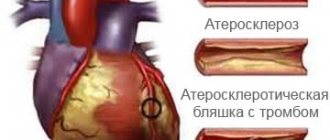
atherosclerosis
(deposition of lipids in the vessel wall, the formation of a lipid atherosclerotic plaque and narrowing of the vascular lumen).
Atherosclerosis. Drawing from the site asosudy.ru
In some cases, under the influence of provoking factors (for example, a hypertensive crisis), damage to the surface layer of the atherosclerotic plaque occurs, which initiates the formation of a blood clot on its surface, followed by occlusion (blockage) of the artery. This, in turn, leads to the cessation of arterial blood supply to this area of tissue, its damage and necrosis.
Tissue necrosis due to cessation of arterial blood supply is called infarction
and can occur in any organ that has an arterial blood supply.
In an adult, the process of atherosclerosis to one degree or another occurs throughout the entire arterial bed - therefore, vasoconstriction, a decrease in arterial blood supply and ischemia can develop in any organ and in any tissue. The digestive organs, urinary system, skeletal muscles, and other systems and organs can suffer from atherosclerosis and ischemia.
However, the most fatal manifestations of ischemia occur in the heart and brain. Cerebral infarction and the principles of post-stroke rehabilitation are discussed in the corresponding section.
Cardiac ischemia
(IHD) is a chronic disease, most often caused by atherosclerosis of the coronary arteries, leading to myocardial ischemia (heart muscle), in some cases complicated by coronary artery thrombosis, which causes necrosis of the heart muscle (myocardial infarction).
One of the chronic manifestations of coronary artery disease is angina pectoris -
syndrome that occurs during physical activity. During exercise, the heart begins to contract more often and stronger (as this is necessary for blood supply to muscles and other organs), which leads to an increase in the myocardium's need for oxygen. In the presence of atherosclerosis and narrowing of the vessel, the level of blood flow remains the same; thus, demand increases, but delivery does not. This leads to an imbalance of demand and delivery, that is, ischemia. Myocardial ischemia causes the development of a typical pain sensation in the heart region (most often behind the sternum). Pain in the heart (which can also radiate to the left half of the chest, left shoulder, arm, lower jaw and other areas), which occurs during physical activity and is associated with myocardial ischemia, is called angina pectoris.
With a stable course of angina, pain in the heart occurs at the same level of physical activity and, as a rule, proceeds in the same way (localization, nature and intensity of pain, irradiation, duration, response to drugs).
Provoking factors (such as increased blood pressure) can lead to damage to the surface of the atherosclerotic plaque, which triggers the process of thrombus formation on its surface. As the clot enlarges and the coronary artery narrows, the usual manifestations of coronary artery disease may worsen: for example, angina pectoris occurs at a lower than usual level of exercise; localization changes; the duration and intensity of pain increases; the effectiveness of the drugs decreases. Such a deterioration in the clinical course is called unstable (progressive) angina.
and is an indication for mandatory hospitalization.
Subsequent expansion of the thrombus can lead to complete occlusion of the vessel, cessation of arterial blood supply to the corresponding area of the myocardium and its necrosis - myocardial infarction (MI).
Myocardial infarction is the most common cause of death in developed countries, as it can grossly disrupt the contractility of the heart muscle (in this case, the pumping function of the heart suffers) and cause electrophysiological instability of the myocardium (which leads to cardiac arrhythmias of varying degrees of severity). Necrosis of muscle tissue can be complicated by its rupture and hemorrhage into the pericardial cavity. The complications listed above (as well as a number of others) can irreversibly disrupt the functioning of the heart and lead to death.
Myocardial ischemia. Photo from cardio.by-med.com
If a person remains alive after a myocardial infarction, then inflammation develops in the area of necrosis followed by the formation of a scar (post-infarction cardiosclerosis)
.
The scar, which is made up of connective tissue, is unable to contract and therefore the overall pumping ability of the heart is reduced. As a result, in the case of a significant area of myocardial damage, a persistent decrease in the pumping function of the heart develops - heart failure
.
Physical exercises occupy an important niche in the treatment and rehabilitation of patients with coronary artery disease. A large meta-analysis of the Cochrane database showed that exercise in patients with coronary artery disease reduces overall mortality by 27% and mortality from cardiovascular events by 31% [1].
Moreover, after an acute cardiovascular event (myocardial infarction or an episode of unstable angina), most patients do not know what level of exercise is necessary and indicated for them; this uncertainty leads the patient to avoid any physical effort and contributes to a sedentary lifestyle [2].
Cardiac rehabilitation programs are developed and applied to apply physical exercises that are appropriate to the patient’s health condition.
(PCR).
To do this, a pre -stress test
(LT) is carried out with a stepwise increase in the load level, ECG registration and blood pressure monitoring. This allows you to determine the optimal and safe level of load, to find out the possible ischemic threshold (the level of heart rate at which ischemia occurs, which is not subjectively felt by the patient). A bicycle ergometer or treadmill (treadmill) is usually used as a load during NT.
Carrying out a load test on a bicycle ergometer. Photo from the site rik-klinika.ru
As a result of a stress test using special methods, a heart rate level is calculated that is safe with respect to the occurrence of ischemia and at the same time provides training therapeutic effects. In the future, when carrying out RCC, the possibility of regular medical monitoring, registration of basic parameters (ECG, blood pressure) and correction of the load level is necessary.
Optimal beneficial effects on the health of patients with coronary artery disease can be achieved only with aerobic endurance training performed for at least 30 minutes a day 3-5 times a week [2]. Interval training is also useful, alternating short episodes of high-intensity exercise (20-30 seconds) followed by 2 times longer recovery episodes. In this case, short episodes of intense exercise stimulate the adaptation of the peripheral vascular system in the leg muscles without the risk of overloading the central circulation. However, the conclusion about the safety and effectiveness of this type of training is only preliminary and must be confirmed in randomized controlled trials [2,3].
Training within the framework of RCC should be carried out under medical supervision and under the guidance of a rehabilitation physician! For certain categories of patients (severe ischemic heart disease, ventricular arrhythmias, heart transplantation), it is recommended to develop cardiac rehabilitation programs in a hospital setting.
As a result of correctly selected training heart rate and a cardiac rehabilitation program, physical endurance increases, the ischemic threshold increases (that is, the heart rate level at which ischemia occurs increases), the frequency and intensity of angina attacks decreases, and the survival period increases [4]. In patients with heart failure, structured exercise programs improve quality of life (reducing shortness of breath and fatigue) and reduce mortality and hospitalization [5].
Hatha yoga is not an aerobic exercise option and cannot provide the full range of positive effects recorded for cardiac rehabilitation programs for coronary artery disease. However, when used as part of comprehensive rehabilitation, yoga practice can bring a number of positive effects.
Thus, a randomized controlled trial showed that yoga classes for 18 months, 5 times a week for 45 minutes, lead to a statistically significant decrease in heart rate, systolic and diastolic blood pressure in patients with coronary artery disease [6]. Another study involving 80 patients with stable coronary artery disease shows an improvement in respiratory function and lung diffusion capacity as a result of 3 months of asana and pranayama practice compared to controls [7].
Numerous studies show modulation of autonomic tone as a result of yoga practice and increased activity of the parasympathetic nervous system, which is important in IHD.
The construction of the practice of hatha yoga for ischemic heart disease (as well as any option for the use of physical exercises for this disease) should be based on the results of stress tests that determine the safety of using physical exercises in a particular patient and threshold heart rate values that should not be exceeded. In addition, current ultrasound data (EchoCG) should be taken into account. It is better if the nature and intensity of the load is determined by a cardiologist-rehabilitation specialist, taking into account all the data.
If we formulate the general principles of yoga practice for ischemic heart disease, then first of all we should mention techniques that should be limited or excluded:
1) Sympathotonic techniques: kapalabhati, bhastrika, surya-bhedana, active dynamic vinyasas, warming techniques with active shortened exhalation. An increase in the tone of the sympathetic system contributes to an increase in heart rate and the strength of myocardial contractions, which, in turn, increases the myocardial oxygen demand and promotes ischemia. One of the directions of pharmacological treatment of coronary artery disease is the prescription of beta blockers - drugs that selectively block adrenal receptors of the heart and thereby reduce sensitivity to sympathetic influences. Therefore, it is advisable to limit the use of techniques that enhance sympathetic influences - either by removing them from the training program altogether, or by using them in combination with parasympathetic compensations. Separate studies show that the use of kapalabhati for 10 minutes twice a day for two weeks by patients with stable coronary artery disease in combination with the practice of anuloma-viloma (alternate breathing) in the same volume did not lead to any negative results; At the same time, a statistically significant improvement in external respiration functions was noted [8]. It can be assumed that the slow type of breathing (anuloma-viloma) played the role of a compensatory, parasympathetic technique in relation to the sympathotonic technique (kapalabhati). It is possible that the use of kapalbhati in patients with stable coronary artery disease receiving standard pharmacotherapy does not lead to undesirable effects even when used in isolation - however, controlled studies are required to clarify this issue; Until then, you should adhere to the precautions mentioned above, excluding sympathotonic techniques or using them with sufficient parasympathetic compensation.
2) Inverted asanas. There are no studies on the effect of inverted body positions on the course of coronary artery disease, however, it can be assumed that long-term fixations in an inverted body position, increasing pressure in the cavities of the heart, will enhance the contractile function of the heart (according to the Frank-Starling law*) and increase the myocardial oxygen demand. In addition, inverted positions in case of organic heart pathology can generally have a negative effect on intracardiac hemodynamics, so the issue of using inverted asanas is best decided with the involvement of a cardiologist.
3) Long-term static fixations involving significant groups of skeletal muscles (parshvakonasana, virabhadrasana 1 and 2, chaturanga-dandasana, etc.). Static muscle contraction increases total peripheral vascular resistance and thereby increases the load on the left ventricle of the heart, increasing the myocardial oxygen demand.
In general, for IHD, the safest mode of practice is with a predominance of parasympathetic elements: practice of asanas without long-term fixations in a relaxation mode, intermediate short shavasanas, ujjayi in the proportion of visama-vritti (1:2), uddiyana-bandha, brahmari, nadi-shodhana (anuloma- viloma), brahmari, savasana and yoga nidra.
At the same time, a more active practice mode is possible, but to build it it is necessary to obtain the results of a stress test
with determination of the threshold heart rate. During exercise, you should monitor your heart rate (by periodically counting your pulse or using sports equipment for monitoring), without exceeding the heart rate that has been determined to be safe during exercise testing.
___________________________________________
*
Frank-Starling law is a physiological law according to which the force of contraction of myocardial fibers is proportional to the initial value of their stretch;
that is, as the filling of the heart chambers increases, the force of myocardial contraction also increases. Bibliography:
1) Jolliffe JA, Rees K, Taylor RS, Thompson D, Oldridge N, Ebrahim S. Exercise-based rehabilitation for coronary heart disease. Cochrane Database Syst Rev Update. 2001; (1):CD001800 Update SoftWare
2) Joseph Niebauer “Cardiorehabilitation. Practical guide" Moscow, Logosphere, 2014
3) Wisloff U, Stiylen A, Loennechen JP, Superior cardiovascular effect of aerobic nervous system training versus moderate continuous training in heart failure patients: a randomized study. Circulation.2007: 115 (24): 3086-3094
4) Fox KF, Nutall M, Wood DA et al. A cardiac prevention and rehabilitation program for all patients at first presentation with coronary artery disease. Heart. 2001; 85:533-538
5) Piepoli MF, Davos S, Francis DP, Coats AJ. Exercise training metaanalysis of trials in patients with chronic heart failure. BMJ. 2004; 328:189-194
6) Pal A, Srivastava N, Narain VS, Agrawal GG, Rani M. Effect of yogic intervention on the autonomic nervous system in the patients with coronary artery disease: a randomized controlled trial. East Mediterr Health J 2013 May;19(5):452-8.
7) Asha Yadav, Savita Singh & KP Singh. Effect of yoga regimen on lung functions including diffusion capacity in coronary artery disease patients: A randomized controlled study. Int Journal of Yoga, 2015, Vol. 8, Issue 1, p. 62-67
 Asha Yadav, Savita Singh & KP Singh. Role of pranayama breathing exercises in rehabilitation of coronary artery disease patients – a pilot study. Indian Journal of Traditional Knowledge, Vol. 8 (3), July 2009
How does yoga help with heart problems?
Is it possible to help yourself if your heart is bothering you? Yes, this is possible thanks to special yogic asanas.
But you should start exercising only after consulting a doctor, who will indicate the reason for the weakening of the heart muscle. It is important that there are no severe pathologies.
Yoga exercises can strengthen both the heart and the muscles of the body. Special asanas gently and delicately strengthen the heart muscle and blood vessels than traditional sports.
If a person wants to improve his health, yoga exercises should become systematic. It does not take much effort and time, but the result will be positive.
Notes
When you gain a little experience, you can add Paschimottanasana after Utkatasana to compensate for the backbends. Be sure to do child's pose after cobra pose (again, to compensate for the backbend), and also lie on your back, similar to Savasana (corpse pose).
These are the relatively simple exercises for the heart and blood vessels that yoga offers. Of course, everything is not limited to them - there are many more yoga poses to improve the health of the entire cardiovascular system and more. But these asanas can be a good basis for regular practice.
Important: These yoga exercises are marketed as heart-strengthening; If you have any pre-existing heart conditions, be sure to consult your doctor before attempting these yoga poses.
Scientific evidence for the productivity of yoga
The California Scientific Institute conducted special studies that proved that systematic yoga will help eliminate the development of diseases of the cardiovascular system. By choosing special exercises, you can lower blood pressure, normalize your pulse, and strengthen your heart muscle without resorting to pills. Asanas will also come to the rescue after cardiac surgery; they will reduce stress and anxiety. The yoga instructor and author of some yogic projects notes that complex exercises have a great effect on all human organs. But to strengthen the cardiovascular system, it is important to use asanas that open the chest. These are the exercises presented in the complex proposed below.
The benefits of yoga for the heart muscle
We will tell you in detail how yoga is beneficial for the heart and why. The University of Rotterdam analyzed the results of several dozen studies in which more than three thousand citizens took part, resulting in interesting conclusions. Researchers have proven that yoga for heart pain lowers blood pressure and reduces cholesterol levels, and also creates many other beneficial effects.
Yoga for heart problems is similar to fast walking or jogging. It is impossible to say unequivocally why asanas are useful. At the same time, they relieve stress and create a calming effect, which is especially important for cardiovascular disorders. Breathing exercises, in turn, ensure full saturation of the body with oxygen.
Features when selecting asanas
There are many asanas that strengthen the heart muscle, but they must be selected taking into account the state of health and how the muscles are developed.
At the initial stage, it is recommended to train under the supervision of an instructor.
When selecting exercises, you need to be aware of priorities and limitations. So, in order to lower blood pressure, it is necessary to perform special breathing asanas and meditations to relieve internal anxiety. If you are overweight, yogic complexes that work special muscle groups help. When a person is sick, asanas are selected that affect the problem and have a beneficial effect on the entire body.
Kumbhaka
This yoga movement for heart pain allows you to control your pulse and calm down during stress. The complex reduces the heart rate, so it can be used even for tachycardia.
To perform the exercise, you must take a standing, sitting or lying position. You must concentrate on the work of the heart muscle. Take a deep and long breath through your nose, then hold your breath for 10-30 seconds and slowly exhale (also through your nose). The Kumbhaka breath-holding technique is useful for a sick heart, but you need to start with 8-10 seconds, gradually extending the hold time by several seconds every day.
Asanas for the prevention of cardiovascular diseases
The complex is designed to strengthen the cardiovascular system. For those with heart problems, it is important to consult a doctor before performing exercises.
All you need for practice is a mat. Repeat each asana 3-5 times.
Mountain Pose (Tadasana)
Performance:
- Stand on the edge of the mat, legs apart, feet parallel to each other.
- Slightly spreading the mat with your soles, point your knees forward.
- Pull your pelvis in.
- Open your chest, moving your shoulders back and down.
- Pull the crown up.
Garland Pose (Malasana)
Execution sequence:
- Place your feet wider and point your toes slightly apart.
- Place your palms in front of you.
- Exhaling, do a full squat, do not move your heels.
- Spread your hips to the sides, helping with your hands. Lower your pelvis low, pull your tailbone in.
- The crown extends upward, stretching the vertebral axis.
- Inhale and stand up.
Extended side angle (Utthita parsvakonasana)
Execution order:
- While in Malasana pose, place both hands on the surface.
- Take your left leg back and straighten it, and turn your foot outward by 45 degrees. The right one is to bend the knee so that a right angle is formed.
- Place your right hand in front of your right foot.
- The shoulder should be in an exact projection above the hand. The right arm and shin are parallel to the surface.
- Inhaling, raise your left arm upward, turning your torso. The upper limbs form an even vertical, look up.
- With subsequent exhalations, try to lower your pelvis lower, and move your left hand back further.
- The back muscles are relaxed, open the chest to the maximum.
- Exhaling, the left hand lowers to the surface. Take a step back with your right foot, and pull your left foot towards your left hand.
- Do similar actions in the opposite direction.
- After this, pull your right leg towards your right hand and sit on the mat, legs bent (feet on the surface).
Table Pose (Goasana)
In a sitting position, take the upper limbs back and lower them to the surface, phalanges forward. Inhaling, push off the surface with your feet and palms. Raise the pelvis, stomach and chest up to the maximum so that the body is parallel to the surface. Look up, tense your buttocks, point your navel inside the body. After staying in the pose for 10–15 seconds, lower your buttocks to the surface and lie on your back. Align the lower limbs, place the upper limbs along the body.
Bow Pose (Dhanurasana)
From the previous position, roll over onto your stomach, legs apart, arms along the body. Bend your lower limbs at the knees and grab the outer part of your ankles with your hands, bringing your feet together. Tighten your buttocks and, while inhaling, lift your lower limbs and chest off the surface. Don't throw your head back, look forward. As you exhale, releasing your hands, smoothly lie down on the surface.
Camel Pose (Ushtrasana)
Execution sequence:
- Sit on your heels, straighten your back and kneel slightly apart.
- The fingers of the lower extremities rest against the surface.
- With tension in the gluteal muscles, exhaling, begin to smoothly bend back.
- Place your hands on your heels one by one. Keep your thighs and upper limbs perpendicular to the surface.
- While inhaling, pull your stomach forward, turning your chest and trying to direct it upward.
- Look up, stretching your neck muscles.
- Exhaling, release your hands, straighten up and sit on your heels.
Beneficial Yoga Exercises for Heart Health
Tadasana (mountain pose, variant)
Stand straight with your feet and heels together, arms down, palms resting on the outer thighs.
Stand calmly, steadily, breathe normally. As you inhale, raise your arms in front of your chest, placing your palms together in the Indian greeting "Namaste" (this hand position is called Anjali Mudra).
As you exhale, rise onto your toes and stay in this position, trying to maintain balance.
As you exhale, lower your heels to the floor.
Now, in this pose there are additional development options : 1) rising on your toes, try to close your eyes, while focusing on maintaining a stable position; 2) rising on your toes, while inhaling, stretch your arms above your head - maintain this position for as long as you can without undue tension, then lower yourself onto your heels, lowering your arms into Anjali Mudra, then along your body. In this version, you don’t have to close your eyes, but direct your gaze either forward or at your palms raised up.
One more thing : perform this pose for 5-15 minutes every day: you can first do several approaches, then try to stay in the final position for as long as possible.
Vrikshasana (tree pose)
We stand in the same position as at the beginning of Tadasana. We fold our hands into Anjali Mudra.
Bend your left leg and place your foot on the inner surface of your right thigh. Try to move the knee of your bent leg as far back as possible; keep your back straight.
Now, while inhaling, raise your arms above your head, stretching them upward; the gaze is directed forward.
In the final position, stay for a while, breathing freely and focusing on maintaining balance. Try to relax.
As you exhale, lower your hands into Anjali Mudra, then your left leg. Repeat with your right leg.
Virabhadrasana (warrior pose)
Stand up straight with your gaze directed forward. As you inhale, raise your arms above your head to your sides, bringing your palms together at the end.
As you exhale, jump with your legs about 90-100 cm apart.
Rotate your right foot 90 degrees to the right, and slightly to the right your left foot, while simultaneously rotating your entire body to the right.
Inhale, and as you exhale, squat down on your right leg, bending it at the knee until your right thigh is parallel to the floor and your shin is perpendicular.
Keep your chest open, your arms above your head, your gaze directed forward, and breathe normally.
As you exhale, return to the starting position and repeat on the other side.
Option: You can place your hands in Anjali Mudra if you find it difficult to stretch them upward.
Utkatasana
Stand straight with your feet slightly apart. Place your hands in front of your chest in Anjali Mudra.
Inhale and exhale, and as you inhale, extend your arms up above your head, while bending your knees so that your hips drop - almost until they are parallel to the floor.
The back arches slightly, the shoulders are straightened, the gaze is directed forward.
Close your eyes, breathe naturally, and focus on maintaining balance.
After some time, return to the starting position. Perform 2-3 approaches per practice.
Important: Avoid overexertion, especially in the lower back area, and if you have not previously exercised on a regular basis.
Bhujangasana (cobra pose)
Lie on your stomach, place your hands in front of you, chin on the floor.
Place your forehead on the floor, place your palms on each shoulder, and close your eyes.
Exhale, and as you inhale, slowly, without using your hands, lift your head, then your shoulders, then, using your hands, lift your body.
In the final position, look in front of you, breathe normally.
As you exhale, lower yourself to the floor and relax. You can perform 2-3 approaches per practice.
Important: When you first start practicing snake pose, do not throw your head back. Also remember that when lifting your torso above the floor, your back muscles always work; do not allow “breaking” in the lumbar region when you bend back.
After Bhujangasana, perform Balasana (child's pose), which is described in the article “6 Yoga Poses to Relax the Muscles of the Back and Neck,” just do it without a blanket or pillow, but simply by placing your head (forehead) on the floor.
Then, at the end of the practice, simply lie on your back for a few minutes to relax.
