Cerebellar stroke is still one of the least studied diseases of the central nervous system. The variability of clinical manifestations, their similarity to symptoms of damage to the cerebral hemispheres and other features significantly complicate diagnosis. This pathology occurs relatively rarely: approximately 2% of patients with acute disorders of cerebral circulation have a localization of the lesion in the cerebellum. Men of middle and retirement age suffer more often. What are the characteristic symptoms of the disease and how to treat such a stroke? The answers to these questions are in our article.
Types of cerebellar stroke
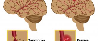
According to the standard classification based on pathogenesis, two types of cerebellar stroke are distinguished:
- ischemic;
- hemorrhagic.
According to statistics, the first type significantly predominates. Ischemic lesions of the cerebellum, according to various sources, account for 80 to 90 percent of cases. However, according to expert forecasts, within 50 years the number of diseases with a hemorrhagic nature is expected to double - the result of aging and changes in the racial composition of the planet's population.
Ischemic cerebellar stroke
The development of ischemic damage to the cerebellum is based on partial or complete blockage of blood vessels bringing arterial blood by a thrombus. In this regard, nerve cells stop receiving oxygen and nutrients. As is known, nervous tissue is very sensitive to hypoxia, and if blood flow is not normalized, neurons begin to die.
Hemorrhagic cerebellar stroke
With the hemorrhagic nature of the disease, a sharp disruption of the blood supply to the cerebellum occurs due to rupture of the artery or capillary feeding this structure. Due to the anatomical features of blood circulation, the consequences of a vascular accident are very difficult to predict, since the brain stem is often involved in the pathological process.
Puncture-aspiration method
| Puncture removal of hematoma with administration of fibrinolytic |
It is advisable to use the puncture-aspiration method for small lateral and medial hematomas accompanied by severe neurological disorders. The method consists of puncturing the hematoma with a catheter and simultaneously evacuating the liquid part of the hematoma. For precise positioning of the catheter, it is recommended to use neuronavigation. In some cases, drainage is carried out within 24 hours. The puncture-aspiration method with the introduction of fibrinolytics is indicated for lateral and medial supratentorial hemorrhages of medium size (from 30 to 60 ml) and for cerebellar hematomas (15–30 ml) provided the patient’s condition is stable. In this case, the technique is supplemented by fractional administration of fibrinolytic drugs at certain intervals. In the case of isolated ventricular hemorrhages, external ventricular drainage is performed with fractional intraventricular administration using fibrinolytics. According to the Research Institute of Neurosurgery, this method can reduce mortality in massive IVH by up to 40%, while in the natural course of this disease it approaches 100%.
Symptoms of cerebellar stroke
The clinical picture of both ischemic and hemorrhagic disorders of the blood supply to the cerebellum is varied, but there are characteristic symptoms that make it possible to suspect damage to the blood vessels in this particular area. These include cerebellar ataxia. This syndrome is associated with the functional activity of the cerebellum and includes:
- Balance imbalance. The patient cannot stand steadily and sways in different directions when walking.
- Changes in speech. Disturbance in the flow of arterial blood into the cerebellum is accompanied by scanned speech. The patient loudly tries to pronounce ordinary words.
- Loss of precision in movements. It is difficult for the patient to control fine motor skills, movements become sweeping.
- Tremor. Trembling of the limbs, head, and torso appears.
- Nystagmus. The eyeballs make involuntary oscillatory movements. Their frequency can reach several hundred per minute.
Among the general symptoms characteristic of acute cerebral circulation disorders are headache, dizziness, spontaneous vomiting, fever, loss of consciousness, difficulty swallowing, and muscle paralysis.
Causes and factors of cerebellar stroke
The ischemic type of cerebellar stroke is usually considered the outcome of atherosclerotic disease. The thrombus travels through the vertebral or basilar artery through the bloodstream. Embolism can also occur with fresh myocardial infarction or atrial fibrillation. Surgical interventions on the neck are very dangerous, since during their implementation it is possible to traumatize the vertebral arteries with further disruption of blood flow.
The main causes of hemorrhagic stroke of the cerebellum:
- high blood pressure or sudden changes;
- cerebral amyloid angiopathy - deposition of amyloid in small blood vessels through which blood enters the central nervous system;
- taking antithrombotic drugs (“blood thinners”);
- neoplasms;
- inflammatory diseases of the walls of blood vessels.
Wrong lifestyle
In addition to the immediate causes, risk factors play a significant role in the development of cerebellar stroke. One of the key ones is the wrong lifestyle. It includes:
- low mobility during the day;
- food based on foods high in fat and sodium;
- smoking, alcohol abuse, taking narcotic drugs;
- constant nervous tension, chronic stress.
These factors contribute to disruption of normal blood circulation in organs, including the nervous system.
Diseases and medications
Diseases of the internal organs can provoke a cerebellar stroke. The relationship between cerebral circulatory disorders and the following somatic diseases has been proven:
- arterial hypertension;
- metabolic syndrome, accompanied by high levels of cholesterol and low-density lipoproteins in the blood;
- diabetes mellitus and other endocrine pathologies;
- cardiac ischemia;
- diseases in which blood clotting increases.
Uncontrolled use of medications that affect the functioning of the heart, change hormonal levels, and thin the blood can also cause acute impairment of cerebral circulation (including in the cerebellum).
Other reasons
Other causes of vascular accident include genetic predisposition and age factor. A number of genes are currently being studied that are potentially associated with an increased risk of cerebral circulatory disorders.
Clinical syndromes of cerebral hemorrhage in hypertension
Hypertensive intracerebral hemorrhages can occur in any patient with hypertension. But more often hemorrhages occur with constant essential arterial hypertension. Hypertensive intracerebral hemorrhages almost always occur while the patient is awake, but are not always necessarily provoked by overexertion. Unlike sudden embolisms of cerebral vessels, hemorrhagic stroke develops within a few minutes, and its symptoms are determined by the location and size of the hemorrhage.
First aid for stroke
When providing first aid, it is necessary to understand what a cerebellar stroke is, otherwise there is a huge risk of harming the patient. The main goal is to prevent further death of neurons, and inept and incorrect actions can lead to extremely negative consequences. Recommend:
- Lay the patient on the bed, if the circulatory disorder occurred at home, or on the ground, slightly elevate the torso.
- Turn your head to the side to avoid vomit getting into your respiratory tract.
- Seek medical attention immediately.
- Try to calm the victim down and calm down yourself.
- Provide sufficient fresh air and avoid tight clothing.
Stroke treatment
The volume, tactics and duration of treatment depend on the type of vascular disorder. Common activities include:
- Ensuring the supply of oxygen to brain tissue.
- Correction of blood pressure indicators.
- Prevention of convulsive syndrome.
- Maintaining water-electrolyte and energy balance, normal body temperature.
Drug treatment of cerebellar stroke
The use of medications is possible only after the cause of the vascular accident has been established. For the treatment of ischemic stroke of the cerebellum, the following is used:
- Thrombolytics. Such drugs are able to dissolve blood clots in blood vessels, relieving areas of nervous tissue from ischemia.
- Neuroprotectors. Used to reduce the sensitivity of neurons to oxygen starvation. The use of these drugs allows you to avoid further death of nerve cells and gradually restore the functions of damaged areas.
- Diuretics are medications that help fight swelling of the brain.
In the treatment of acute circulatory disorders in the cerebellum of the hemorrhagic type, neuroprotectors and diuretics are also used, but the key drugs are hemostatic agents.
The amount of drug therapy is selected individually for each patient. It expands in the presence of paralysis, concomitant somatic or endocrine pathology.
Surgery
In parallel with drug treatment, the issue of surgical intervention is being resolved. Its functions:
- Removal of a blood clot or hematoma.
- Redistribution of blood flow to supply affected areas of nervous tissue.
- Stopping bleeding when drug therapy is ineffective.
Traditional surgery is performed openly or using an endoscope.
Direct surgical removal of hematoma
| Direct surgical removal of hematoma |
Direct surgical intervention is used for subcortical hematomas of medium and large sizes, for large hematomas of lateral or mixed localization, accompanied by increasing edema and dislocation of the brain, deteriorating condition of the patient, for cerebellar hematomas. The operation consists of removing the hematoma by encephalotomy, aspiration of blood, removing dense clots with fenestrated tweezers and washing the wound with saline solution. After removing the hematoma, it is necessary to examine its walls and perform thorough hemostasis using coagulation and hemostatic agents. Better results can be achieved by using microsurgical techniques, which can significantly reduce the size of the encephalotomy and thereby minimize surgical brain trauma. For large VMH, accompanied by edema and dislocation of the brain, a wide osteoplastic craniectomy is performed with plastic surgery of the dura mater with periosteum or artificial materials. In case of cerebellar hematomas, it is advisable to supplement direct removal of the hematoma with the installation of external ventricular drainage.
Recovery and rehabilitation after cerebellar stroke
Early initiation of rehabilitation treatment makes it possible to more effectively activate the relationship between neurons, returning lost functions to nerve structures. Rehabilitation measures should be varied and include both medications and physical procedures, special physical exercises, and occupational therapy. The main goals of rehabilitation treatment:
- regaining control over motor activity;
- normalization of speech;
- restoration of lost self-care skills;
- improvement of muscle tone after paralysis;
- prevention of complications (contractures).
Stroke in the cerebellum: possible consequences, prognosis
Despite its low prevalence, cerebellar stroke has a poor prognosis for the patient and is accompanied by high mortality, reaching thirty percent or more. Common consequences of cerebellar stroke:
- Disability. According to statistics, only 20% of patients who have suffered an acute cerebellar circulatory disorder are able to care for themselves independently by six months from the onset of the disease. The rest of the victims need help from others.
- Paresis and paralysis. Restoration of motor activity in damaged limbs is slow and not always complete.
- Speech impairment. Some patients still have scanned speech.
Often such severe consequences of a cerebellar stroke cause despair in the patient. However, you should not give up: daily rehabilitation activities can partially or even completely restore lost skills.
Stroke Prevention
To prevent acute cerebral circulatory disorders, WHO specialists have developed simple recommendations:
- Control and correction of blood pressure.
- Prevention of heart rhythm disturbances.
- Minimizing risk factors.
- Annual examination of the condition of the great vessels.
- Monitoring blood parameters.
Cerebellar stroke is an extremely serious disease, but a number of modern diagnostic and treatment methods increase patient survival and improve their quality of life in the future.
Diagnostics
To confirm the diagnosis of cerebellar stroke, an ultrasound is performed. The study allows you to determine the displacement, see the area of edema, and detect a damaged artery.
An informative diagnostic method is magnetic resonance imaging or computed tomography. Both methods provide an accurate picture of the changes, the size of the damage, the depth, and detect the cause of the disease.
A blood test is taken for general formula and biochemistry. The data will allow you to identify the level of sugar, cholesterol, and other substances that are signs of the diseases that provoked the attack.