Causes and course of the disease
Currently, the causes of this disease are not fully understood, but most researchers are of the opinion that it is an autoimmune disease, in the occurrence of which viruses such as cytomegalovirus and herpes virus play an important role.
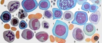
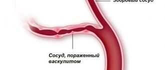
An equally important factor is the genetic peculiarity of the immune system, especially since with this disease in the blood of patients there is an increased content of the HLA-A8 antigen, which is responsible for the genetic predisposition to the development of autoimmune diseases. In the pathogenesis of Wegener's granulomatosis, immunological disorders such as deposition of immune complexes in the walls of blood vessels and impaired cellular immunity are of great importance. Patients experience necrotizing vasculitis, which affects medium- and small-caliber arteries and is accompanied by the formation of polymorphic cell granulomas that contain giant cells.
Diagnostics
In patients with a developed clinical picture, the diagnosis is usually simple, but due to the variety of forms and course options, difficulties arise in the early stages of the disease. Approximately 25% of patients in the initial stage have no signs of lung or kidney damage.
Classification criteria for the diagnosis of Wegener's granulomatosis
| Criterion | Definition |
| 1. Inflammation of the nose and mouth | Ulcers in the mouth. Purulent or bloody discharge from the nasal cavity |
| 2. Changes in X-ray examination of the lungs | Nodules, infiltrates or cavities |
| 3. Changes in urine | Microhematuria (>5 red blood cells per field of view) or accumulations of red blood cells in urine sediment |
| 4. Biopsy | Granulomatous inflammation in the arterial wall or in the perivascular and extravascular spaces |
If two or more criteria are present, the sensitivity of diagnosis is 88% and the specificity is 92%. To confirm the diagnosis, the presence of classical antineutrophil cytoplasmic antibodies (cANCA) in the blood is determined.
Clinical picture
Clinical manifestations of Wegener's granulomatosis can be divided into the following stages:
- Damage to the upper respiratory tract, and in some cases the eyes and ears;
- Generalization of the disease with damage to internal organs. The lungs and kidneys are most often affected;
- The terminal stage in which pulmonary, renal and heart failure develops.
The duration of the first period is 1-2 years. Most patients with signs of early Wegener's granulomatosis have manifestations of acute sinusitis and acute rhinitis. At the initial stage of the disease, patients complain of nasal congestion, dryness and scanty mucous discharge, which soon becomes purulent, and then an admixture of blood appears. Some patients with granulations in the nasal cavity and destruction of the nasal septum experience nosebleeds. One of the characteristic symptoms of Wegener's granulomatosis is the formation of purulent-bloody crusts of a brown-brown color. They are removed in the form of casts, while the mucous membrane becomes thinner and acquires a bluish-red color, and in some places necrotization (death) of tissue is observed. With the development of the inflammatory process, the number of crusts increases, and they acquire an unpleasant, putrid odor.
In some cases, granulation tissue is observed in the nasal passages, which has a bright red color. Most often it is located on the turbinates, as well as in the upper cartilaginous parts of the nasal septum; somewhat less frequently, the posterior part of the nasal septum becomes its location. If you touch it, it bleeds, and because of this it is often mistaken for a tumor of the nasopharynx.
Wegener's granulomatosis is characterized by ulceration of the mucous membrane in the anterior parts of the nasal septum. At the beginning of the disease, the ulcer is on the surface, but gradually deepens and reaches the cartilage. With further progression of the disease, the ulcer necrotizes the cartilage and a perforation (hole) of the nasal septum is formed, at the edges of which granulation tissue is located. With further development of the process, necrotization of the bony part of the nasal septum occurs and the external nose, having lost its support frame, becomes saddle-shaped deformed.
With this disease, in the first 1-3 years other organs may not be involved in the process, but very often after 3-4 months intoxication appears and the process generalizes with the involvement of other organs and systems. Most often, one sinus is involved in the process and occurs against the background of ulcerative necrotic rhinitis. In case of exacerbation of the process, a deterioration in the general condition is observed. With further development of the disease, the outer (lateral) wall of the nose, which is the wall of the maxillary sinus, is gradually involved in the ulcerative-necrotic process. In case of its necrosis, a single communication is obtained between the nasal cavity and the sinus. The maxillary sinus is most often affected, and the frontal and ethmoid sinuses are somewhat less common. Quite often, despite the bright manifestations of sinusitis, no purulent discharge is detected during sinus puncture. Quite rarely, simultaneous destruction of the sphenoid sinus and nasal septum occurs.
In the later stages of the development of Wegener's granulomatosis, a necrotic mucous membrane is observed in the nasal cavity, covered with a large number of crusts, which are difficult to remove in the form of an impression. Near the bone walls of the nasal sinuses there are specific granulomas that affect the muperiosteum and disrupt the nutrition of the bone. Before the bone begins to break down, a process of demineralization occurs.
Friends! Timely and correct treatment will ensure you a speedy recovery!
With Wegener's granulomatosis, the most striking and logical primary manifestation of the disease is damage to the upper respiratory tract. A third of patients have ear lesions, but otitis media is only rarely the first sign of the disease. In some cases, complications arise such as paresis of the facial nerve, spread of the pathological process to the labyrinth, with the development of labyrinthitis.
Make an appointment right now!
Call us by phone or use the feedback form
Sign up
Systemic damage in Wegener's granulomatosis quite often manifests itself as a combination of rhinological and ophthalmological symptoms, manifested as keratitis (inflammation of the cornea of the eye). If granulomatous infiltrates are located deep in the cornea, they can ulcerate, leading to the formation of deep ulcers that have undermined, raised edges. The sclera may also be involved in the pathological process. If the process affects the superficial layers of the sclera, then episcleritis occurs, and if the deep layers become inflamed, then scleritis develops. In some cases, a more severe disease develops - uveitis (inflammation of the choroid of the eyeball). With the development of keratoscleritis and keratosclerouveitis, swelling of the conjunctiva of the eye occurs, and patients complain of pain in the eye and blurred vision, the appearance of lacrimation and photophobia, and the occurrence of blepharospasm (persistent spasmodic closing of the eyelids). If such symptoms occur, you should consult an ophthalmologist.
The pathological process in the eye area is most often one-sided. In the later stages of Wegener's granulomatosis, exophthalmos (bulging eyes) or enophthalmos (recession of the eyeball) often develops. Exophthalmos often occurs if there is granulomatous tissue in the orbit and may recur. Enophthalmos appears as a late symptom of Wegener's granulomatosis. It occurs due to scar formation in the orbital tissues and optic nerve atrophy.
Ulcerative-necrotic changes in the larynx, pharynx and trachea are much less common.
In this case, the mucous membrane is hyperemic (pronounced reddened), and tubercles appear on the palatine arches, tonsils, soft palate and posterior wall of the pharynx, which quickly ulcerate. The eroded surface is covered with plaque, which is gray-yellow in color and difficult to remove, and the surface underneath bleeds. Patients complain of hoarseness, sore throat, stridorous (noisy, wheezing) breathing. Then the pain intensifies and profuse salivation (salivation) is observed. As the symptoms increase, weakness, headache, fatigue occur, and the temperature rises.
At stages 2 and 3 of the development of Wegener's granulomatosis, the most common clinical manifestation is lung damage. This causes a cough, which is sometimes accompanied by shortness of breath and hemoptysis. In the lungs you can find rounded infiltrates, which can be single or multiple. When they disintegrate, cavities with thin walls are formed, which are sometimes filled with liquid.
The classic sign of stages 2 and 3 of Wegener's granulomatosis is kidney damage. Characteristic pathologies are proteinuria (protein in the urine), microhemoturia (red blood cells in the urine), and progressive renal failure.
In Wegener's granulomatosis, joint damage and the development of ulcerative skin lesions are rare. Much more common are general symptoms such as weakness, fever, and weight loss.
The disease can occur in both acute and chronic forms. But the more acute the onset of the disease, the more severe its further course and the faster the generalization of the pathological process occurs.
Differential diagnosis
For the purpose of correct diagnosis, diseases that also occur with pulmonary-renal syndrome should be excluded: microscopic polyangiitis, Churg-Strauss syndrome, periarteritis nodosa, Goodpasture syndrome, hemorrhagic vasculitis, systemic lupus erythematosus; rarely - streptococcal pneumonia with glomerulonephritis. A differential diagnosis is also carried out with other diseases: lymphoid granulomatosis, angiocentric malignant lymphoma, malignant tumors, median granuloma of the nose, sarcoidosis, tuberculosis, berylliosis, systemic mycoses, syphilis, leprosy, AIDS, etc. With a predominantly renal course, differential diagnosis is carried out with idiopathic rapidly progressive glomerulonephritis .
Local form
In the local form, an inflammatory process is observed in the nasal mucosa, in the paranasal sinuses, in the pharynx and larynx, in the trachea, in various parts of the visual and auditory analyzers. Patients suffer from constant rhinorrhea and periodic nosebleeds. In addition, nasal breathing is impaired, bloody crusts form in the nose, and the voice becomes hoarse. Many scientists consider the local form of granulomatosis as an independent disease, which is characterized by dominant lesions of the skin and respiratory organs.
Generalized form
In the generalized form, internal organs are exposed to granulomatous inflammation, pulmonary and coronary vasculitis develops, and quite often glomerulonephritis. Granulomas can disintegrate, resulting in the formation of bleeding cavities in the tissues of the lungs and kidneys. Very often, pulmonary and renal vasculitis can be accompanied by lesions of the skin, pericardium, and peripheral NS. In this case, characteristic symptoms are observed: chills, fever, myalgia, hemorrhagic rash, arthralgia, arrhythmia, debilitating cough, cardialgia, renal dysfunction, shortness of breath. Symptoms of Wegener's disease may vary from person to person.
There is a borderline state between these two forms of the disease, which manifests itself in the form of pulmonary and extrapulmonary vasculitis without the occurrence of glomerulonephritis.
conclusions
Thus, therapy for Wegener's disease (granulomatosis) is complex and takes a long time until stable remission is achieved. If the case is severe, then immunosuppressive treatment should be combined with combined pulse therapy in the presence of glucocorticoids. Severe complications of drug therapy can be avoided by extracorporeal hemocorrection. Cryomodification of autoplasma is currently popular among rheumatologists and hematologists. This procedure allows you to avoid further progression of the disease, and also allows you to reduce the dose of cytostatics taken by the patient.
Such methods of therapy, together with traditional treatment, can quickly improve the condition of patients and achieve remission.
Medicines
When treating the disease use:
- "Cyclophosphamide", "Methotrexate" and "Azathioprine", which are part of the group of cytostatics.
- "Dexamethasone" and "Prednisolone" are part of the group of glucocorticoids.
- Anticytokine therapy.
- Hemodialysis, cascade plasma filtration, extracorporeal pharmacotherapy, plasmapheresis.
- Antibiotics, which are prescribed to prevent the occurrence of infectious complications.
- Immunoglobulins. May be prescribed if there is a relapsing course of the disease.
Description of the pathology
In the walls of blood vessels, Wegener's disease (photo in the article) is characterized by giant cell granulomatous-necrotizing inflammation. First of all, the formation of granulomas occurs in the vessels and tissues that surround the respiratory tract and eyes, after which they begin to develop in the kidneys, skin, heart and nervous system.
Granulomatosis received its name thanks to its discoverer, who identified this systemic vasculitis as a separate nosology. This happened in the thirties of the last century.
There are several forms of Wegener's disease (we'll look at the symptoms later).