An enlarged heart can be diagnosed at any age. An enlarged organ on fluorography does not always mean the presence of heart disease. In healthy people, this size is individual. This depends on many factors: from the structure of the chest to the power of physical activity.
For a person with a large build and short stature, an enlarged heart to the left will be the norm, as well as its vertical expansion for a thin person with tall stature.
Reasons for the development of pathology
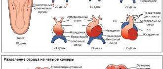
An enlarged heart in an adult is also called myocardial hypertrophy syndrome. Occurring against the background of an underlying cardiac disease, this condition can aggravate the overall clinical picture and worsen the prognosis of the underlying pathology. Enlargement of the right ventricle of the heart is less common than the left; in rare cases, there are changes in the size of both ventricles. According to statistics, against the background of concomitant cardiac pathologies, myocardial hypertrophy leads to mortality in 75-80% of cases. Therefore, timely detection and treatment of diseases of the cardiovascular system will be the key to a stable state of health.
The average heart weight in adult men is about 330 grams, in women - about 250 grams; to determine the external size of your own heart, just look at your clenched fist - the volume of the heart is about the same.
This pathology can be detected both in childhood and in adulthood. Athletes are susceptible to myocardial hypertrophy, since during active training the heart is subjected to an inappropriately increasing load, actively pumping blood. The heart muscles become overstrained, an enlargement of the ventricle or even two occurs, and a state of myocardial hypertrophy occurs.
With coronary heart disease, reduced blood flow to the organ occurs. The cells of the main muscle do not receive sufficient nutrition, as a result of which they begin to actively contract, and the process of their degeneration into connective tissue begins. It no longer has the ability to stretch or contract, so only the cavities of the heart can be enlarged.
This phenomenon is most pronounced during myocardial infarction - a certain part of the muscle tissue of the wall becomes inactive, which leads to the formation of a scar in this place.
The most common factors leading to an increase in myocardial size are excessive stress on the heart during heavy physical work, too much activity during sports, bad habits, as well as concomitant pathologies of the cardiovascular system. To summarize, the main reasons can be identified:
- heart defects (both congenital, hereditary, and acquired during life);
- physical labor and various strenuous sports that place excessive stress on the heart;
- ischemia;
- arterial hypertension;
- abuse of tobacco products and alcoholic beverages;
- excess body weight;
- sudden, unexpected stress on the heart muscle, against the background of an inactive, sedentary lifestyle.
This condition may have a hereditary etiology, that is, transmitted through generations from relatives suffering from cardiovascular diseases. In the presence of such an acquired “inherited” disease as aortic stenosis, arterial hypertension, mitral valve stenosis, a large heart is not such a rare occurrence. Enlargement of the ventricle, or both, occurs under excessive loads, when the organ triggers a compensation mechanism in order to protect itself. As a result, the size of the myocardium is increased.
Carditis, rheumatic carditis are diseases of the heart, which can also lead to the fact that it becomes enlarged against the background of flabbiness of the myocardial muscles, while the ventricles grow in size, and the heart dilates transversely. There have been cases when such a hypertrophied organ occupied both sides of the chest.
Myocarditis is a heart pathology that occurs as a consequence of a severe viral infection. When the disease worsens, the patient feels shortness of breath, becomes lethargic, a picture of general malaise appears, and the heart beats at an accelerated pace. There is an enlargement of the right ventricle of the heart against the background of acute heart failure. This condition also causes myocardial hypertrophy.
Septic endocarditis is considered a dangerous disease because it occurs against the background of infection of the inner surface of the heart by the microbial environment. As a result of infection, the valves, muscles, and left and right ventricles undergo significant changes. The patient suffers from fever, aching joints, increased body temperature (which is often confused with cold symptoms), and the myocardium increases in size.
Cardiomegaly or enlarged heart?
Every year, hundreds of thousands of citizens die from cardiovascular pathologies around the world. In most cases, the reason for this is untimely consultation with a doctor and deterioration of cardiac function.
Enlargement of the organ is associated with the development of ventricular hypertrophy, accumulation of metabolic products and neoplastic processes. Cardiomegaly often occurs in healthy people, this includes athletes and pregnant women.
The volume of the heart varies within different limits for each person. If we talk about gender differences, then in men this organ is larger than in women. So for the age category from 20 to 30 years, the approximate heart volume will be the following values:
- women – 580 cm3;
- men - 760 cm3.
This figure also depends on body weight. A diagnosis of cardiomegaly should be made only after a thorough examination, because in some cases a slightly enlarged heart is the norm, which is strictly individual for each person.
Symptoms
Any manifestations of pathology may be absent for a long time, which is why this syndrome is considered very dangerous. The patient may not be aware that he has a large heart, as well as the reasons that caused this condition. Pathology can be detected on an X-ray, during a routine preventive examination, or in worst cases, during an autopsy after sudden death. Myocardial hypertrophy can be manifested by the following symptoms:
- Shortness of breath of unknown etiology, occurring both during physical exertion and at rest.
- Pain in the chest, similar to the symptoms of angina pectoris.
- Heart rhythm disturbances.
- Swelling of tissues, deterioration in the removal of fluid and salts from the body.
- Dizziness, fainting.
The listed symptoms should be the reason for a mandatory visit to a therapist, who, after a thorough examination and examination of the patient’s complaints, will send him to a cardiologist for examination.
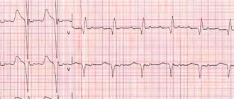
Myocardial hypertrophy is diagnosed using ultrasound; it shows itself to be the most informative in terms of identifying cardiac pathologies.
If they are detected, it is recommended to conduct an ECG (recording of an electrocardiogram), and to clarify concomitant diseases, magnetic resonance imaging is recommended.
The heart is expanded in diameter
I ask you, if it is possible to provide an explanation of the medical term “the heart is expanded in diameter,” what does it mean? This is written on the results of my husband’s chest fluorography. He is 54 years old, second-degree hypertension, coronary heart disease, varicose veins in the legs, overweight - this is an incomplete list of diseases accompanying his life.
I ask you to give recommendations on how to improve his health. What is the reason for the enlargement in the heart, is it life-threatening, and if so, how to protect your husband from possible consequences?
Sincerely, Lyudmila Reutskaya
Unfortunately, everything you wrote about your husband’s health condition is very dangerous. The heart enlarges from constant stress caused by high blood pressure and excess weight. At the same time, it cannot adequately perform its function, working as a pump, and tries to cope with this by increasing muscle mass (myocardium). But at the same time, the cavities of the heart expand, the valves stretch, the hole between the ventricles and the atrium becomes larger, and so-called pseudo-defects develop, in which blood flows from the ventricle into the atrium. This leads to heart failure and various heart rhythm disturbances. It is not the diagnosis “enlarged heart” itself that is dangerous, but the fact that these changes can lead to myocardial infarction or cerebral stroke.
Consequences of the disease
In case of increased myocardial activity and its subsequent increase, it is necessary to be observed by an experienced cardiologist and undergo regular examination courses. An important part of drug therapy will be a review of your lifestyle. In particular, you need:
- Give up bad habits - alcohol and cigarette abuse.
- Get rid of obesity and extra pounds to reduce the load on the heart muscle.
- Reduce the amount of salted, smoked, cholesterol-rich foods you consume.
- Balance the diet, enrich it with microelements and substances to normalize heart function.
- Reduce inadequate stress on the heart.
If measures are not taken to maintain the myocardium in a healthy state, this can lead to the development of stroke, heart attack, heart failure and even death.
People with alcohol addiction are at particular risk. Against the background of constant intoxication, the heart of a drinker sometimes reaches very large sizes. Restoration of myocardial size can only occur if alcohol is completely abstained.
Medicine offers modern drugs and treatment methods that, if used in a timely manner, will help reduce blood pressure, normalize blood flow to the myocardial muscle and return the heart to its original size. The main thing for successful treatment is to detect the pathology in time.
Fluorography
Enlarged heart in the picture
To avoid complications, it is necessary to undergo fluorography regularly (2 times a year). The specialist will pay attention to the shadow in the space between the lungs. Expansion occurs due to an increase in the main organ in one direction or another.
An enlarged heart on fluorography is not a diagnosis. It is impossible to assess the condition of an organ based on the results of a fluorography image. The image description may contain the following terms: “mediastinal expansion to the left,” “cardiac expansion to the left,” or simply “expansion.”
This will be one of the auxiliary studies and does not play a major role for the cardiologist in determining the exact diagnosis.
Fluorography of the heart has a certain degree of error, but with regular examination it is possible to notice any deviations in time. In this case, it is recommended to consult a cardiologist as soon as possible.
Clinical manifestations
To understand what an enlarged heart means, it is necessary to identify its manifestations. The most common symptom indicating changes inside the left ventricle is angina. It develops against the background of compression of blood vessels that provide nutrition to the myocardium. As a result, its expansion in size and its consumption of a significant amount of oxygen in parallel with nutrients are noted. In addition to these symptoms, atrial fibrillation appears, signs of atrial flutter and starvation of the heart muscle are observed. Often a pathology occurs during which the organ freezes for 3-5 seconds and the beat is not visible, which leads to an unconscious state. In certain situations, shortness of breath may indicate the presence of cardiomegaly. The following symptoms are considered additional:
- high blood pressure;
- unstable blood pressure;
- headache;
- disturbances in heart rhythm;
- disturbed sleep;
- poor condition and general malaise;
- painful discomfort in the heart area;
- painful discomfort in the chest.
Dilatation of the right ventricle is not accompanied by any complaints. Only its causes and adverse effects can be demonstrated in the laboratory:
- dilatation of the right ventricle;
- disturbances in heart rhythm;
Right ventricular hypertrophy
The right ventricle allows blood to pass through and pushes it deep into the vessels connecting to the lungs. It is enriched with oxygen. Due to the fact that the right side of the heart and lungs are connected, various difficulties with the respiratory system lead to cardiomegaly.
The right ventricle is 3 times smaller in volume than the left, therefore the electrical activity of the latter is higher. The increase can be pronounced when the mass of the right one exceeds the left one. With moderate expansion, the sizes of the ventricles do not vary. Insignificant excitement is noted. At the initial stage of the disease, symptoms are mixed or absent at all. If there is a tendency towards stable expansion, symptoms are expressed in difficulty breathing, a feeling of heaviness in the chest, and the appearance of painful discomfort.
In addition to these signs, the patient experiences fluttering or freezing and delayed heartbeat. Possible dizziness and unconsciousness.
Left ventricular hypertrophy
This pathology is a typical heart lesion for those suffering from hypertension. Dilatation of the left ventricle is considered a rather unsafe phenomenon, which is characterized by high mortality. This process involves structural adaptation of the organ regarding metabolism and changes that occur in hemodynamic data.
Cardiomegaly causes significant thickening of the walls. Often it leads to a significant change in the septum, which is located between the 2 ventricles. Due to the expansion that occurs, the elasticity of the walls is lost. Thickening can be either uniform or in certain areas of localization. This specificity directly affects the course of the disease.