Home » Articles » How to place an IV at home for a patient with “bad” veins?
An IV is considered a simple medical procedure, but requires certain knowledge and experience. It is strictly not recommended to carry out the procedure of intravenous infusions on your own due to the high risk of complications.
To install an IV at home with the help of a nurse, you don’t need a lot of money: a specialist will do everything professionally, in compliance with the rules. True, it is not always possible to avoid difficulties during infusion, especially if the patient has inconspicuous, thin and fragile veins that are not easy to reach.
Content
- Why are IVs placed in the veins in the arms?
- What is poor venous access?
- Causes of “bad” veins in the arms
- 5 proven ways to find a vein Physical activity
- Squeezing
- Warming
- Gravity
- Fluid replacement
- Tools and accessories
Diagnostic capabilities
Triplex scanning of the veins of the upper extremities is an expert study in three modes:
- grey-scale scanning mode (B-mode),
- color Doppler mapping mode (CDC),
- spectral Dopplerography mode (ultrasound Dopplerography, Doppler ultrasound).
The study allows you to evaluate the veins of the upper extremities in real time - the geometry of the veins, their patency, determine the presence of thrombosis and occlusions, identify varicose veins, anomalies in the development of veins, and valvular insufficiency.
The purpose of triplex scanning of the veins of the upper extremities is:
- diagnosis of congenital (angiodysplasia) and acquired venous pathology.
- assessment of the functioning of the valve apparatus of the venous system of the upper extremities (functional tests are used for this purpose).
- determination of indications for surgical intervention.
Why are IVs placed in the veins in the arms?
It is no coincidence that the veins in the arms are chosen for medical procedures, since they have the most suitable structure for this. On the upper limbs there are deep and superficial vessels. The latter lie close to the surface of the skin, making it easy to draw blood from them.
The pressure in them is only 5-10 mmHg (in the arteries in the arms it can be 139 mm), the venous wall contains a small number of muscle cells, is pliable and stretches well. In addition, the skin on the hands is much thinner than on the feet, so it can be pierced without causing severe pain to the person.
After a puncture, a small scar appears on the wall of the vein - the same as after damage to the skin in any other place. It can persist for a long time - months and even years. The integrity of damaged venous walls is never completely restored, but changes (if there are not too many of them) do not have a significant impact on the functioning of the body.
Intensive care nurse: venous catheterization
One of the main responsibilities of an intensive care nurse is organizing venous access for a patient, or, more simply, catheterizing a vein. A catheter is placed in different cases: to take blood for analysis (sometimes tests are taken several times a day, and pricking again each time is painful and traumatic for the patient) or to administer drugs and nutritional solutions to the patient. Placing a catheter is almost no different from a regular injection - you just need the vein to be straight in a certain area.
Where you can inject and where you can’t
To select a vein, you need to roughly estimate why the catheter is being placed in the first place. For emergency patients with severe injuries, bleeding, or those who are being prepared for abdominal surgery, a large-diameter catheter is placed, and a large vein is selected accordingly. For a grandmother of a very elegant age, whose diagnosis is not so serious, a smaller catheter is selected - the veins at this age are most often no longer the same.
How to choose the injection site? Nurses start with the distal veins, i.e. those that are “on the periphery”: the veins of the hand, forearm. The principle of ascending from the hand above also works (bad veins on the hand - move to the forearm).
The veins on the elbow are clearly visible, but this place is not very suitable for catheterization. The person bends and straightens the arm, sometimes unintentionally, and the catheter can become kinked or broken. Although, if you don’t need to insert a catheter, but only one or two injections are expected, the elbow bend is what you need. The vein on it is thick enough and will withstand all this.
Elsewhere, the vein may not be visible, but with a tourniquet applied, it can be easily felt with the fingers (palpated). In this case, an experienced nurse will be able to give an injection without even seeing the vein. In some patients, the median ulnar vein is not visible, but you can feel another one, which is a little closer to the outside of the arm, the lateral saphenous vein (it is also not visible with the eyes). I have had cases when I fell into such veins almost blindly - my veins also have this feature, so I am well aware of it.
What is poor venous access?
The situation when a nurse cannot get into a vein is familiar to about a third of patients who have received intravenous injections or an IV at home or in a hospital setting at least once in their lives. The reason is not always the inexperience of the medical worker. Sometimes even specialists with extensive experience do not get to the right place the first time - most often this happens when venous access is difficult.
Normally, the veins should protrude slightly above the skin between the muscles and have a bluish or blue tint. But for some people, they are too thin or almost invisible, making it difficult to insert the needle. In nursing practice, there are cases when a vein is difficult not only to see, but also to palpate. Then experienced specialists use proven methods to find a vein and place an IV in the place where this is easiest to do.
Causes of “bad” veins in the arms
Inconspicuous, thin or fragile veins are a serious problem for those who perform medical procedures. There can be several reasons for this phenomenon, ranging from natural causes to taking medications and frequent injections:
- Features of the body. The pattern and location of the veins are individual for each person, therefore they are the same unique biometric characteristic as a fingerprint or iris pattern. Accordingly, “hidden” vessels may be a feature of the body.
- Genetic predisposition. The strength of venous valves is genetically determined. According to statistics, if both parents suffer from varicose veins, the probability of their children inheriting it is about 80%. The same goes for fragile or thin veins - they are often a family problem.
- Aging. Venous access worsens with age - this is due to a decrease in elastin and collagen content, as well as thinning of subcutaneous fat. The skin becomes thin, “papery”, and the veins become thin and fragile, which increases the risk of injury and bleeding.
- Excess weight. In people with a high BMI (body mass index), the veins are hidden under fat. With obesity, it is difficult to perform not only intravenous injections and drips, but also some diagnostic procedures - ultrasound, MRI.
- Frequent injections. Any injection represents an injury, and if there were too many of them, scars form in place of healthy tissue. The veins “hide” and harden, so it becomes more difficult to place an IV or give an injection.
- Regular drug administration. In addition to frequent injuries, venous access in drug addicts deteriorates due to the administration of caustic, aggressive drugs. They literally burn the venous walls, making it extremely difficult for long-time drug addicts to carry out medical procedures.
- Exposure to ultraviolet radiation. Ultraviolet rays can also destroy elastin and collagen, which is why venous access is often difficult in people who are frequently exposed to the sun.
- Taking medications. Some drugs worsen the condition of the veins and increase the risk of complications during catheterization. Corticosteroids cause atrophy of the epidermis, and anticoagulants increase the risk of bleeding.
Poor venous access may be a transient phenomenon. If a person is stressed (for example, afraid of injections) or is cold, finding a vein to place an IV can be difficult.
Indications for examination of the veins of the upper extremities
- Signs of impaired venous blood flow in the upper extremities
- Symptoms of acute thrombosis of the veins of the upper extremities, thrombophlebitis and/or phlebothrombosis of injection, bacterial or other origin. Most often it is associated with varicose or postthrombophlebitic disease
- Condition after upper limb injuries
- Condition after paralysis of the upper limbs
- Preparation for surgical treatment
- Preventive examination at the request of the patient
How to properly place an IV: procedure algorithm
In order to administer an intravenous infusion, appropriate knowledge, experience and strict adherence to technique are required. Correct placement of a drip is not only about inserting a needle without consequences for the patient, but also about choosing a system, infusion rate, etc.
Tools and accessories
To carry out the infusion, it is necessary to prepare the medications prescribed by the doctor (check the expiration date, volume and other important indicators), as well as instruments and consumables:
- Dropper stand. It is a portable stand with hooks onto which containers with solutions are placed. Its height is 1.5-2 m, which provides sufficient pressure for insertion.
- Infusion system. Droppers differ in the diameter of needles, tubes and the speed of infusion of drugs. It is very important to decide on the size of the needle, which is inversely proportional to the number: twenty-second is the thinnest, fourteen is the thickest. It depends on the anatomical features of the patient and the characteristics of the drug.
- Related accessories. In addition to a tripod and a system for placing an IV, you will need a tourniquet (it helps to find the vein), a patch to fix the needle and cotton wool.
In addition to the prescribed medications, you need to prepare an antiseptic liquid. Most often, alcohol is used; if a person has allergies, they take alcohol-free solutions (for example, Chlorhexidine).
Preparation for the procedure
Before manipulations, be sure to wash your hands and wipe them with an antiseptic. Failure to comply with hygiene rules can lead to infection and sepsis, even if the health worker has perfect infusion technique.
- Connect the system to the container or package with the drug. To do this, you need to wipe the needle with alcohol and pierce the cork - this is not difficult, since they are made of soft rubber or other materials.
- Hang the containers on the racks and secure them well. Be sure to check that there is no air in the system - fill the tubes and drip chamber about a third with liquid. Small bubbles are usually not dangerous - they will stick to the walls of the container and will not enter the bloodstream.
If sterility is violated at one of the stages, infusion is strictly prohibited. This can lead to blood poisoning and other dangerous consequences.
Placement of the IV
Before the procedure, it is necessary to familiarize the patient with the features of its implementation. Each drug has a special effect on the body, and some sensations may frighten a person. It is important to distinguish the side effects of drugs from the alarming symptoms that develop when infusions are performed incorrectly. Pain, dizziness, nausea, weakness and other discomfort should be reported to a healthcare professional immediately. The procedure algorithm is as follows:
- the patient takes a comfortable position - it is best to do the IV while lying down, but it is also possible in a semi-sitting or sitting position;
- the arm in the biceps area is tied with a tourniquet, the needle insertion site is thoroughly disinfected;
- the needle is inserted into the darkest, well-filled vein parallel to the surface of the arm at an angle of 35-45 degrees;
- after blood appears in the catheter, it is positioned as parallel to the limb as possible and fixed with an adhesive tape;
- connect the system tube to the catheter, make sure the connection is tight, and additionally secure it with a bandage;
- The rate of supply of the solution is adjusted using a dial with a wheel - sometimes the number of drops is marked on it to make counting easier.
During the procedure, it is necessary to monitor the patient and the needle insertion site. If the fluid flows too quickly, the person may become dizzy. In such a situation, it is better to simply reduce the infusion rate by turning the wheel in the desired direction.
After completing the procedure, it is necessary to close the system, carefully remove it, apply cotton wool soaked in alcohol to the injection site and press well. You need to keep it for at least 10-15 minutes, and preferably 20-25 minutes. During this time, a blood clot will form, which will clog the wound. If you remove the cotton too quickly, drops of blood will get under the skin, resulting in a bruise or bruise. Do not rub or touch the injection site, as this can also lead to the accumulation of blood clots.
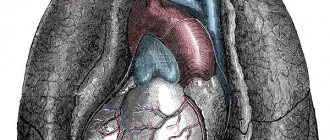
Vessels of the lower and upper extremities (arteries, veins)
Ultrasound examination (ultrasound) of the vessels of the lower extremities and veins
Recently, vascular diseases have begun to occupy one of the first positions among all diseases of middle-aged and older people. At the same time, we note that the age limit of middle age has begun to steadily shift from the 40-year mark to the 30-year mark. What contributes to this?
This is facilitated by factors such as:
- negative impact of the environment;
- smoking, especially if it started in adolescence;
- unhealthy diet with increased consumption of carbohydrates and fats;
- sedentary lifestyle.
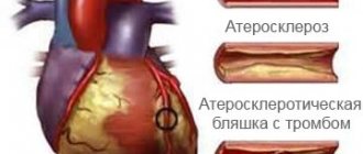
Each of the above conditions provokes the development of hypertension, obesity, atherosclerosis, and diabetes.
In the case of all these diseases, the vessels are primarily affected. They gradually begin to lose their elasticity and patency due to excessive load, as well as the deposition of atherosclerotic plaques in their walls. Changes can occur not only in arteries, but also in veins. Note that in practice, venous insufficiency of the lower extremities – varicose veins – is quite common. According to statistics, every 5 women and every 15 men suffer from varicose veins of the lower extremities.
It is important to say that the risk group for this disease includes those people whose profession involves being on their feet for a long time (for example, hairdressers). In addition, pregnancy can also provoke varicose veins.
Signs of vascular pathology may include heaviness in the legs and arms, numbness, cramps, pain in the limbs, the appearance of dilated saphenous veins and spider veins.
Currently in medicine there are various methods for treating vascular pathology. But, before starting treatment, even a conservative one, it is worth undergoing a high-quality and detailed diagnosis.
One of the safest and most effective methods for studying blood vessels is ultrasound scanning (ultrasound). Vascular ultrasound techniques are the result of the development of modern equipment that allows, under the control of a monitor, to examine a vessel in real time, see its lumen, determine valvular insufficiency of the veins, evaluate and measure blood flow parameters. It should be noted that it is already becoming possible to diagnose vasoconstrictions, assess valvular insufficiency of the veins, in the case of a blood clot, it is possible to accurately determine the size of the blood clot and monitor its changes during the prescribed treatment.
Ultrasound of vessels (veins and arteries) of the upper and lower extremities, as well as vessels of the neck and head, is carried out in triplex Doppler scanning mode (under visual control of the monitor, with the determination of any blood flow parameters).
So, if at your job you are forced to stand on your feet for a long time, which is why you feel discomfort, leg cramps, swelling, and notice the appearance of spider veins on the skin, then in this case, be sure to check the condition of your blood vessels with an ultrasound.
Complications from improper infusion
When carrying out the procedure, it is necessary to strictly follow all the rules - placement technique and asepsis. An incorrectly placed IV carries the risk of complications, which include:
- bruise or hematoma at the injection site;
- vessel damage;
- bumps under the skin due to the accumulation of the drug;
- unintentional introduction of a small (infiltration) or significant (extravasation) amount of solution into surrounding tissues;
- vein spasm;
- inflammation of the vein with the formation of a blood clot;
- tissue infection;
- air embolism.
Complications also include allergic reactions to the drug. They can be minor (itching and rash in the area where the solution was administered) or severe - Quincke's edema and anaphylactic shock. In any case, the patient should be left under the supervision of a doctor - sometimes minor allergic reactions lead to serious consequences.
What to do if complications develop?
The most dangerous complication of an incorrectly placed IV is an air embolism, or air bubbles entering the bloodstream. It rarely causes death - according to experts, in order to cause death, at least 200 ml of air must be injected into the vein. A small bubble simply dissolves in the cells, but sometimes it can impair the functioning of vital organs, so it is better not to take risks.
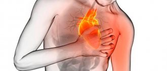
Symptoms of an air embolism depend on the size of the vessel affected and the amount of air trapped. The affected area swells, becomes painful, the skin turns blue or red, after which trophic ulcers and necrotic areas form. Tachycardia is possible, as well as manifestations from the affected organs. With pulmonary embolism, hemoptysis and pulmonary edema are observed, if the coronary or cerebral arteries are affected, heart attacks and strokes are possible with all the ensuing consequences. If signs of embolism appear, the patient should be hospitalized immediately.
Dangerous complications when intravenous infusion technique is violated include infiltration and extravasation, which can lead to unpleasant consequences. Injecting large amounts of medication past the veins can lead to severe tissue damage and necrosis. Treatment is carried out in a hospital under medical supervision.
The appearance of hematomas and bruises does not always depend on the experience or qualifications of the nurse. In some patients, the vessels are so fragile that they burst when the needle is inserted, resulting in a bruise. Such complications are not dangerous to health, but can sometimes lead to unpleasant consequences requiring medical intervention. You should consult a doctor if you have the following symptoms:
- painful sensations that increase and do not disappear for 2-3 days;
- increase in size of the bruise;
- muscle spasms;
- difficulty moving your hand;
- deterioration in general health (weakness, fever, headache);
- increased temperature at the site of the bruise.
Such signs indicate that an infection has entered the body, and sometimes the onset of necrotic processes. Normally, bruises disappear within 2-3 weeks, gradually changing color from bluish-purple to greenish and yellow. A bruise that does not go away for a long time is also a reason to consult a doctor, even if it is not accompanied by pain or malaise.
Phlebitis, or inflammation of the venous walls, usually develops after prolonged treatment with intravenous infusions, but when infected or administered caustic, irritating drugs, it can develop after several procedures. The vein area becomes dense and painful, body temperature rises to 38-39 degrees. Phlebitis is treated conservatively (anticoagulants, anti-inflammatory drugs) in combination with physiotherapy.
Headache and venous congestion
Venous stagnation (or disturbances of venous outflow from the cranial cavity) is a syndrome that often occurs with headaches of various types. Unfortunately, it is a common situation when venous outflow disorders are not taken into account in the treatment of tension headaches and migraines; as a result, patients suffer for years.
Even though venous outflow disorders are often described during head studies (MRI, vascular ultrasound), they are practically not taken into account in combination with other signs and complaints of the patient.
Manifestations of venous stagnation
Venous pressure inside the cranial cavity normally changes constantly - it increases with straining, coughing, sneezing, and decreases with rest. If venous outflow becomes difficult, characteristic complaints may appear:
- Heaviness in the head and pain in the morning.
- Swelling of the face (especially the eyelids) after sleep.
- Bursting headache.
Disturbances of venous outflow and nature of pain
Disturbances in venous outflow can change the nature of pain in patients with tension-type headaches. For example, pressing pain in the temples is accompanied by heaviness in the head after sleep.
There are even situations when disturbances in venous outflow are the main and only cause of headaches, and tension headaches come later.
The situation is no easier for migraine patients. In addition to the migraine itself, many also experience tension in the neck muscles and impaired venous outflow. In such patients, headaches almost never stop.
In practice, it turns out that an incorrect diagnosis (without taking into account venous outflow disorders) leads to long-term and ineffective treatment of headaches.
Difficulties in diagnosis
How can a doctor suspect the presence of a “venous factor”? After all, as we see, complaints with venous stagnation are not so specific. Only on the basis of symptoms such as swelling of the face and heaviness in the head in the morning, a correct diagnosis cannot be made.
The patient should be asked about changes in pain patterns and headache frequency. Since the tone of the veins can change with changes in atmospheric pressure, weather or cycle phase in women, all this must be taken into account.
Venous congestion can be confirmed using the following methods:
– Consultation with an ophthalmologist - dilated veins will be visible in the fundus. – Ultrasound examination of blood vessels - a decrease in the speed of blood flow through the veins will be noted. – Magnetic resonance imaging in phlebographic mode - dilated venous sinuses and veins will be noticeable.
However, the conclusion “impaired venous outflow” in itself does not carry much value - what is more important is the full clinical picture with the specifics of your complaints.
How to treat?
With regard to drugs for disrupting venous outflow from the cranial cavity, the situation is complex. There are currently no effective drugs with proven action against veins inside the skull. In practice, vascular drugs (for example, Cavinton) and metabolic drugs (Mexidol) are often prescribed, which may cause some improvement (although their effect has not been proven). Sometimes venotonics (for example, Detralex) are prescribed with a positive effect
Among other methods of treating headaches with impaired venous outflow, the methods of manual therapy, massage, and physiotherapy (if there is tension in the neck muscles or the effect of osteochondrosis on the blood vessels) have proven themselves to work well in practice.
Timely detection of venous outflow disorders and consultation with a neurologist can protect against progression and chronicity of pain.
Be healthy!
Maria Meshcherina
Photo istockphoto.com
How to safely place an IV in case of “difficult” veins?
When inserting a needle into a person with thin, fragile, or hard-to-see veins, additional precautions must be taken. They allow you to avoid unpleasant consequences in the form of injuries, hematomas and bruises, and also make the procedure as painless as possible:
- Do not stretch the skin excessively. If the veins are pronounced enough to get to the right place, you can do without a tourniquet. Otherwise, it is better to take a soft tourniquet and not tighten it too much - this is especially true for older people, as well as patients with thin skin and fragile veins.
- Reduce the number of procedures. If possible, it is necessary to reduce the number of vein punctures - administer several drugs in one dropper or one after another. Do not forget that mixing medications yourself can lead to undesirable consequences - this issue should be discussed with your doctor.
- Use a needle with a small diameter. To administer most solutions, a needle with a minimum diameter is sufficient. Exceptions include viscous, thick preparations; extreme caution should be exercised when administering them.
- Do not press the needle too hard. If the needle is sharp enough, there is no need to press too hard. For insertion, a smooth, gentle movement with slight pressure is sufficient.
- Insert the needle parallel to the skin. This is done in order to reduce the risk of through vein puncture. The skin over the vessel should be slightly stretched and secured so that it does not slip.
- Do not rush. When placing an IV, haste can lead to unpleasant consequences - you need to act slowly and carefully.
When the needle is inserted correctly, the patient feels slight discomfort when applying a tourniquet and performing a puncture. Unpleasant sensations during the infusion of the solution, including dizziness, nausea, weakness and darkening of the eyes, indicate that the infusion is not going properly.
How can you tell if the medicine is going past the veins?
To prevent infiltration and extravasation, the patient must be closely monitored during the procedure. List of warning signs:
- unnaturally shiny, dense, stretched skin at the site of needle insertion;
- tension and the appearance of swelling (rapidly growing swelling is especially dangerous);
- change in the shade and temperature of the hand - redness, blueness, too cold skin;
- self-slowing or stopping the flow of medication;
- continuation of the infusion after applying a tourniquet to the vein;
- leakage of solution around the injection site;
- burning or discomfort in the area where the needle is located.
If such symptoms occur, stop the infusion immediately and remove the needle. The injection site must be carefully examined, and if a large amount of solution is injected past a vein (especially if it is viscous or caustic), you must immediately consult a doctor. To eliminate the consequences in such cases, the introduction of an antidote is required.
If the amount of the drug that gets into the tissue is small, you need to monitor the affected area for 2-3 days. At the first manifestations of necrosis, immediately contact a medical facility.
If a vein is damaged, it is necessary to press it for a few minutes, and then apply a dry sterile bandage; if a hematoma appears, apply a compress. If the patient's condition has not worsened, the IV can be reinserted into another vein.
Where do they put an IV if the veins in the arms are bad?
If it is not possible to place a drip into a vein in the bend of the elbow, the nurse chooses another location. Inserting a needle into small vessels is strictly not recommended. It is extremely difficult to get into them, and problems subsequently arise much more often - hematomas and phlebitis are possible. The examination begins from the back of the hand, where the vessels are also quite large and well defined. A catheter is usually placed there - the bend of the elbow is not suitable for this purpose, since the device can be easily damaged when bending the arm.
Next, the veins of the forearm are examined, and a place is chosen as low as possible. The vessels that run along the outside of the forearm near the wrist are rarely used - they run close to nerves, so there is a high risk of damage. IVs are also practically not placed in the veins of the inner part of the arm below the elbow (the so-called antecubital fossa). When the solution gets into nearby tissues, compression of important anatomical structures occurs, which threatens damage to the veins and necrosis.
Droppers are placed in the veins of the legs extremely rarely - only when it is impossible to administer the drug in any other way. The vessels are more prone to inflammation and phlebitis, and the procedure is extremely painful - the skin on the lower extremities is rough, so it is much more difficult to pierce it. There are no large veins other than the central femoral ones, and the popliteal ones are inconvenient due to their location.
In the absence of other options, doctors choose the “veins of last resort” for infusions, that is, the central ones. These include:
- subclavian (under the right or left collarbone);
- internal jugular on the sides of the neck;
- femoral, which are located in the groin, are used extremely rarely due to the high risk of complications.
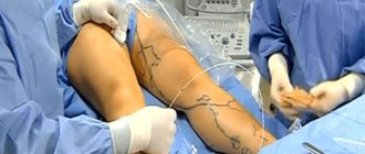
Such intravenous infusions are a minor surgical operation - first, catheterization is performed, after which medicinal solutions are administered. Catheterization is carried out under anesthesia and ultrasound control - some vessels are invisible to the eye, so it is impossible to determine without appropriate landmarks.
It is especially difficult to give IVs to newborns or infants. The veins in their arms are very thin, so not every experienced nurse can get into them. In addition, young children are unable to lie completely still for long periods of time, which increases the risk of injury. For such patients, IVs are often placed in the legs or veins in the head - such a procedure often shocks parents, but significantly reduces the risk of complications.
Carrying out the procedure on your own can lead to serious complications, so it is better to consult an experienced healthcare professional. To install an IV in Moscow at prices lower than in commercial clinics, you need to call a qualified nurse by calling the phone number on our website. An experienced specialist will perform the procedure quickly, painlessly and absolutely safely!
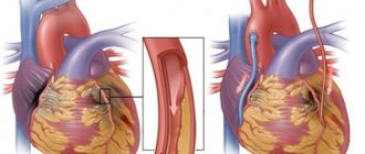
Vein thrombosis (phlebothrombosis) - symptoms and treatment
Treatment tactics for superficial and deep vein thrombosis differ from each other.
Superficial thrombophlebitis is most often treated on an outpatient basis under the supervision of a surgeon or cardiovascular surgeon. Antiplatelet therapy that prevents thrombus formation (acetylsalicylic acid preparations, etc.) and non-steroidal anti-inflammatory drugs (for severe pain) are usually prescribed.
Local treatment is of particular importance. The most popular are heparin-based ointments and gels; semi-alcohol compresses are also used. Heparin has a local anticoagulant anti-inflammatory effect and also improves microcirculation. The mechanism of action of semi-alcohol compresses is based on the decongestant effect of alcohol and its antiseptic properties. Compression hosiery plays an important role in the favorable outcome of treatment of superficial thrombosis of the veins of the lower extremities. Thanks to moderate elastic compression, a frame is created around the limb, which stabilizes the venous walls, preventing increased stretching. In addition, the risk of a blood clot breaking off and migrating to the heart and lungs is reduced, since it becomes fixed to the vessel wall.
If it is impossible to provide outpatient medical observation, the patient should be hospitalized in a hospital [15].
Deep vein thrombosis requires more serious approaches to treatment. Anticoagulant therapy is mandatory, which significantly reduces the risk of pulmonary embolism. It must be remembered that timely and adequate prescription of anticoagulants can minimize the consequences of thrombosis and significantly reduce the severity of postthrombophlebitis syndrome [1][2].
Modern anticoagulants are divided into two groups: direct and indirect. Direct drugs include heparin and its low molecular weight analogues. Direct anticoagulants are prescribed in the acute period of deep vein thrombosis of the lower extremities. The duration of use is about two weeks, depending on the specific clinical situation and the general condition of the patient.
Indirect anticoagulants act indirectly and are used for a long period of time (4 months or more). The mechanism of action of indirect anticoagulants is to block the synthesis of blood coagulation factors in the liver, as well as a number of biochemical reactions. This group of drugs is completely safe. A number of patients with cardiovascular pathology take them for life. However, taking some drugs is necessarily accompanied by laboratory monitoring of blood coagulation parameters (international normalized ratio - INR, prothrombin index - PTI, fibrinogen, etc.).
Modern medicine and pharmacology have in their arsenal such drugs that do not require laboratory control. Their dosage is calculated per kilogram of the patient’s body weight. Important! Anticoagulants are taken under the mandatory supervision of the attending physician.
The main risk of anticoagulant therapy is gastrointestinal bleeding. Patients should monitor the condition of the gastrointestinal tract: carry out timely drug therapy for gastric ulcers and chronic gastritis, and also, if necessary, undergo fibrogastroscopy (only as prescribed by a doctor).
If signs of gastrointestinal bleeding appear (black stools, vomiting “coffee grounds”), you should immediately seek medical help in a surgical hospital.
Despite the fact that a number of deep vein thromboses, depending on the location of the clots, can be treated on an outpatient basis, groups of patients who have:
- signs of pulmonary embolism (shortness of breath, chest pain, cough, sometimes with hemoptysis, drop in blood pressure, rapid pulse, cyanosis of the skin);
- iliofemoral (iliofemoral) and more extensive thrombosis;
- thrombosis of the superior and inferior vena cava;
- primary localization of thrombophlebitis on the thigh with damage to the great saphenous vein;
- primary localization of thrombophlebitis in the upper third of the leg with damage to the small saphenous vein;
- ascending thrombophlebitis - superficial thrombophlebitis, spreading to the above segments, despite adequate therapy;
- a floating thrombus in the superficial or deep vein system, identified by duplex ultrasound scanning at the prehospital stage.
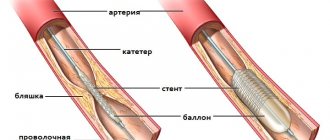
Surgical treatment for venous thrombosis depends on the location of the clot.
In case of thrombophlebitis of the superficial saphenous veins above the knee joint, it is necessary to raise the question of performing a Troyanov operation - ligation of the great saphenous vein to prevent the spread of a blood clot into the deep vein system.
For deep vein thrombosis, surgical approaches may be as follows:
- deep vein thrombectomy - surgical removal of a blood clot;
- thrombolysis - the introduction of special drugs into the vascular bed that dissolve blood clots;
- placement of vena cava filters (clot traps);
- a combination of these methods.
Rehabilitation of patients who have suffered venous thrombosis is always complex. It is aimed at preventing relapse of the disease and includes:
- preventing the action of provoking factors;
- active motor mode;
- body weight control;
- taking pharmaceutical drugs;
- wearing elastic knitwear;
- pneumomassage of the limbs;
- physiotherapeutic and sanatorium-resort treatment.
Bibliography
- Mukhina S. A., Tarnovskaya I. I. Practical guide to the subject “Fundamentals of Nursing”: textbook. - M.: Rodnik, 2005.
- www.who.int World Health Organization Publications WHO WHO/GSBI: Injection Safety and Related Procedures Toolkit.
- Savelyev N. “Injections, IVs, dressings and other medical procedures and manipulations”: - M.: AST, 2021.
- Bikkulova D.Sh. Venous access protocols are a comprehensive solution to CVC problems. //Journal Polyclinic 1(2)/2014
- Briko N.I., Bikkulova D.Sh., Brusina E.B., et al., Prevention of catheter-associated bloodstream infections and care of the central venous catheter (CVC). //Clinical recommendations. — M.: “Remedium Privolzhye”, 2021