Cardiovascular diseases (CVD) are one of the leading causes of mortality in the modern world. In 2021, according to the World Health Organization, 17.9 million people worldwide died from these diseases, accounting for about 30% of all deaths. At the same time, 7.4 million people died from coronary heart disease (CHD) and 6.7 million people died as a result of stroke, one of the causes of which is arterial hypertension (AH).
The most dangerous manifestation of IHD is myocardial infarction (necrosis of the heart muscle), which occurs due to blockage of blood vessels supplying the heart by a blood clot. A thrombus forms on the surface of an atherosclerotic plaque, which narrows the lumen of a blood vessel and consists of cholesterol and other compounds of fat, calcium, and connective tissue fibers.
If an atherosclerotic plaque forms in the vessels supplying blood to the brain, there is a risk of thrombosis of the brain vessels and death of part of the brain tissue (ischemic stroke).
What are cardiovascular diseases?
Cardiovascular diseases are a group of diseases of the heart and blood vessels that include:
- coronary heart disease - a disease of the blood vessels that supply blood to the heart muscle;
- cerebrovascular disease - a disease of the blood vessels that supply blood to the brain;
- Peripheral artery disease - disease of the blood vessels that supply blood to the arms and legs;
- rheumatic carditis - damage to the heart muscle and heart valves as a result of a rheumatic attack caused by streptococcal bacteria;
- congenital heart disease – deformations of the structure of the heart existing from birth;
- Deep vein thrombosis and pulmonary embolism - the formation of blood clots in the leg veins that can dislodge and move towards the heart and lungs.
Heart attacks and strokes are usually acute illnesses and occur primarily as a result of blockages in blood vessels that prevent blood from flowing to the heart or brain. The most common cause of this is the formation of fatty deposits on the inner walls of the blood vessels that supply blood to the heart or brain. Bleeding from a blood vessel in the brain or blood clots can also cause a stroke. Myocardial infarction and stroke are usually caused by a combination of risk factors such as tobacco use, unhealthy diet and obesity, physical inactivity and harmful use of alcohol, high blood pressure, diabetes and hyperlipidemia.
Hereditary predisposition
Our body is made up of trillions of cells. Every cell has a nucleus that contains the genetic information that makes you unique. This information is contained in chromosomes. Think of a chromosome as like a ball of wool, and you can stretch it into one long strand called DNA. There are regions of DNA called genes. Each of us has about 20,000 different genes. Since each chromosome has two copies, there are also two copies of each gene (one from each parent).
What is the pattern of inheritance of cardiovascular diseases in families? What is the risk of recurrence of the disease in a family if there is already one patient in it?
Today it is known that hereditary factors are involved in the development of most diseases. From this point of view, diseases of the heart and blood vessels are no exception. Many of them (coronary heart disease, atherosclerosis, hypertension) are multifactorial
.
This means that both environmental factors (for example, ecology) and a person’s lifestyle (for example, the type of diet and amount of physical activity), as well as genetic (hereditary) factors, equally contribute to their formation. However, for children, the most relevant are the so-called monogenic
diseases, for the occurrence of which a mutation (“breakage”) in just one single gene is sufficient. These include, for example, many heart rhythm disorders, such as long QT syndrome (Romano-Ward or Jervell Lange Nielsen syndrome) or short QT syndrome and others. Monogenic heart diseases also include hypertrophic cardiomyopathies.
Gene
(Ancient Greek γένος - genus) is a structural and functional unit of heredity.
A gene is a section of DNA that carries information about the sequence of a specific protein. Genes are located on human chromosomes, each of which is a long DNA molecule. All chromosomes, as well as human genes, are paired: each gene is present in every cell of the body in two copies - one copy on each of the identical (so-called homologous) chromosomes (Fig. 1). Figure 1. DNA, genes and mutations (“breakages”) in genes.
Humans have 23 pairs of chromosomes, 22 of which are called autosomes
and are the same in men and women.
And one pair of chromosomes - the sex chromosomes
- differs between the sexes: in men these are XY chromosomes, and in women they are XX.
In human germ cells there is a half set of chromosomes - not 46, but 23, each chromosome and every gene in the germ cell is present in a single copy. Men have 2 types of germ cells: either with the X chromosome or with the Y chromosome. In women, all germ cells, in addition to 22 autosomes, contain the X chromosome.
Genotype
(genotype, from the Greek genos - genus and typos - imprint, form, sample) - a set of genes of an organism.
In contrast, the totality of all external characteristics of an organism (anatomical, functional, etc.) constitutes a phenotype
.
Hereditary pathology of the cardiovascular system can be either isolated or part of complex genetic syndromes.
Genetic syndrome
– this is a combination in a patient of signs of damage to many organs, united by a common genetic cause.
For example, the rare genetic disorder Marfan syndrome is caused by mutations in the fibrillin 1 (FBN1) gene, affecting multiple organ systems. (Marfan syndrome) includes anomalies (structural features) of the skeletal system in the form of an asthenic physique with long “spider” fingers, a funnel-shaped or keeled chest, anomalies (structural features) of the eyes - in the form of dislocation of the lens with loss of visual acuity and cardiovascular system in in the form of prolapse of heart valves, enlargement of the aortic root and pulmonary artery
.
If a child has a hereditary disease in the family, the question arises about the risk of recurrence of the same disease in future children of these parents. In resolving this issue, a geneticist who provides medical and genetic counseling
couples, during which they collect a family pedigree, examine a family member with the disease, examine family members using genetic tests, and determine the risk of birth of a recurrence of the disease in the family, as well as identify possible options that will avoid the increased risk.
An examination of family members may show that in a sick child, a “breakdown” in a gene arose for the first time in this family (de novo). In this case, the risk of recurrence of the disease in future children of this parental couple is minimal. However, there are families in which the disease occurs in several generations. In these families, pathology is transmitted according to certain patterns that determine the risk of having a child with the disease. It is not possible to consider all possible inheritance options within the framework of this brochure, but we will still analyze the most common types of inheritance in cardiology. Autosomal dominant
diseases appear in people even if only one of the two copies of the gene is “broken.”
Autosomal dominant diseases and traits can be traced through many generations of the vertical pedigree (Figure 2). Figure 2. Pedigree with an autosomal dominant type of inheritance: vertical inheritance of the disease, from a parent of any sex to a descendant of any sex.
Circles are female, squares are male. Designations of family members with the disease are shaded. How to determine genetic risk with autosomal dominant inheritance? Since a family member with the disease has two types of germ cells - with a healthy and a “broken” copy of the gene, the risk of transmitting the disease to his child is 50% (Fig. 3).
Figure 3. Inheritance of dominantly inherited heart disease.
Autosomal dominant inheritance is the most common type of transmission of the disease in families for cardiac diseases.
Autosomal recessive
diseases and symptoms only appear if both copies of the gene are “broken.” Family members who have one copy of the gene “broken” and the other copy healthy do not have clinical manifestations of such a disease, but are carriers of it. In a pedigree, the disease can be traced horizontally in one generation through brothers or sisters (Fig. 4).
In autosomal recessive diseases, it is often possible to identify family ties between parents.
How to calculate genetic risk for autosomal recessive inheritance? Possible combinations of germ cells from parents who are carriers of an autosomal recessive disease (25% of healthy offspring with two healthy copies of the gene, 50% of clinically healthy carriers of one “broken” and one healthy copy of the gene, and 25% of offspring with the disease and two “broken” copies of the gene) lead to a 25% chance of having a sick child.
Figure 4. Pedigree for autosomal recessive inheritance: horizontal inheritance of the disease. A related marriage in a pedigree is depicted by a double solid line connecting the symbols representing the spouses. Sick individuals are shaded. The mother and father of sick children are healthy carriers and have one healthy and one mutant copy of the gene. Each parent passed on a mutant copy of the gene to sick children; the probability of such a simultaneous transfer of a “broken” copy of the gene to the child from both parents is 25%.
Diseases linked to chromosome X
and their nature of inheritance is usually considered separately due to the peculiarities of the manifestation of the disease only in individuals of a certain sex; it is also usually divided into the so-called X-linked recessive and X-linked dominant. Here we look at the most common X-linked recessive inheritance.
Since men have only one X chromosome, all X-linked genes are present in only one copy. Those. their cells do not have a second copy of the gene located on the X chromosome. If the breakdown is located in this single copy of the gene, then in men, there is no second copy of the gene on the X chromosome that could compensate for the lack of function of the single “broken” copy. X-linked recessive diseases therefore appear in boys and are transmitted in the pedigree by healthy female carriers. Female carriers have two X chromosomes - one with a “broken” one, the other with a healthy copy of the X-linked gene. This type of transmission in the pedigree is sometimes called “diagonal” (Fig. 5).
Figure 5. Pedigree for X-linked recessive inheritance: diagonal inheritance of the disease. With this type of inheritance, only men are affected (shaded). The symbol “circle with a dot inside” indicates a female carrier of an X-linked gene mutation. All female carriers of the disease are clinically healthy because they have one healthy copy of the X chromosome gene, which compensates for the work of the second - mutant - copy of the gene. All men have the only chromosome X, and if it carries a mutant copy of the gene, then they are definitely sick. Female carriers pass the disease along with a mutant copy of the X chromosome to their sons with a 50% chance; they also have a 50% chance of passing on a healthy X chromosome to their son.
Genetic risk for X-linked recessive inheritance: if a carrier of an X-linked recessive mutation marries a healthy man, then each of their sons will have a 50% risk of getting the disease, and each daughter will have a 50% chance of being a carrier.
What are the risk factors for cardiovascular disease?
The main risk factors for heart disease and stroke are poor diet, physical inactivity, tobacco use and harmful use of alcohol.
The effects of behavioral risk factors on an individual may include increased blood pressure, increased blood glucose, increased blood lipids, and overweight and obesity. These “intermediate risk factors” can be assessed in primary care settings and may indicate an increased risk of myocardial infarction, stroke, heart failure and other complications.
Quitting tobacco use, reducing salt intake, consuming fruits and vegetables, regular physical activity and avoiding harmful use of alcohol have been shown to reduce the risk of developing cardiovascular disease. In addition, drug therapy may be necessary to reduce the risk of CVD and prevent heart attack and stroke in people with diabetes, high blood pressure, and elevated lipid levels. To increase people's motivation to make and maintain healthy behaviors, health policies are needed to create an enabling environment for healthy choices to be made and afforded to them.
In order for people to choose and maintain healthy behaviors, policies are needed to create an environment conducive to healthy choices, their accessibility and affordability.
There are also a number of factors that influence the development of chronic diseases, or underlying causes. They reflect the main driving forces leading to social, economic and cultural changes - globalization, urbanization and population aging. Other determinants of CVD include poverty, stress and genetic factors.
Dyslipidemia
Numerous studies have established that one of the main reasons for the development of vascular atherosclerosis is a violation of fat or lipid metabolism, namely an increase in the level of cholesterol (C) in the blood. In one long-term study that followed its participants for many years in the United States, it was found that people with cholesterol levels from 6.2 to 6.8 mmol/l had a twice the risk of developing coronary artery disease than people with cholesterol levels below 5.2 mmol/l. If the cholesterol level was above 6.8 mmol/l, then the risk increase was 3-fold.
The main lipids found in human plasma are free fatty acids, triacylglycerides, phospholipids and cholesterol. All lipids are found in the blood in protein-bound form in the form of lipoproteins. Chylomicrons are the largest lipoprotein particles. In addition, there are very low density lipoproteins, low density lipoproteins, lipoproteins (a) - a protein complex rich in cholesterol, and high density lipoproteins.
Strong evidence of the connection between the state of blood lipids and atherosclerosis came from the results of international studies, which established that the degree of development of fatty streaks and plaques in the coronary arteries of the heart was associated with the level of total cholesterol and very low-density lipoproteins.
A high level of cholesterol in the blood can be a manifestation of a hereditary disease - familial hypercholesterolemia. The frequency of this disease in the heterozygous form is high and amounts to 1 case per 500 people in the population (with a cholesterol level of 7.5 -12.5 mmol/l). And the frequency of familial hypercholesterolemia in the homozygous form is much less common and amounts to 1 case per 1,000,000 people in the population (with a cholesterol level of 15 - 30 mmol/l). The homozygous, most severe form of this disease is characterized by the development of severe atherosclerotic vascular damage and its complications already in childhood, namely in the first decade of life. Cholesterol is deposited not only in the blood vessels, but also under the skin, on the tendons, and the cornea of the eye.
In general, lipid metabolism is a complex process that plays a leading role both in maintaining the vital functions of the body and in the development of a number of pathological conditions, one of which is atherosclerosis. If your family members under the age of 45 have already suffered myocardial infarctions, you should determine the blood cholesterol levels not only in yourself, but also in your children. Normally, the cholesterol level of a healthy person should not exceed 5.2 mmol/l.
Methods for correcting blood lipid disorders include: a low-fat diet, normalizing body weight, increasing physical activity, and, if necessary, taking special medications.
What are the common symptoms of cardiovascular disease?
Symptoms of heart attack and stroke
Often the underlying blood vessel disease is asymptomatic. A heart attack or stroke may be the first warning sign of the disease. Symptoms of a heart attack include:
- pain or discomfort in the middle of the chest;
- pain or discomfort in the arms, left shoulder, elbows, jaw or back.
In addition, the person may experience difficulty breathing or shortness of breath; nausea or vomiting; feel dizzy or faint; break out in a cold sweat and become pale. Women are more likely to experience shortness of breath, nausea, vomiting, and back and jaw pain.
The most common symptom of a stroke is sudden weakness in the face, most often on one side, arm or leg. Other symptoms include sudden numbness of the face, especially on one side, arms or legs; confusion; difficulty speaking or difficulty understanding speech; difficulty seeing in one or both eyes; difficulty walking, dizziness, loss of balance or coordination; severe headache for no specific reason, as well as loss of consciousness or unconsciousness.
People experiencing these symptoms should seek medical help immediately.
What is rheumatic carditis?
Rheumatic heart disease is damage to the heart valves and heart muscle due to inflammation and scarring caused by rheumatic fever. Rheumatic fever is caused by an abnormal response of the body to a streptococcal infection. The disease usually first manifests itself as a sore throat or tonsillitis in children.
Rheumatic fever primarily affects children in developing countries, especially in settings where poverty is widespread. Worldwide, rheumatic carditis is associated with almost 2% of all deaths from cardiovascular diseases.
Symptoms of rheumatic carditis
- Symptoms of rheumatic heart disease include: shortness of breath, fatigue, irregular heartbeats, chest pain and loss of consciousness.
- Symptoms of rheumatic fever include: fever, joint pain and swelling, nausea, stomach cramps and vomiting.
How can the burden of cardiovascular disease be reduced?
For the prevention and control of cardiovascular disease, WHO has identified a number of “best buys,” or highly cost-effective interventions that are feasible even in low-resource settings. They include 2 types of interventions—population-based and individual-level interventions—that can be used in combination to reduce the high burden of cardiovascular disease.
Examples of interventions that can be implemented to reduce CVD at a national level include the following:
- comprehensive tobacco control policies;
- taxation to reduce consumption of foods high in fat, sugar and salt;
- building walking and cycling paths to increase physical activity levels;
- strategies to reduce the harmful use of alcohol;
- ensuring proper nutrition for children in schools.
To prevent first myocardial infarctions and strokes, individual health interventions should target individuals with moderate or high overall cardiovascular risk or those with individual risk factors such as diabetes, hypertension and hypercholesterolemia above recommended levels. carrying out treatment.
The former (an integrated approach that takes into account all risk factors) is more cost-effective than the latter and can significantly reduce the incidence of cardiovascular events. This approach is feasible in low-resource settings, including the use of non-physician medical personnel.
For secondary prevention of CVD in people with pre-existing disease, including diabetes, treatment with the following medications is necessary:
- aspirin;
- beta blockers;
- angiotensin-converting enzyme inhibitors;
- statins.
The positive results obtained are largely unrelated, but when used in combination with smoking cessation, almost 75% of recurrent vascular events can be prevented. There are currently significant gaps in the implementation of these measures, especially at the primary health care level.
In addition, treatment of CVD sometimes requires expensive surgery. These include:
- coronary artery bypass grafting;
- balloon angioplasty (in which a small balloon catheter is inserted through an artery to restore lumen to a blocked vessel);
- plastic and valve replacement;
- heart transplant;
- operations using an artificial heart.
Some CVDs require medical devices to treat. These devices include pacemakers, artificial valves, and patches to close holes in the heart.
High cardiovascular risk: main components
Lecture transcript
XXV All-Russian Educational Internet Session for doctors
Total duration: 29:10
00:00
Oksana Mikhailovna Drapkina, Secretary of the Interdepartmental Scientific Council on Therapy of the Russian Academy of Medical Sciences, Doctor of Medical Sciences, Professor:
- We continue. Next lecture of the master class. It will be read by Doctor of Medical Sciences Igor Vladimirovich Sergienko. "Cardiovascular risk stratification." Please.
Igor Vladimirovich Sergienko, Doctor of Medical Sciences:
— Dear Vladimir Trofimovich, Oksana Mikhailovna, thank you for your introduction.
We currently use two risk stratification systems. These are the SCORE system and the Framingham scale. However, you all know that these risk stratification systems have certain disadvantages. What do these scales take into account? What do we consider when using the SCORE risk stratification scale?
We take into account the patient's gender. We take his age into account. We take into account whether the patient smokes or not. Of course, we take into account the level of blood pressure. What is very important is to take into account your cholesterol level. As soon as this system appeared, doctors (ordinary therapists) immediately began to ask questions: does the system work or not for patients with diabetes? If a patient (I know for sure) has atherosclerosis, and the SCORE scale shows a low risk, what then should be done?
We realized that this system is not entirely perfect, and that it needs to be improved and optimized.
At the moment, for risk stratification, risk factors are used, which can be divided into, you know, large (or classical) and new (or non-classical).
Large ones are the patient’s age and gender, family history, arterial hypertension, dyslipidemia, diabetes mellitus, smoking, obesity (at the moment, waist circumference rather than body mass index is used), physical inactivity and an atherogenic diet.
However, now new or non-classical risk factors are becoming very important, which can be divided into lipid, non-lipid and subclinical atherosclerosis, which we will talk about today. It should be noted that non-classical risk factors (for example, low LpA) can already be considered classical.
02:23
What risk factors does the European Society of Cardiology and the Russian Society for the Study of Arterial Hypertension recommend that we use? This is the value of pulse pressure. This is age. This is smoking. This is dyslipidemia. This is the change in fasting plasma glucose. Impaired glucose tolerance. Unfavorable family history. Abdominal obesity.
On the slide you see the risk stratification that the Russian Society for the Study of Arterial Hypertension proposed to use in 2010. What is very important? We focus not only on the level of arterial hypertension (that is, pressure numbers), but also look at whether the patient has risk factors, target organ damage, diabetes mellitus and so-called associated conditions.
If you look, you will see that even in patients with a slight increase in blood pressure, if there are three or more risk factors, or if the patient has end-organ damage, or if the patient has diabetes mellitus, then we immediately classify this patient as at least into the high additional risk category. If the patient has previously suffered a heart attack or stroke, then it no longer matters what his blood pressure level is. This is already a very high risk patient.
This is very good, it should be used, but the slide shows the Federal Register of Arterial Hypertension for 2009. It shows the frequency of assessment of risk factors for cardiovascular diseases and target organ damage in real outpatient practice. What do we see? Risk factors for cardiovascular diseases are taken into account by 9% of doctors, and target organ damage by 13% of doctors. We see that the system in real practice does not work as well as we would like.
What methods does a doctor use when a patient comes to him to determine his risk? This is data from a survey and physical examination. He will talk with the patient, find out whether his relatives were sick, determine gender, height, measure body mass index or waist circumference, and ask if the patient smokes. In the best case, the doctor will have a biochemical blood test in front of him and can even see the patient’s lipid spectrum. All. This will already give the doctor the opportunity to stratify the risk.
05:09
Is it correct? Why do we not take into account the results obtained using instrumental methods? For example, 10 years ago it was probably not necessary to talk about duplex examination of the carotid arteries in widespread practice for risk stratification. But now the devices are available in many medical institutions.
The slide presents a rather interesting study that lasted three years. It took place in the USA. We looked at the incidence of complications of coronary disease in patients with different levels of initial risk. Conventionally, they can be divided into conditional risk, low and moderate risk. We see that in patients whose risk was determined to be low, the incidence of complications was 75%, and in patients whose risk was determined to be high, it was 12%.
What is it? It turns out that we have identified the risk incorrectly. This is partly explained by the “Glagov” phenomenon (Professor Arutyunov spoke well about it today). It turned out that in 60-70% acute coronary syndrome develops with angiographically slightly altered arteries of the heart (coronary arteries).
Why? When an atherosclerotic plaque appears, the vessel begins to remodel. This is aimed at maintaining its lumen. This situation may last for a long time. The plaque volume in this patient may not be different from the plaque volume in a patient who significantly stenoses the coronary artery. But this patient may also experience tearing, rupture of the fibrous capsule, and thrombosis. This patient's risk of developing a myocardial infarction is no less than that of a patient with significant coronary artery disease.
But we will actively treat a patient with significant damage to the coronary arteries and prescribe medications. If we see, let’s say, uneven contours during coronary angiography, we may not prescribe any medications to the patient, thereby sharply increasing the likelihood of him developing some adverse cardiovascular events. We understand that a search for new risk criteria is needed.
The slide presents an interesting study that included 222 patients who had previously suffered a myocardial infarction. These are men and women of the middle age group. In these patients, the first diagnosis was myocardial infarction. These patients and the doctors who had previously seen them did not know that the patients had coronary heart disease.
07:54
Seventy percent of these patients had a low risk of developing coronary heart disease according to the Framingham risk scale. Moreover. Already when it became known that the patients had suffered a myocardial infarction, the risk in these patients was re-evaluated, including using lipid profile data. It turned out that 75% of these patients according to this risk stratification do not need to take any medications.
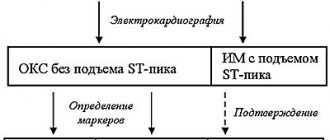
What factors are now mainly considered as candidates for inclusion in a risk stratification system? These are heredity and genetic markers. These are biochemical markers of inflammation. If I think everyone knows about high-sensitivity C-reactive protein, then maybe not everyone knows about lipoprotein-associated phospholipase A2.
I would like to draw your attention to the fact that there are different types of phospholipases – there are several of them. We know, for example, secretory phospholipase, there is LpA-small associated phospholipase A2. However, there is a separate protein - this is LP FLP A2. It is considered as a possible marker of atherosclerotic plaque instability.
It is believed that if its level is elevated, then the patient most likely has an unstable plaque. It is urgent to take measures to stabilize it. I must say that in Israel this marker is already used as a standard screening method. A patient with ABS comes for examination. They suggest to him - would you like to see if you have an unstable atherosclerotic plaque? To do this, you need to conduct this analysis.
Maybe they are getting a little ahead of themselves. This protein still needs to be studied, but we see that we will be able to diagnose unstable atherosclerotic plaque in the future.
We must take into account signs of organic vascular damage, and we can take this into account at the moment. This is, first of all, preclinical atherosclerosis, that is, thickening of IMT, the presence of atherosclerotic plaques. An increase in the stiffness of elastic vessels, which we can determine by an increase in the speed of the pulse wave. This is the ankle-brachial index, which is very easy for any doctor to measure with only an ankle tonometer. This is the level of coronary calcium, which is not very easy to measure.
10:19
The slide shows the 2010 recommendations of the American Heart Association. They can be used to identify additional cardiovascular risk criteria in asymptomatic patients with respect to atherosclerosis. We see that this is a family history in the first place, and the class of evidence is one (very high). Genetic studies show only third class evidence.
Thickening of the MI complex in high-risk patients is class IIa evidence. This is the ankle-brachial index, this is the pulse wave velocity, this is the coronary calcium index, the level of evidence of which varies depending on the risk determined by the Framingham scale. This is the level of C-reactive protein, this is the level of lipoprotein-associated phospholipase A2.
A very interesting SHAPE algorithm was also proposed to clarify the risk in men over 45 years of age and in women over 55 years of age, in whom the risk was initially determined to be low or moderate. This is the so-called test for preclinical atherosclerosis. The test can be considered negative or positive.
What is a test for preclinical atherosclerosis? This is a multispiral computed tomography to assess the coronary calcium index and a duplex study of the carotid arteries, which takes into account the thickness of the MI complex, as well as the presence or absence of an atherosclerotic plaque.
We see that if the test is negative, then the patient remains in the low to moderate risk group. However, if the test is positive, the patient immediately moves into at least the moderately high risk category. Next, we look at what specific values were obtained from the patient during the examination. We focus specifically on the numbers, on the quantitative determination of the coronary calcium index, the quantitative assessment of the thickness of the MI complex and whether atherosclerotic plaques significantly narrow the arteries or not.
We see that if the plaques narrow by more than 50%, then the patient already falls into the very high risk category. But this is not something abstract that we wrote to the patient and forgot about it. No. When we classify a patient into one category or another, we must achieve target levels of cholesterol or low-density protein. The doses of drugs and the drugs we use will be different. This is our strategy for treating the patient.
12:58
The slide presents the prognostic significance of the thickness of the MI complex in different parts of the arterial bed for the diagnosis of stenotic atherosclerosis of the coronary arteries. We looked at which arteries are best examined with ultrasound, so that these values can be used for risk stratification.
We see on the slide in green that the best vascular basin is the carotid artery basin, since the sensitivity and specificity are higher here. It is best to look at the carotid arteries.
The CAFES-CAVE study is presented in a slide that shows the likelihood of developing cardiovascular complications in patients whose baseline risk is determined to be low, depending on what we find on the carotid duplex study. We see that if we find a completely normal picture, then the risk will be minimal.
Increasing the thickness of the MI complex slightly increases the risk of complications. The presence of an atherosclerotic plaque without stenosis increases the risk by almost 40%. The presence of a plaque that significantly stenoses the lumen of the carotid artery increases the risk by 80%, that is, it completely changes our understanding of the patient.
The slide also shows the relative risk of development in individuals who do not have complaints, depending on physical and instrumental examination. In first place is the positive stress test. If, during a stress test, we see data in a patient that indicates myocardial ischemia, most likely he will have damage to the coronary arteries. However, we see that if we listen to a bruit on the femoral artery in a patient or identify atherosclerotic plaque in the carotid or femoral artery, then that patient also has a high probability of coronary artery disease.
15:04
The thickness of the “intima-media” complex as a risk factor, that is, how can this be displayed quantitatively? Quantitatively, we can display this as follows. An increase in the thickness of the MI complex by 0.2 millimeters increases the risk of developing myocardial infarction by 33% (by a third), and stroke by 28%.
A meta-analysis of 8 studies was also conducted, which showed that each increase in the thickness of the MI complex by 0.1 millimeter increases the risk of myocardial infarction from 10% to 15%, and stroke from 13% to 18%. We have no right to ignore this indicator.
LP FLPA A2 as a risk factor for complications. What? Coronary heart disease, stroke, death from cardiovascular causes and death not related to cardiovascular causes. We see the relative risk assessment. In all cases, LP FLPA A2 plays an important role. We should, in theory, take its level into account when we stratify a patient's risk.
How can we take this into account? We have to measure the patient's phospholipase A2 level and see if it is more than 200 nanograms per milliliter, then the moderate-risk patient will move into the high-risk group. If a high-risk patient has more than 200 nanograms per milliliter, then that patient goes straight into the very high-risk category. I once again emphasize that it will not just be a formal transition, but we must change our therapy.
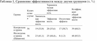
Allow me to report the results of our research, which was carried out in our medical institution - the Russian Cardiology Research and Production Complex - together with 12 clinics in the Western Administrative District of Moscow. It included 2400 patients. These patients were divided into three groups. Group one is healthy people who have less than two risk factors. The second group is patients with risk factors who have more than two risk factors. The third group is patients who have coronary heart disease.
17:25
What did we get? As would be expected, in patients with coronary heart disease, the level of LP FLPA A2 was the highest. In healthy individuals (group one) it was the smallest. Pulse wave speed. Here we got results that were not entirely clear to us at first. We see that in patients with coronary artery disease, the pulse wave speed is in the middle, and the highest pulse wave speed is found in patients with high risk.
Why is this happening? We had the right to expect something different, however, after analyzing how these patients were treated, what treatment was prescribed to them, we saw that patients who have coronary heart disease took medications. In particular, it was mainly a combination of Diroton and Amlodipine. Some used this combination, some used the equator drug right away, which, as you know, consists of amlodipine and diroton.
In patients, we reduced the pulse wave speed in this way. It became closer to the indicators of healthy individuals. This, colleagues, is very important. This shows that we treat patients who have coronary heart disease correctly and treat them well. But we do not pay any attention at all, excuse the vulgarity, to patients who have a high risk of developing cardiovascular diseases, but who do not yet have them.
We are talking in this case about primary prevention, although, as you know, Professor Nelson said that there is no primary prevention. When we treat a patient, this is always secondary prevention. In this case he is right. We are already beginning to treat subclinical atherosclerosis. We must do this.
Oksana Drapkina : I would like to ask. The question is practical. This combination - an ACE inhibitor and a calcium blocker - in clinical practice, it suggested itself. Then - if we remember this octagon, hexagon, then the triangle of rational antihypertensive therapy, then the lines were always not dotted, but solid, connecting ACE inhibitors and calcium blockers.
In my opinion, it is very successful to combine Amlodipine, a rather powerful drug in terms of lowering blood pressure, and plus lisinopril. Indeed, the experiment proved some antiatherogenic effects of amlodipine. Vascular cell proliferation decreased and endothelial function improved.
You talked about pulse wave speed. Calcium blockers are the drugs of choice. For example, for elderly patients with isolated systolic hypertension. This is a pretty successful combination (I just want to share), which is in one tablet (fixed combination), of course, it greatly helps us retain patients and maintain their compliance. Do you feel this too in the clinic?
20:44
Igor Sergienko : Of course. Clinic doctors - local therapists - are advanced doctors. Don't underestimate it at all. Without our help, they treated patients correctly and correctly, if I can, of course, evaluate the results of their work.
We see very concrete results. We see that the people for whom they made efforts, who were prescribed the drug... I very much agree with compliance, because when several tablets are prescribed, we know that some patients refuse. Moreover, one visit to the clinic, and after that...
When the patient is with us, it’s easier for us (in the hospital). We can prescribe several medications, the patient can take them and feel better during hospitalization. Then he won’t refuse. The patient came here once. Okay, he came to the appointment twice. He needs to be prescribed the drug immediately. Equator was prescribed, the patient began taking it and felt better. When we inform him of the results we get...
Oksana Drapkina : About the forecast.
Igor Sergienko : About the forecast. Here the patient, of course, will not refuse this drug.
Oksana Drapkina : About its metabolic neutrality, because hypertensive people in our country, in general, are obese.
Igor Sergienko : Yes, as a rule.
Oksana Drapkina : You especially showed a picture with a high risk - it’s quite difficult. Sorry for the interruption!
21:59
Igor Sergienko : No, no. Thank you. On the contrary, it's very nice.
Oksana Drapkina : Please, continue.
Igor Sergienko : The value of the coronary calcium index. Everything is clear here. In patients with coronary heart disease it is higher than in other groups. This is completely understandable: calcium is deposited, and, unfortunately, we cannot dissolve it with any medications.
Very interesting slide. Duplex study of the carotid arteries in patients with low and moderate risk. What does it mean? Roughly speaking, this patient came to close his sick leave for ARVI, for example. He was asked to participate in our study. The patient agreed. He underwent a duplex study of the carotid arteries.
We see that only 40% of women and 30% of men have arteries without atherosclerotic plaques. However, 20% of men have two atherosclerotic plaques in the carotid arteries, and 16% have four. These are people who considered themselves and whom doctors considered to be completely healthy, without, of course, prescribing anything to them. We see on the slide what kind of picture they have, however. The patients were clearly not thoroughly examined.
The slide also shows low- and moderate-risk women. The frequency of detection of atherosclerotic plaque depending on age and the presence or absence of arterial hypertension is shown. We, of course, see that in patients with arterial hypertension, the likelihood of identifying atherosclerotic plaque in the carotid arteries is higher than in patients without arterial hypertension. This is completely understandable.
We also see the distribution by age: in patients of an older age group, the likelihood of detecting plaques is increased compared to patients of average age and women under 45 years old, because I cannot say that this is a younger age group.
This allowed us to draw an interim conclusion from our study. Women over 55 years of age, regardless of the presence or absence of arterial hypertension, should undergo a duplex examination of the carotid arteries in all cases.
24:07
The presence of arterial hypertension increases the likelihood of detecting an atherosclerotic plaque, but its absence does not at all exclude a significant likelihood of the presence of an atherosclerotic plaque in women with low and moderate risk. We set a task - to find criteria for an increased likelihood of the presence of atherosclerotic plaques in the carotid arteries in women who initially have a low or moderate risk.
We searched for differences in the studied parameters in women with low and moderate risk under the age of 45 years without arterial hypertension, and did not find significant differences. The same is true for women under 45 years of age, but with arterial hypertension. Also, no significant differences were found.
This allowed us to make an interim conclusion that in women with low and moderate risk under the age of 45, without taking into account the presence of arterial hypertension, it is not advisable to conduct a duplex examination of the carotid arteries as a screening method.
What about the 45-55 age group? We assessed the likelihood of having an atherosclerotic plaque in this age group, regardless of blood pressure level. They showed that there is a relationship with body mass index, with uric acid levels greater than 260 µmol/liter, and with PL A2 levels greater than 315 ng/ml. With these indicators, the likelihood of having an atherosclerotic plaque in this age category of women is higher.
We concluded that in women 45-55 years old, regardless of the presence of arterial hypertension, it is advisable to conduct a duplex study of the carotid arteries when the BMI is more than 30 or the uric acid level is more than 260. We do not take into account the FL level for now, since this indicator is not determined everywhere. Rather, this is an exception for now.
26:13
A study of low- and moderate-risk men found that men under 40 years of age had the same findings as a subgroup of women under 45 years of age, and men over 50 years of age had the same findings as women over 55 years of age.
Let me not repeat the same calculations, not show the same slides, but simply make an intermediate conclusion that it is always advisable for men over 50 years old to conduct a duplex study. For men under 40 years of age, duplex examination of the carotid arteries is not advisable. I mean, of course, only as a screening.
What happens in the middle age category among men aged 40-50 years with a moderate initial risk? We have shown that the likelihood of detecting an atherosclerotic plaque is associated with the presence of metabolic syndrome and a uric acid level greater than 350 µmol/liter.
An interim conclusion was made that men aged 40 to 50 years, regardless of blood pressure, should undergo a duplex examination of the carotid arteries if they have metabolic syndrome or a uric acid level of more than 350 µmol/liter.
The slide shows the algorithm that we developed after conducting this research. The research is ongoing. It will end at the beginning of 2012, but for now we can propose an algorithm of indications for duplex examination of men and women of low and moderate risk in various age categories.
Let me present a potential algorithm for moving men and women at low and moderate SCORE risk to high risk.
So, if we have a woman over 45 years of age or a man over 40 years of age, and they have a blood pressure of 130-139 systolic and 85-80 diastolic, or if patients are taking antihypertensive drugs and it doesn’t matter what their blood pressure is if we If we find an atherosclerotic plaque in these patients (I repeat, it does not matter whether it significantly or insignificantly narrows the lumen), then these patients can immediately be included in the high-risk category.
For the third time, I draw your attention to the fact that they are not only classified as high risk, but we are obliged to change their treatment.
28:40
The objectives of the next stage of the examination will be to study the growth rate of atherosclerotic plaque in the carotid arteries. Study of the prognostic significance of the number of atherosclerotic plaques in the carotid arteries, since we have not yet taken this into account. Of course, the search for markers of instability of the atherosclerotic plaque, which, it seems to me, would be very promising with LP PLA A2.
I would like to thank 70 local therapists in 12 clinics in the Western Administrative Circle of Moscow for this study.
Thank you.
29:10
WHO activities
Under WHO's leadership, in 2013, all Member States (194 countries) agreed on global frameworks to reduce the burden of preventable NCDs, including the Global Action Plan for the Prevention and Control of NCDs 2013–2020. The plan aims to reduce premature deaths from NCDs by 25% by 2025 through 9 voluntary global targets. 2 of these global goals directly address the prevention and control of CVDs.
Goal six of the Global NCD Action Plan aims to reduce the global prevalence of high blood pressure by 25%. High blood pressure is one of the main risk factors for cardiovascular disease. The global prevalence of high blood pressure (defined as systolic and/or diastolic pressure ≥140/90 mmHg) among people aged 18 years and over in 2014 was about 22%.
To achieve this goal, it is necessary to reduce the incidence of hypertension through national policies that address behavioral risk factors, including harmful use of alcohol, physical inactivity, overweight, obesity and high salt intake. Early detection and cost-effective management of hypertension to prevent myocardial infarction, stroke, and other complications requires a risk-based approach.
Goal eight of the Global NCD Action Plan aims to ensure that at least 50% of eligible people receive drug therapy and counseling (including glycemic control) to prevent myocardial infarction and stroke. Preventing heart attack and stroke using a comprehensive approach that takes into account overall cardiovascular risk is more cost-effective than treating based solely on individual risk factor thresholds and should be part of the core package of services to achieve universal health coverage. sanitary assistance. Achieving this goal will require strengthening key components of the health system, including financing health care services to ensure access to essential health technologies and essential medicines for NCDs.
In 2015, countries will begin setting national targets and measuring progress against the 2010 baselines set out in the Global Noncommunicable Disease Status Report 2014. The UN General Assembly will hold its third high-level meeting on NCDs in 2021 to review countries' progress towards achieving the voluntary global targets by 2025.