What it is?
Heart cells (cardiomyocytes) that die as a result of hypoxia (after a heart attack) are replaced by scar tissue.
It is significantly different from healthy. Having sufficient mechanical strength, the scar does not exhibit elastic properties and cannot contract. Blood passes through the heart chambers under high pressure. The dead area of the heart is not immediately replaced by durable scars, so the weakened tissue in this area stretches and swells under blood pressure. This is how a protrusion appears in the cavity of the heart - an aneurysm. The pathological formation can quickly increase and pose a threat of rupture of cardiac tissue. Aneurysms most often occur in the ventricles, the left side of the wall being mainly affected by this anomaly. This happens because in this part of the heart the blood pressure is higher than in all the others.
An enlarged heart is a consequence of other pathologies that weaken or damage the heart tissue. An additional factor contributing to protrusion in the heart area is the forced increased work of the heart.
Diagnostics
It is impossible to diagnose an aneurysm without an instrumental examination. Therefore, after collecting anamnesis and examining the patient, the doctor issues a referral to:
- ECG;
- Echo-CG;
- MRI;
- MSCT of the heart (multispiral computed tomography);
- Heart PET (positron emission tomography);
- myocardial scintigraphy (isotope scan) - intravenous administration of radioactive isotopes (radionuclides) to assess absorption by the heart muscle;
- EPI (electrophysiological study) - stimulation of various parts of the heart to detect life-threatening arrhythmias;
- coronary angiography and cardiac probing - X-ray contrast examination (catheterization) - insertion of a special catheter through the artery.
Types of cardiac aneurysms
Cardiac aneurysms can differ from each other, depending on:
- Protrusion forms.
- Method of occurrence.
- Dimensions of the neoplasm.
- Location in the heart area.
- Duration of the education period.
- Structural changes in tissues.
Form
The appearance of the aneurysm will indicate to the doctor how quickly the formation can develop. In accordance with this, diagnostics will be carried out and the method of treatment will be determined.
- Bag form. The protrusion is quite voluminous, with a wide base and a large cavity, reminiscent of a hanging bag. The tissue at the site of expansion is subject to significant stretching. Consequences: increased risk of violating the integrity of the aneurysm, the risk of blood stagnation in the “bag” and the development of blood clots.
- Mushroom shape. The base of the bulge is narrowed, the cavity area is expanded under the influence of blood. The shape resembles a mushroom. The tension of the walls is significant, the tissue structure is damaged. The source of aneurysms is small areas of dead tissue or scar formation. Consequences: very high risk of thrombosis and rupture of the heart walls.
- Diffuse (flat) shape. Such an aneurysm develops from a wide area of the damaged surface, but is characterized by a small volume. It is almost flat (does not protrude from the cavity of the heart). Occurs with extensive heart attacks. Neoplasms are most often localized in the area of the anterior wall, on the side of the left ventricle. Consequences: development of heart failure, arrhythmia, prospect of increasing organ size.
- Double aneurysm (one inside the other). The most dangerous protrusion of the heart wall. One aneurysm develops in the cavity of another. The first bulge has a diffuse or saccular shape. The wall of this formation is additionally stretched in one of the areas. A new protrusion forms there. That is, a pathological stratification of the structure of the cardiac tissue occurs. Consequences: “aneurysm within an aneurysm” ruptures most often.
Method of occurrence
- True aneurysm. It develops in the layers of the heart wall, which is partially replaced by connective tissue.
- False aneurysm. A cavity is formed between the pericardial tissue and the fibrous adhesion. There is no stretching of the cardiac cavity. There is a micro-hole in the wall through which blood flows into the newly formed cavity.
- Aneurysm in the heart of functional origin. A very rare type of pathology. Protrusion of the muscular layer of the myocardium occurs without changing the myocardium itself. Reason: the area of the heart stops participating in contractile activity due to any pathological disorders.
Aneurysms are also classified by size. This will help the doctor more accurately predict the further development of the pathology.
- Small aneurysms. Such a cardiac aneurysm can be noticed when the organ contracts. Damaged areas are unable to contract.
- Medium aneurysms. They expand slightly (up to several centimeters). Do not protrude beyond the pericardium.
- Giant aneurysms. They are large in size, which means the shape of the heart is noticeably changed. Some are equal in volume to the cavity of the left ventricle.
Location
Protrusions prefer to develop in the cavity of the left ventricle. It is this part of the heart that needs oxygen most, so during an attack of acute hypoxia, the cells of the left ventricle are the first to die. Rarely, aneurysms are located in the posterior wall or in the septum between the ventricles. More often - in the upper or front part.
An interventricular aneurysm is represented by a shift of the septum into the right ventricular zone. The consequences of such deformation include the development of heart failure, since the left ventricle increases and the right one decreases.
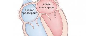
On the right side, the heart is almost not subject to pathological stretching. Typically, dilatation of the right ventricle occurs only in the case of injuries received in the heart area. Another way for the development of protrusions in the right ventricle is the consequences of certain operations on the heart muscle: correction of a heart defect acquired at birth (tetralogy of Fallot, pulmonary stenosis). Aneurysms are almost never found in the atria.
Moment of formation
With the help of such clarification, aneurysms that arise after a heart attack are differentiated. To distinguish, determine the amount of time passed after the onset of tissue necrosis until the formation of a protrusion.
- Acute education. The heart is already enlarged within the first two weeks. The scar tissue has not yet fully formed. In this case, the aneurysm behaves unpredictably: it can grow quickly, it can rupture, and its shape can also change.
- Subacute aneurysm. It forms a little later (after the 3rd week and before the 8th). At this time, the connective tissue is already quite strong, which reduces the risk of its opening or further growth. Blood clots may form in the aneurysm cavity.
- Chronic aneurysm. The expansion of the heart in diameter is formed almost two months after the heart attack. The scar has already completely hardened, so its stretching cannot be rapid or significant. In this case, almost no ruptures of the heart wall are recorded, but the risk of thrombosis and arrhythmia is high.
Bulge structure
If an aneurysm formed after a heart attack, then its structure will contain a large amount of connective tissue.
When an infection penetrates the myocardium and damages it, the muscle layer predominates in the structure.
Prediction of the further state of the neoplasm depends on the classification of aneurysms according to their composition.
- Fibrous bulge. It contains predominantly connective tissue in its structure, which cannot participate in the contraction of the heart. Under blood pressure it stretches and becomes thin. Occurs in the first time after a heart attack.
- Fibromuscular bulge. The structure contains muscle tissue and scar tissue. This phenomenon often occurs when a parietal infarction develops. Not all layers of cardiac tissue undergo necrosis.
- Muscle bulge. It consists almost entirely of muscle cells. Muscle strains occur due to a congenital abnormality, poor blood flow, or impaired transmission of nerve impulses. This part of the heart does not contract with all the others, so the internal pressure on it increases. A muscular aneurysm develops. The pathology occurs without symptoms.
Surgical methods
Today, the following types of interventions are known to be used for cardiac aneurysms:
- An operation to strengthen the walls of the aneurysm with autografts (used only for diffuse aneurysms).
- Closed surgery for resection of an aneurysm (the base of the aneurysm is covered with special clamps).
- Open surgery to remove an aneurysm.
- Diaphragmoplasty (the heart wall is strengthened with diaphragmatic tissue).
The choice of treatment method depends on the shape, location and size of the aneurysm, as well as the health status and age of the patient. Despite the fact that surgery is associated with some risks, without surgical treatment most patients die within the first years.
Paths of aneurysm development
Aneurysm, in most cases, occurs in the post-infarction state. Usually the upper part of the left ventricle or the front part protrudes; in rare cases, the posterior part of this part of the heart swells.
The following conditions can accelerate the formation of a cardiac aneurysm after a heart attack:
- second heart attack;
- accelerated heart rate;
- the appearance of heart failure;
- attacks of high blood pressure;
- violation of recommendations on bed rest.
But a heart attack is not the only way pathology develops. There are also other causes of abnormal stretching of cardiac tissue.
. When a child is in the womb, he may be exposed to various negative influences (nicotine or alcohol poisoning, intrauterine infections, the effects of medications). These factors influence the formation of heart structures. After birth, as the baby breathes independently, the pressure in the heart area increases, and the deformed areas of the heart protrude. As the baby grows older, pathological phenomena may disappear. But there will always be a risk of developing new aneurysms throughout your life.
Congenital pathology- Infectious organ damage . With the penetration of infection into the heart, myocarditis develops. Inflammation covers the walls of the heart muscle, which leads to partial cell death and their replacement with connective tissue. In such areas, the risk of developing an aneurysm increases.
- Postoperative complications . After operations aimed at eliminating heart defects, complications are possible in children and the elderly. The cardiac sutures may not heal properly, leaving a dense scar, which will serve as the site for the formation of an aneurysm. Another reason: the heart, having survived the operation, contracts at an increased rate, which leads to an increase in pressure inside it. The operated areas suffer from this first of all; the weakened tissue begins to stretch.
- Myocarditis of toxic origin . This is a fairly rare reason for the development of an aneurysm. Toxic substances enter the bloodstream for various reasons. Toxins penetrate the heart and damage its tissues, sometimes necrosis develops. Severe manifestations of allergic reactions also lead to damage to the endocardium (the inner lining of the heart). All these factors create conditions for deformation of the heart wall.
- Cardiosclerotic changes of an idiopathic nature . In some cases, the patient is diagnosed with cardiosclerosis of unknown etiology. Cardiomyocytes begin to be replaced by the connective layer; over time, this process leads to the formation of an aneurysm.
- Closed and open organ injuries . As a result of an open injury, a scar (scar) forms in the heart at the site of injury. When closed, the development of false aneurysms or myocarditis, accompanied by cardiosclerosis, is possible.
- Irradiation . If the heart has been exposed to strong radioactive radiation, cardiosclerosis develops in its cells. This phenomenon can occur during the treatment of a cancerous tumor in the mediastinum. The rays entering the heart trigger the process of slow destruction of heart cells. An aneurysm can develop over a long period of time.
Long-term inflammatory process . Dysfunction of the immune system leads to rheumatism of the heart muscle. The inflammatory process occurs due to the struggle of heart cells with their own antibodies. Prolonged inflammation weakens the heart, myocarditis and cardiosclerotic changes develop.
Treatment of aortic aneurysm
A cardiac aortic aneurysm, the diameter of which exceeds 5 cm, is subject to surgical treatment due to the fairly high risk of its rupture. Surgery is performed under conditions of hypothermia and artificial circulation. In addition, surgical intervention is performed if the specialist observes too rapid growth of the aneurysm. The surgical mortality rate of this disease is about 15%. Contraindications to surgery are present if the patient suffers from severe heart disease.
Rehabilitation after treatment
During rehabilitation, it is important to follow a diet (eat as little fatty foods as possible, include apples, garlic, strawberries, and legumes in the diet). In addition to proper nutrition, it is very important to avoid stressful situations, control blood pressure, and monitor your weight (it should not exceed the norm). After the surgical intervention, an individual daily routine is drawn up for the patient. It is necessary to set aside time for rest. In this case, the patient undergoes regular examination by the attending physician.
Symptoms
Pain in the chest does not always accompany the appearance of an aneurysm. The tumor does not have nerve receptors, so there cannot be pain in this area. Unpleasant sensations occur in healthy tissues due to poor circulation.
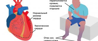
A feeling of loss of strength indicates the development of heart failure. As a result of this condition, the heart cannot send the required volume of blood to the muscles, so general weakness occurs.
The rhythm of contractions is disrupted - this is the most typical sign of an aneurysm. Interruptions in heart rate occur from time to time, but do not last long. They usually occur due to emotional stress or significant physical activity.
The pale complexion of the face and body is explained by the phenomenon of heart failure. The heart does not supply blood well to the skin, which causes them to turn pale. You may experience numbness in the limbs and low skin sensitivity.
Palpable heart pulsation. A person with an aneurysm constantly feels their heart beating, even in a state of absolute rest. This occurs due to the increased volume of the left ventricle; it moves closer to the ribs, which creates the sensation of an increased heartbeat.
Congestion associated with heart failure causes deterioration of lung function. Oxygen escapes from the lungs more slowly, causing shortness of breath.
Coughing attacks are a very rare occurrence with an aneurysm in the heart. Cough occurs in the case of large formations when the lungs are compressed. When you take a deep breath, the pleura in the compressed lung becomes irritated, and a coughing attack begins. There is no wheezing and no sputum is produced. Cough can occur for the same reason as shortness of breath.
Prevention
Prevention of cardiac aneurysm consists of timely diagnosis of myocardial infarction and its treatment literally from the first minutes of diagnosis and hospitalization in an intensive observation unit. After discharge from the hospital, it is necessary to observe a cardiologist and follow the doctor’s recommendations and prescriptions. The cardiology center of the Federal Scientific and Clinical Center of the Federal Medical and Biological Agency has developed several heart research programs. Seek advice from our specialist to promptly identify the disease.
In parallel with drug treatment and observation by a doctor, it is recommended to adjust your lifestyle and diet:
- remove fatty, smoked and salty foods from the diet;
- stop drinking alcohol and smoking;
- exclude heavy physical activity and emotional stress.
Features of expansion of the right ventricle of the heart
The cause of this pathology is called “cor pulmonale”. This phenomenon occurs when a person develops lung diseases that make breathing difficult. In this case, the pressure in the artery passing through them increases. This affects the work of the right ventricle; it is overloaded, pumping large volumes of blood. Its cavity expands. Provoking factors:
- Respiratory diseases: the presence of bronchial asthma, chronic bronchitis, tuberculosis and so on.
- Pathologies of the thoracic part of the body: scoliosis, poliomyelitis.
- Diseases of blood vessels localized in the lungs: embolism, arteritis, thrombosis, tumor effects on blood vessels.
Signs of an enlarged right ventricle are:
- dyspnea;
- weakness;
- fainting;
- arrhythmia;
- bloody cough;
- chest pain;
- cold sweat.
In children, congenital defects may be identified that lead to enlargement of the right ventricle. They are accompanied by arrhythmias, cyanosis, shortness of breath, and rapid heart rate. Such children do not grow well.
Cause
Usually the cause of the formation of a cardiac aneurysm is a heart attack, most often of the left ventricle of the heart. When they talk about the development of myocardial infarction, the damaged parts of the heart are weakened, and under the influence of blood pressure they can bulge. Blood trapped in them forms a blood clot. Aneurysms occur in patients with atherosclerotic cardiosclerosis and cardiosclerosis of other origins. Much less common are aneurysms that arise due to other reasons: infections, injuries, and congenital defects.