Oleinik Evgeniy Mikhailovich
, cardiovascular surgeon, phlebologist.
Phlebology
is a branch of medicine that studies the structure and function of veins, as well as developing methods for diagnosing, treating and preventing their diseases.
Frequent vein diseases found in modern people are:
- Chronic venous insufficiency
of the lower extremities -
varicose veins
of the lower extremities; - Phlebopathy;
- Spider veins;
- Thrombosis of superficial and deep veins, thrombophlebitis;
- Trophic ulcers.
The main reason for the development of chronic venous insufficiency of the lower extremities is human upright posture, and therefore, evolution provided for the special functioning of the veins of the lower extremities with the help of valves.
The operation of the valves resembles the operation of a system of sluices in a river; with each “suction” of blood to the heart, the valves open and raise venous blood.
The relevance of the problem of venous disease of the lower extremities is beyond doubt. With the development of civilization, unfortunately, the level of physical activity decreases, and as a counterbalance to this, static loads, stressful situations, and nutritional disorders increase. Risk factors
development of chronic venous disease (chronic venous disease of the lower extremities) are:
- Sedentary lifestyle;
- Orthopedic problems (flat feet, wearing the wrong shoes);
- Increased static loads on the legs (standing and sitting work);
- Gender (yes, yes, exactly gender, in the fair half of humanity, unfortunately, this disease is more common);
- Use of contraceptives;
- Diabetes;
- Increased weight;
- Age;
- Heredity;
- Pregnancy;
- Smoking.
Main symptoms
chronic venous insufficiency are:
- Heaviness in the legs;
- Swelling of the legs;
- Protruding veins and stars on the skin of the legs;
- In severe cases, the appearance of long-term non-healing wounds on the legs.
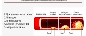
Development of chronic venous insufficiency
has a staged, gradually developing character. In the initial stages, the main manifestations of the disease are heaviness and swelling in the legs. This is due to disruption of the normal functioning of the valve system, which causes stagnation of venous blood in the tissues. Subsequently, with the progression of insufficiency of the function of venous valves, blood stagnation intensifies, so-called spider veins appear, and then varicose veins. The increase in stagnation of venous (“bad”, full of “waste”) blood leads to changes at the level of tissues and cells: their nutrition is disrupted, the color of the skin in these places changes, microcracks appear, sensitivity is impaired and trophic ulcers develop.
Types and purposes of ultrasound of the veins of the lower extremities
In recent decades, ultrasound diagnostics has become a simple, inexpensive, and most importantly, accessible research method.
The quality and capabilities of ultrasonic devices are constantly being improved, their classification is constantly expanding. The great advantage of the technique is its non-invasiveness. Vein ultrasound is no exception. Modern ultrasound scanners allow a thorough diagnosis of the venous system.
The main goals of ultrasound of the veins of the lower extremities are to assess the condition of the vessels (their diameter, course, relationship with other structures, wall condition), examine blood flow parameters, and identify blood clots. The article will talk about the types and purposes of ultrasound examination of blood vessels, as well as how ultrasound of the veins of the lower extremities is performed.
Due to the constant improvement of devices, many terms have appeared to denote various components of ultrasound examinations and their combinations.
Let's try to figure out what ultrasound, DS, ultrasound, ultrasound of veins are.
- Doppler ultrasound (Doppler ultrasound) is a method based on the Doppler effect. This effect consists of changing the frequency of an ultrasonic wave when reflected from a moving object. In the lumen of the vessel, such objects are formed elements of blood, for example, red blood cells. Computer processing of reflected signals allows one to obtain a curve of the speed characteristics of blood flow. Currently, ultrasound scanning in its pure form is not used, but is included in the structure of other studies.
- DS (duplex scanning (USD)) - allows you to visually evaluate the vessel - its wall, course, lumen, as well as conduct an ultrasound scan of any part of the vessel - i.e. in addition to examining the vessel, show the characteristics of blood flow at any point. Modern devices also use coloring of blood flows in blue and red colors, based on the Doppler effect (the so-called color Doppler or color Doppler mapping (CDC)). Combining duplex scanning with color flow is also called triplex scanning.
- USAS (ultrasound angioscanning) - a synonym for DS, is the same.
- Ultrasound of veins (ultrasound examination of veins) - this term refers to the entire combination of the above methods. Modern devices provide all the components of the study - ultrasound, DS, and color Doppler mapping (CDC).
Currently, when a doctor prescribes an ultrasound scan of the veins of the lower extremities, an ultrasound scan of the veins of the lower extremities, he means that the patient will undergo an ultrasound scan of the veins according to all possible parameters.
How is a vein ultrasound performed?
Ultrasound examination of the vascular system of the legs means a procedure for examining the anatomy of veins and arteries, assessing their functioning, the condition of the valves and the characteristics of blood flow. The technique makes it possible not only to find out why blood circulation is affected in a particular area of the vessel, but also to identify inflammation or blood clots. Ultrasound of the vessels of the lower extremities is an important procedure for diagnosing existing problems and helps in planning the correct treatment.
Types of sonography
To study the vessels of the lower extremities, a procedure based on the Doppler effect is used. In medicine, it means the reflection of ultrasound radiation from red blood cells flowing through the vessels.
As a result of a Doppler examination, the doctor gets an idea of the characteristics of the passage of the vascular cord, the speed of blood flow and other nuances of the functioning of the veins and arteries of the legs.
So what types of research are there, and what do they show?
USDG
The term “USDG” means “ultrasound Dopplerography”. This is a technique usually used to: – establish the patency of deep vascular collectors; – assessing the condition of superficial veins; – diagnostics of the condition of valves, including valves of typical main components of the venous system, that is, perforating veins.
UZDS
Duplex scanning combines the principles of Doppler and traditional examination methods and performs the following functions: – studies the operation of vein valves in real time; – makes it possible to assess the condition of the vascular walls; – allows you to analyze the patency of deep and superficial veins; – calculates the presence and location of blood clots.
Technologically, the method remains the most popular and accurate way to assess the performance of the venous system in all respects.
Online scanning
Online scanning is a complex that combines ultrasound and ultrasound scanning. It is intended to determine: – the condition of the walls of arteries and veins; – assessment of valve health; – patency of vascular collectors; – the condition of the veins connecting superficial vessels with deep ones; – the presence of blood clots and their characteristics, including size and location; – degree of blockage of the vessel.
Ultrasound with color mapping
The most modern method of studying the veins and arteries of the legs is distinguished by color identification of the speed of blood flow in different areas. For example, shades of red characterize the flow of blood to the sensor, and blue tones characterize the flow of blood directed away from the sensor.
Important! The brighter the color, the higher the speed of blood movement.
In modern diagnostics of arterial-venous pathologies, the popularity of this method is growing due to its high information content and simplicity.
Preparation
When referring veins and arteries of the legs for ultrasound (as well as other soft tissues, as a rule), there is no need to carry out preliminary preparation for the procedure - it is not required.
How do they do it?
How is the deep veins and arteries of the legs diagnosed?
Ultrasound examination of blood vessels looks like this:
The patient enters the ultrasound diagnostic room, provides the doctor with a referral and frees the veins of the lower extremities. That is, he takes off tights or trousers, remaining in his underwear. The diagnostician applies a little conductive gel to the legs one by one, which will ensure better adhesion of the sensor to the surface of the skin.
During the manipulation, the doctor can change the frequency of the sensor for better visualization of deep vessels, but the patient will not feel this in any way.
The difference between an ultrasound scan is that during the examination the doctor will measure the pressure in both the upper and lower extremities. During the procedure, the patient will change his position from sitting to lying down and back.
During the examination of the leg veins, they are first examined with the patient lying down, then he will be asked to stand up. In addition, as part of the examination, special tests are carried out to determine the blood flow between the superficial and deep vessels. The test consists of taking a deep breath, without interrupting it, you need to make a strained effort.
The deep veins of the legs are examined in traditional and color modes. For a better examination, the patient is asked not only to perform stress tests, but also the areas where the veins are located are palpated with different intensities. Such tests (like the ultrasound itself) are performed in different positions: lying on your back or stomach, as well as standing.
Contraindications
In addition to the indications for ultrasound of the blood vessels of the legs, there are also contraindications.
Contraindications to duplex vascular examination are associated with the relatively long duration of the procedure - it takes about 40 minutes.
It may turn out that this examination turns out to be secondary and the study of adjacent tissues or internal organs comes to the fore.
Important! You should not perform an ultrasound in the area of the veins of the lower extremities if the skin is broken or damaged.
The main contraindications to studying vessels using the duplex method are:
– acute infectious processes; – any diseases accompanied by open wounds on the skin at the location of the sensor; – burns; – emergency conditions; – mental illnesses that make it impossible to carry out the procedure.
There are also conditions in which it is not recommended to scan blood vessels due to possible discomfort for the patient during prolonged immobility. These conditions include:
- excessive fullness;
- bloating;
- cystitis and some diseases of the genitourinary system;
- lymphostasis, causing severe swelling of the limbs.
Advantages and disadvantages
The advantages of the ultrasound method of studying limbs are:
- no pain;
- non-invasiveness, that is, the absence of punctures and other damage to the skin;
- economic accessibility of the procedure;
- ease of implementation;
- absence of radiation or ionizing load;
- linking the research picture to real time;
- the ability to perform a biopsy under ultrasound guidance;
- good visibility of all soft tissue features;
- the possibility of repeated repetition (for example, during treatment therapy to monitor its effectiveness).
Minuses:
- there may be insufficient data for a complete diagnosis;
- Adequate assessment of small vascular formations is not always possible;
- with atherosclerotic changes, the patency of the ultrasound wave may be impaired;
- is not a replacement for angiography;
- If carried out on old equipment or with insufficient qualifications of the doctor, the procedure may have low diagnostic value.
How much does it cost, where is the best place to do it?
It is best to contact your attending vascular surgeon, who will recommend a good specialist or tell you where and at what time he himself conducts ultrasound examinations.
In the Department of Vascular Pathologies, ultrasound can be performed free of charge, as prescribed by the surgeon. For a fee, in the absence of a referral, ultrasound examination of the extremities is carried out in phlebological clinics or multidisciplinary medical centers. You can find out the cost of the examination from the administrator by phone or by making a personal appointment. But it is better to refuse to perform vascular ultrasound in the offices of small diagnostic centers where any organs are examined.
Reference! The price of an ultrasound scan will depend on the specifics of the procedure and which vessels the patient needs to examine.
For example, for an ultrasound scan of the legs, the price will be from 1300 to 3500 rubles, and duplex angioscanning will cost 800–5000 rubles, color scanning using the duplex method will cost from 900 to 6500 rubles. On average, the cost of examining the vascular sections of the legs using ultrasound will cost about 2 thousand rubles.
There are also portable ultrasound machines, so ultrasound can be performed at home: this route naturally requires specific knowledge. Still, it is better to entrust this matter to a specialist.
Conclusion
Any patient, if indicated, can safely undergo angioscanning of blood vessels, because this manipulation is safe and painless. It does not require preparation and allows you to know exactly how healthy the vessels are an hour after the start of the examination.
Who needs to undergo Doppler ultrasound of the extremities?
An ultrasound examination of the veins is usually prescribed by a doctor (surgeon, phlebologist, therapist). Indications for it can be a wide variety of conditions. Most often, the doctor prescribes a study if there are complaints and suspicions of diseases of the venous system. In other cases, ultrasound of the veins is prescribed to exclude hidden pathology of the venous system (for example, before operations, during pregnancy).
The main indications for prescribing ultrasound examination of veins are:
- Swelling of one or both legs
- Leg pain
- Feeling of heaviness and fatigue in the legs
- Night cramps in the legs
- Varicose veins, spider veins
- Before any operations
- Late pregnancy
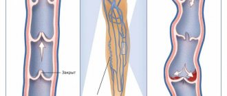
Deep vein thrombosis
Thrombosis in the early stages may be asymptomatic.
The main classic symptoms of deep vein thrombosis are: increased temperature in the affected area. The general body temperature may rise to 39 degrees. Bursting pain and heaviness in the legs appear, the color of the skin is bluish. During the first two days after the formation of thrombosis, the symptoms are mild. As a rule, this is not pronounced pain in the calf muscle, intensifying during movement and when touched, and swelling. Subsequently, the condition worsens and swelling increases. The risk of blood clot rupture increases. Within a few days after the formation of a blood clot, superficial veins appear. Indications for ultrasound scanning for the following symptoms: - Presence of swelling of the legs; -Cold feet; -Pain in the calf and feeling of “heavy legs”; - Convulsions; - Suspicion of changes in the blood vessels of the legs (especially during pregnancy); - Periodic numbness of the legs; -Itching in the legs, without external skin changes; -Diabetes; -Decreased vascular tone; - Suspicion of atherosclerosis; - Suspicion of varicose veins or other changes in venous blood flow; - Trophic changes in the skin; -Heart problems (angina or heart attack); - Migraines and fainting; -Pain when walking or exercising; -Increased cholesterol levels.
How is the examination done?
Many patients are concerned about the question of how ultrasound examination is performed, whether there are any contraindications or painful sensations during the examination.
Ultrasound diagnostics is non-invasive, painless, does not cause any discomfort, and has no contraindications. An ultrasound can be performed either by an ultrasound diagnostics doctor or by a surgeon or phlebologist who knows this method and has undergone appropriate training.
The examination of the veins of the lower extremities is carried out from the inguinal fold to the ankles. The patient completely frees his legs (panties can be left on) and lies down on the couch. The doctor applies a special gel to the lower limbs, along which the sensor will be moved during the examination. The gel is needed to ensure good contact between the skin and the sensor so that there is no obstacle to the passage of ultrasonic waves. The examination can be carried out both lying down and standing. At certain moments, the doctor may ask you to hold your breath, strain, bend your leg, etc.
The study lasts from 10 to 40 minutes (depending on the structural features of the veins and the identified pathology). For example, detected varicose veins increase the examination time, because it is necessary to carry out a number of additional tests. At the end of the study, the doctor gives the patient paper napkins to remove any remaining gel. The gel is absolutely harmless and does not leave stains on clothes.
Then the doctor writes a conclusion and hands it to the patient. It should be remembered that the conclusion itself is not a diagnosis, but only describes the identified ultrasound signs. The diagnosis can only be made by a clinician (surgeon, phlebologist), based on a comparison of the ultrasound picture, examination data and other examinations. In many cases, an ultrasound of the veins is performed not by an ultrasound diagnostics doctor, but by a phlebologist who is proficient in vein ultrasound. Then a clinical diagnosis can be made immediately.
Magnetic resonance angiography
Electromagnetic waves in a magnetic field help detect occlusions, aneurysms, stenoses, and atherosclerosis. Contrast agents are not needed for this type of diagnosis.
Multislice computed tomography
This method is used to study the vessels of the brain and lower extremities. To detect pathologies, X-rays are used. The tomograph displays the scanned vessels layer by layer on the screen. This produces a three-dimensional image, which clearly shows the condition of the veins and arteries and what the results of their regeneration are after the treatment.
How to decrypt data
Only a doctor can determine what an ultrasound examination of the veins of the lower extremities shows.
The doctor evaluates:
- Vein diameter
- Condition of the vascular wall (irregularities, thickening)
- The condition of the veins of the right and left legs is compared
- Valve condition
- Presence of pathological blood flows (refluxes)
- Vessel lumen (presence of blood clots, their structure, level of extent, apex)
- Presence of varicose veins
- Presence of incompetent perforators (veins connecting superficial and deep veins)
If during the examination the doctor notices changes in other structures that are not related to the veins (for example, pathology of the arteries, Baker's cyst, altered lymph node), he may note this in the protocol or recommend additional research.
Based on the data received, the doctor writes a conclusion; if necessary, photographs are attached to the conclusion.
Example of a conclusion: “Echo signs of varicose veins of the lower extremities. Insufficiency of the great saphenous vein on the left. Incompetent perforator of the left leg." It should be noted that the classification of varicose veins according to this conclusion is impossible - the stage is determined by the attending phlebologist based on a comparison of the ultrasound picture with clinical signs.
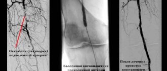
Angiography
Angiography is used to examine arterial dysfunction. This type of diagnosis will be called arteriography. If the goal is to determine the condition of the veins, then they talk about phlebography.
In any case, the procedure takes place in a hospital where there is an X-ray angiography room.
In order to determine the condition of the veins and arteries, it is necessary to inject a contrast agent and take x-rays of its progress through the body. Atherosclerosis, thrombosis, dilation of the vessel, aneurysm, thrombophlebitis will be clearly visible.
Where to take the study
Currently in Moscow there are many medical centers that offer ultrasound diagnostics. What should you look for in order to get to a good specialist?
The doctor must have extensive experience, work experience, and have positive reviews from other patients. It is very good to have an ultrasound of the veins done by a doctor who not only knows ultrasound diagnostics, but is also a surgeon or phlebologist. Such a doctor has the most complete understanding of the anatomy and functioning of the venous system, knows well how ultrasound of the veins of the lower extremities is performed, and can also make a clinical diagnosis and prescribe treatment immediately after the ultrasound.
Dr. Elshansky Igor Vitalievich fully meets these requirements, is a phlebologist surgeon, and also has advanced training in ultrasound diagnostics in angiology, has been involved in the diagnosis and treatment of vascular diseases of the lower extremities for many years, Ph.D.
Phlebectomy
This is an operation to remove pathologically dilated veins. Currently, laser techniques for vein surgery have been developed and introduced into clinical practice. The surgical intervention is performed under local anesthesia and is absolutely painless. 2-3 hours after the procedure, the patient is discharged for outpatient observation. In some cases, a so-called classic operation to remove a pathologically dilated vein and its tributaries is indicated. The most reliable in some cases is a combination of 2 methods.