What are varicose veins?
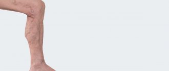
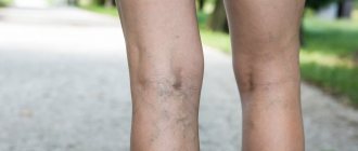
Varicose veins (in common parlance - varicose veins ) are overstretched, irregularly shaped, tortuous blood vessels that have lost their elasticity. They are increased in length and width and look like thick, convoluted blue strands that are visible under the skin. Veins become this way when the venous valves are missing or for some reason cannot perform their functions. If the valves do not work properly, blood flows through the veins in the opposite direction, downwards, accumulating in the lower sections of the veins and bursting their walls. As a result, the veins lose their natural shape, and a pathological chain of various complications begins.
Prevention
Measures to prevent deep vein thrombosis include the following:
- Don't sit still. If you have had surgery or were on bed rest for other reasons, try to get back to work as soon as possible. If you sit for any period of time, do not cross your legs as this can block blood flow. If you are traveling long distances by car, stop every hour or so and take a walk. If you're on an airplane, stand or walk occasionally. If you can't do this, stretch your shins. Do some exercises. Try raising and lowering your heels while keeping your toes on the floor, then lifting your toes while pressing your heels into the floor.
- Do not smoke. Smoking increases the risk of developing DVT.
- Do exercises and control your weight. Obesity is a risk factor for DVT. Regular exercise reduces the risk of blood clots, which is especially important for people who sit a lot or travel frequently.
How common are varicose veins?
Varicose veins are one of the most common diseases of the vascular system. According to some statistical estimates, from varicose veins . The number of people who have varicose veins increases with age, and women are affected much more often than men. According to statistics, in the age group under 25 years only 8% of women suffer from varicose veins, and in the older age group - 55 years and older - 64% of women are affected by varicose veins.
How can you recognize varicose veins in yourself?
The most common sign of varicose veins is fatigue, dull pain, a feeling of heaviness and fullness in the legs after sitting or standing for a long time. Often these symptoms appear or worsen in the evening. However, it is usually impossible to determine exactly where it hurts. And if these unpleasant symptoms - fatigue, heaviness, pain - go away after resting with your legs elevated, then they are really caused by varicose veins (unless some other cause is reliably identified).
However, do not rush to blame everything on varicose veins, especially if there are no external signs in the form of dilated veins. Some other painful conditions may also exhibit the same symptoms.
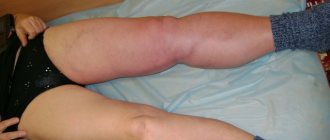
Leg cramps
With varicose veins, painful nighttime cramps in the leg muscles can actually occur (in other words, “leg cramps”). Most often, cramps appear in the calf muscles and can sometimes be so painful that the patient wakes up. Moreover, night cramps usually occur after a hard day, when the patient had to stand or sit a lot.
Complications
Complications of DVT may include:
- Pulmonary embolism (PE). PE is a potentially life-threatening complication associated with DVT. This occurs when a blood vessel in your lung is blocked by a blood clot that travels to the lung from another part of your body, usually your leg. If you have signs and symptoms of PE, it is important to seek medical help immediately. Sudden shortness of breath, chest pain when inhaling or coughing, rapid breathing, rapid pulse, feeling weak or faint, and coughing up blood may occur with PE.
- Postphlebitic syndrome. Damage to the veins by a blood clot reduces blood flow to the affected areas, causing leg pain and swelling, skin discoloration, and skin ulcers.
- Complications of treatment. Complications may arise from blood thinners used to treat DVT. Bleeding is a side effect of anticoagulants. It is important to have regular blood tests when taking these medications.
Is varicose veins inherited?
It is now known that varicose veins are hereditary. Scientists even believe that they were able to isolate a separate gene responsible for the development of varicose veins. It is not yet clear whether this gene causes malformations of the venous valves or malformations of the vein walls themselves. But there is no doubt that these studies will help develop a gene therapy technique - perhaps the most promising way to prevent and treat varicose veins. Unfortunately, this is still a matter of the rather distant future, and gene therapy is not yet available to patients with varicose veins.
Varicose veins during pregnancy
Pregnancy does not cause varicose veins, but it is often a trigger for the appearance of varicose veins in those women who are predisposed to it. For example, in people with congenital insufficiency or even absence of venous valves. This fact has already been established quite definitely, because many pregnant women do not develop any varicose veins. Sometimes varicose veins appear only during the fourth, fifth or tenth pregnancy.
And in some women, they appear during pregnancy and disappear immediately after the birth of the child. Pregnancy acts as a triggering factor for varicose veins due to the fact that during pregnancy the content of sex hormones - estrogen and progesterone - in a woman’s blood increases sharply. These hormones, in high concentrations, help soften the venous walls, the veins stretch, and the valves cannot close normally because of this.
Other causes of varicose veins
Such a widespread prevalence of varicose veins in highly developed Western countries is probably associated with the lifestyle of the population. For example, we spend a lot of time sitting on chairs. From kindergarten until graduation, a person sits for at least 40 hours a week (counting approximately 5 hours during the day in class, 3 hours in the evening doing homework, watching TV, and so on 5 days a week). Now let's multiply these hours by 10 months a year, and so on - up to 17 years. Then - work in some institution where you have to sit even longer. When a person sits in a chair, the veins running along the back of the thighs are compressed, and the calf muscles (the rhythmic contractions of which help move venous blood to the heart) do not work.
Another important factor is nutrition. In Western countries, people prefer a low fiber diet. With such a diet, fecal matter becomes denser, and constipation often occurs. When straining to move hard stool, the abdominal muscles tense and the pressure in the abdominal cavity increases significantly. High pressure spreads to the veins running along the back of the abdominal cavity and to the veins of the legs, which dilate, causing the venous valves to leak.
Anatomy of the veins of the lower extremities
The veins of the lower extremities are divided into superficial and deep.
Superficial veins of the lower limb
The superficial venous system of the lower extremities begins from the venous plexuses of the toes, forming the venous network of the dorsum of the foot and the cutaneous dorsal arch of the foot. From it originate the medial and lateral marginal veins, which pass into the greater and lesser saphenous veins, respectively. The plantar venous network anastomoses with the deep veins of the fingers, metatarsals and the dorsal venous arch of the foot. Also, a large number of anastomoses are located in the area of the medial malleolus.
The great saphenous vein is the longest vein in the body, contains from 5 to 10 pairs of valves, and its normal diameter is 3-5 mm. It originates in front of the medial epicondyle and rises in the subcutaneous tissue behind the medial border of the tibia, bends around the medial femoral condyle behind and passes to the anteromedial surface of the thigh, parallel to the medial border of the sartorius muscle. In the area of the oval window, the great saphenous vein pierces the ethmoidal fascia and flows into the femoral vein. Sometimes the great saphenous vein on the thigh and leg can be represented by two or even three trunks. From 1 to 8 large tributaries flow into the proximal portion of the great saphenous vein, the most constant of which are: the external genital, superficial epigastric, posteromedial, anterolateral veins and the superficial vein surrounding the ilium. Typically, tributaries flow into the main trunk in the area of the fossa ovale or somewhat distally. In addition, muscle veins can flow into the great saphenous vein. The small saphenous vein begins behind the lateral malleolus, then it rises in the subcutaneous tissue, first along the lateral edge of the Achilles tendon, then along the middle of the back surface of the leg. Starting from the middle of the leg, the small saphenous vein is located between the layers of the fascia of the leg (N.I. Pirogov’s canal) accompanied by the medial cutaneous nerve of the calf. That is why varicose veins of the small saphenous vein are much less common than the large saphenous vein. In 25% of cases, the vein in the popliteal fossa pierces the fascia and flows into the popliteal vein. In other cases, the small saphenous vein can rise above the popliteal fossa and flow into the femoral, large saphenous vein, or into the deep vein of the thigh. Therefore, before the operation, the surgeon must know exactly where the small saphenous vein flows into the deep one in order to make a targeted incision directly above the anastomosis. The constant estuarine tributary of the small saphenous vein is the fenopopliteal vein (vein of Giacomini), which flows into the greater saphenous vein. Many cutaneous and saphenous veins flow into the small saphenous vein, most in the lower third of the leg. It is believed that the small saphenous vein drains blood from the lateral and posterior surface of the leg.
Deep veins of the lower limb
The deep veins begin as the plantar digital veins, which become the plantar metatarsal veins, which then drain into the deep plantar arch. From it, blood flows through the lateral and medial plantar veins into the posterior tibial veins. The deep veins of the dorsum of the foot begin with the dorsal metatarsal veins of the foot, which drain into the dorsal venous arch of the foot, from where blood flows into the anterior tibial veins. At the level of the upper third of the leg, the anterior and posterior tibial veins merge to form the popliteal vein, which is located lateral and somewhat posterior to the artery of the same name. In the area of the popliteal fossa, the small saphenous vein and the veins of the knee joint flow into the popliteal vein. Then it rises in the femoral-popliteal canal, now called the femoral vein. The femoral vein is divided into the superficial vein, located distal to the deep vein of the thigh, and the common vein, which is located proximal to it. The deep vein of the thigh usually flows into the femoral vein 6-8 cm below the inguinal fold. As you know, the femoral vein is located medial and posterior to the artery of the same name. Both vessels have a single fascial sheath, while doubling of the trunk of the femoral vein is sometimes observed. In addition, the medial and lateral veins surrounding the femur, as well as muscular branches, flow into the femoral vein. The branches of the femoral vein widely anastomose with each other, with the superficial, pelvic, and obturator veins. Above the inguinal ligament, this vessel receives the epigastric vein, the deep vein surrounding the ilium and passes into the external iliac vein, which merges with the internal iliac vein at the sacroiliac joint. This section of the vein contains valves, in rare cases, folds and even septa, which causes thrombosis to be frequently localized in this area. The external iliac vein does not have many tributaries and collects blood mainly from the lower limb. Numerous parietal and visceral tributaries flow into the internal iliac vein, carrying blood from the pelvic organs and pelvic walls. The paired common iliac vein begins after the confluence of the external and internal iliac veins. The right common iliac vein, somewhat shorter than the left, runs obliquely along the anterior surface of the 5th lumbar vertebra and has no tributaries. The left common iliac vein is slightly longer than the right and often receives the median sacral vein. The ascending lumbar veins flow into both common iliac veins. At the level of the intervertebral disc between the 4th and 5th lumbar vertebrae, the right and left common iliac veins merge to form the inferior vena cava. It is a large vessel without valves, 19-20 cm long and 0.2-0.4 cm in diameter. In the abdominal cavity, the inferior vena cava is located retroperitoneally, to the right of the aorta. The inferior vena cava has parietal and visceral branches, which supply blood from the lower extremities, lower torso, abdominal organs, and pelvis. Perforating (communicating) veins connect the deep veins with the superficial ones. Most of them have valves located suprafascially and thanks to which blood moves from the superficial veins to the deep ones. About 50% of the communicating veins of the foot do not have valves, so blood from the foot can flow from deep veins to superficial ones, and vice versa, depending on the functional load and physiological conditions of outflow. There are direct and indirect perforating veins. Direct ones directly connect the deep and superficial venous networks, indirect ones connect indirectly, that is, they first flow into the muscular vein, which then flows into the deep vein. The vast majority of perforating veins arise from tributaries rather than from the trunk of the great saphenous vein. In 90% of patients, there is incompetence of the perforating veins of the medial surface of the lower third of the leg. On the lower leg, incompetence of the perforating veins of Cockett, which connects the posterior branch of the great saphenous vein (vein of Leonardo) with the deep veins, is most often observed. In the middle and lower thirds of the thigh there are usually 2-4 most permanent perforating veins (Dodd, Gunter), directly connecting the trunk of the great saphenous vein with the femoral vein. With varicose transformation of the small saphenous vein, incompetent communicating veins of the middle, lower third of the leg and in the area of the lateral malleolus are most often observed. In the lateral form of varicose veins, the localization of perforating veins is very diverse.
Varicose veins in older people
Why varicose veins more common in older people, and especially common in women?
1. To answer briefly - because their vascular system wears out with age and, sooner or later, fails. However, there are still many objective reasons why older women suffer from varicose veins more often than younger men and women. Firstly, since women generally live somewhat longer than men, there are correspondingly more elderly women than elderly men, and their veins have been working harder for a longer period of time.
2. Men don't get pregnant. Even if varicose veins that appeared in a woman during pregnancy disappear soon after the birth of the child, these veins were still abnormally enlarged within a few months. And with age, all the muscles of the human body, including the smooth muscles of the vascular walls, become less elastic than in youth. And the veins, which had already expanded once, during pregnancy, in old age again become a little wider than normal.
3. Nowadays, many women over the age of 30 are resorting to hormone replacement therapy, which was originally intended to relieve the unpleasant symptoms of menopause. There is no doubt that hormone replacement therapy helps women look younger, feel better, and generally cope with the menopause years more easily. Doctors' observations also confirm that hormone replacement therapy to some extent reduces the frequency of angina attacks and prevents a decrease in bone strength due to osteoporosis.
However, hormonal supplements at the same time soften the vein walls in the same way as increased levels of estrogen and progesterone during pregnancy. This side effect of hormonal pills is all the more dangerous because the walls of the veins are already becoming weaker - due to natural age-related changes in the muscle layer. So, additional clinical studies are needed to definitively clarify this issue.
What types of vessels are there?
All vessels in the human body can be divided into arteries, veins and capillaries
.
Despite the difference in size, all vessels are constructed approximately the same. The inside of their walls are lined with flat cells - endothelium. With the exception of capillaries, all vessels contain tough and elastic collagen fibers and smooth muscle fibers that can contract and dilate in response to chemical or nerve stimuli. Arteries
carry oxygen-rich blood from the heart to tissues and organs.
This blood is bright red
, which is why all the arteries appear red.
Blood moves through the arteries with great force, which is why their walls are thick and elastic. They are composed of a large amount of collagen, which allows them to withstand blood pressure. The presence of muscle fibers helps turn the intermittent blood supply from the heart into a continuous flow to the tissues. As they move away from the heart, the arteries begin to branch, and their lumen becomes thinner and thinner. The thinnest vessels that deliver blood to every corner of the body are capillaries
.
Unlike arteries, their walls are very thin, so oxygen and nutrients can pass through them into the cells of the body. This same mechanism allows waste products and carbon dioxide to move from cells into the bloodstream. The capillaries through which oxygen-poor blood flows are collected into thicker vessels - veins
.
Due to the lack of oxygen, venous blood is darker
than arterial blood, and the veins themselves appear bluish. Through them, blood flows to the heart and from there to the lungs to be enriched with oxygen. Vein walls are thinner than arterial walls because venous blood does not create as much pressure as arterial blood.
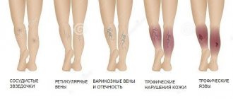
Types of varicose veins
Varicose veins are divided into two main groups:
- The first group includes primary varicose veins, caused by a hereditary predisposition to varicose veins.
- The second group includes varicose veins that appear after damage to the venous walls as a result of injury with the formation of blood clots in the veins or thrombosis.
When a clot or thrombus passes through a vein, the integrity of the venous valves is disrupted and secondary varicose veins are formed.
Varicose veins
Varicose veins are bundles of thin, purple or red veins that appear around the knees or ankles. (Sometimes such vascular “webs” can appear on the face, near the nose.) These vessels cannot be called varicose veins, since, by definition, varicose veins are veins that are increased in length and in diameter. In fact, these are slightly dilated venules (vessels connecting capillaries to the veins themselves), which are located close to the surface of the skin.
Such dilated venules appear due to increased levels of female sex hormones in the blood and are often found in women taking oral contraceptives. But venules can expand even in the presence of varicose veins of larger veins that do not appear externally. However, women with varicose veins often experience symptoms very similar to those of varicose veins.
Vascular diseases of the extremities and their causes
Among diseases of the blood vessels of the legs, atherosclerosis and inflammatory lesions of the arteries are common. Atherosclerotic lesion of the vessels of the lower extremities is a disease that affects older people (over 45 years old), mainly men. With atherosclerosis of the vessels of the extremities, the arteries of the legs narrow, preventing the passage of blood.
Another type of disease that affects the vessels of healthy-looking legs is inflammatory lesions of the arteries. They also cause vasoconstriction, which occurs as a result of the development of the inflammatory process. When inflamed, the walls of the artery thicken evenly along their entire length, complicating the flow of blood. Smoking increases the risk of arterial disease; thromboangiitis obliterans is more often diagnosed in young men who smoke.
Complete blockage or occlusion of the vessels of the legs can occur with any of the listed pathologies. Its sad consequences are gangrene and amputation. To avoid this, consult a doctor promptly when the first characteristic symptoms appear:
- Intermittent claudication;
- Pain in the legs that occurs when walking;
- Feelings of fatigue and weakness in the legs (or one leg);
- Erectile dysfunction in men;
- Cold feet, numbness, tingling sensation.
These and other symptoms of vascular diseases of the lower extremities are a reason to urgently consult a doctor.
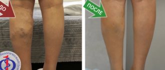
Treatment of varicose veins
Treatment depends on the severity of the disease. If the disease does not manifest itself too strongly, then conservative treatment is best:
- regular rest with your feet up,
- elastic bandaging (or special elastic stockings),
- physical exercises for leg muscles.
If these measures are not enough, the veins affected by varicose veins must be surgically removed at the Phlebology Center. Or using new, experimental methods - elastic strengthening of the venous walls is carried out surgically. That is, a special elastic plastic cover is placed on the outer surface of the affected veins in places of varicose veins, where incompetent venous valves are located. And finally, to treat dilated venules or varicose veins of small veins remaining after surgery, sclerotherapy - that is, the introduction of sclerosing substances into the areas of dilation, which causes clogging of the pathological vein. Blood returns to the heart through normal venous vessels.
Central venous catheterization
Just about central venous catheterization.
Puncture and catheterization of veins, in particular central ones, are widespread manipulations in practical medicine. Experience shows that this manipulation is not safe enough. Therefore, knowledge of the topographic anatomy of the subclavian vein and the technique of performing this manipulation is extremely important.
In one year, more than 15 million central venous catheters are installed worldwide. Among the venous tributaries available for puncture, the subclavian vein is most often catheterized.
Historical reference.
The first puncture of the subclavian vein was performed in 1952 by Aubaniac. He described the technique of puncture from the subclavian approach. Wilson et al. in 1962, a subclavian approach was used to catheterize the subclavian vein, and through it, the superior vena cava. Since that time, percutaneous catheterization of the subclavian vein has become widely used for diagnostic studies and treatment. Yoffa introduced the supraclavicular approach into clinical practice in 1965 to insert a catheter into the central veins through the subclavian vein. Subsequently, various modifications of the supraclavicular and subclavian approaches were proposed in order to increase the likelihood of successful catheterization and reduce the risk of complications. Thus, the subclavian vein is currently considered a convenient vessel for central venous catheterization.
Puncture catheterization of the central vessels is a medical procedure. The subclavian vein, jugular and femoral veins can be punctured, both on the left and on the right. The central venous catheter can function and remain uninfected for many weeks. This is achieved through strict adherence to the rules of catheter care, including adherence to aseptic rules during its installation, precautions when performing infusions and injections.
General view of the set for central vein catheterization:
Indications and contraindications
The following indications for central venous catheterization are distinguished:
- Complex operations with possible massive blood loss
- Open heart surgery with AIK and in general on the heart
- Need for intensive care
- Parenteral (intravenous) nutrition
- Possibility of measuring CVP (central venous pressure)
- Possibility of taking multiple blood samples for control
- Insertion of a cardiac pacemaker
- X-ray and contrast examination of the heart
- Probing of the heart cavities
Contraindications
Contraindications for central venous catheterization are:
- Bleeding disorder
- Inflammatory at the puncture site
- Clavicle injury
- Bilateral pneumothorax and some others
However, you need to understand that contraindications are relative, because if a catheter needs to be inserted for health reasons, then this will be done under any circumstances, because To save a child's life in an emergency, venous access is needed.
For catheterization of central (main) veins, one of the following methods can be selected:
- Through the peripheral veins of the upper limb, usually the elbow. The advantage in this case is the ease of execution; the catheter is passed to the mouth of the superior vena cava. The disadvantage is that the catheter can remain in place for no more than two to three days.
- Through the subclavian vein on the right or left
- Through the internal jugular vein, also on the right or left
For puncture catheterization of the central veins: jugular, subclavian (and, by the way, arteries), the Seldinger method (with a guide) is used, the essence of which is as follows:
- A vein is punctured with a needle, a guidewire is passed through it to a depth of 10–12 cm
- Next, the needle is removed and the catheter is passed through the guidewire.
- After this, the guide is removed, the catheter is fixed to the skin with a plaster
If the catheter is in place for a long time, the following complications may occur:
- vein thrombosis
- catheter thrombosis
- thrombo- and air embolism
- infectious complications (5 - 40%), such as suppuration, sepsis (general inflammation), etc.
That is why central venous catheterization requires careful adherence to the rules of care and monitoring of the catheter:
- Before all manipulations, you should wash your hands, dry them and treat them with 70% alcohol, and put on sterile rubber gloves. The skin around the catheter is inspected daily and treated with 70% alcohol and 2% iodine solution or 1% brilliant green solution.
- The dressing is changed daily and when soiled.
- After completion of infusion therapy, it is necessary to place a heparin lock (special dilution of heaprin).
- It is prohibited to bend the catheter, place clamps on the catheter that are not intended for the design, or allow air to enter the catheter.
- If problems associated with the catheter are detected: pain, swelling of the arm, the bandage gets wet with blood, exudate or infusion medium, fever, catheter kinks, immediately inform your doctor.
- The catheter is removed by the attending physician or anesthesiology service staff, followed by a note in the medical history.
- It is prohibited to leave the hospital premises with a catheter! If referred to another medical institution, the patient must be accompanied by a health worker; In the discharge summary, a note is made about the presence of a subclavian catheter in the patient.
General appearance of the patient:
Complications during treatment
The main danger with conservative treatment (elastic stockings, exercise and resting with legs elevated) is its possible ineffectiveness.
Surgical treatment of varicose veins of the lower extremities should currently be performed by experienced vascular surgeons and phlebologists . Often complications and relapses after surgical treatment are caused by the fact that the operation was not performed by a specialist from the phlebology center.
With sclerotherapy, the main nuisance is small dark spots that can remain at the injection sites for several months, or in some cases forever.
Angioplasty and stenting
The chronic form of NVC syndrome is more difficult to treat. When the venous outflow is decompensated, it becomes necessary to restore the patency of the vessel. Open operations involving the isolation of the IVC and its replacement with a vascular prosthesis are feasible, but very traumatic and ineffective. The artificial vena cava prosthesis often thromboses again and the complex operation becomes completely useless. With the advent of new composite materials for large-diameter stents, our clinic began to perform endovascular methods for restoring the patency of the vena cava. Angioplasty and IVC stenting are performed by experienced endovascular surgeons at the Innovative Vascular Center. The point of the intervention is to restore the patency of the closed segment of the inferior vena cava with a special conductor, a high-pressure balloon and the installation of a metal frame - a stent.
Dilated veins after surgical treatment
If varicose veins have been removed, varicose veins will no longer appear in their place. However, sometimes varicose veins are found after surgery - in veins that were not previously affected, or in small veins that were not identified during the preoperative examination. Varicose veins after surgery appear because the blood is forced to find new outflow paths. At the same time, a larger volume of blood is redistributed to the remaining veins than before, and if there were any defects in the valves or walls, then new problems arise. New varicose veins, as a rule, bring cosmetic inconvenience and can be easily eliminated by a phlebologist using modern sclerotherapy techniques.