From the point of view of modern European phlebology, five main stages of a disease such as varicose veins can be distinguished.
Stages of varicose veins or varicose veins of the lower extremities
According to the latest international data, tens of millions of Europeans suffer from various venous diseases, including manifestations of venous insufficiency. Only a small fraction of this enormous number seeks medical attention in a timely manner. On the territory of Russia, even in progressive Moscow and the Moscow region, a similar situation is observed. Without treatment, varicose veins inevitably progress and turn into a serious pathology with the development of dangerous complications.
Ultrasound diagnostics to identify the early stages of varicose veins
For good modern diagnostics in Moscow you need only 15-20 minutes. It is timely diagnosis that is necessary for good and correct treatment. Innovative technologies can also cope with the medical complications of varicose veins, but it is much better to prevent them.
Let's take a closer look at all 5 stages of varicose veins. It is better to take them seriously to avoid the development of serious complications. Today, millions of residents of our state, both urban and rural, are familiar with these stages of venous pathology first-hand.
Telangiectasia (spider veins) and reticular veins
The first stage of venous disease is reticular veins and telangiectasias. These veins are called arachnid veins in European medical literature. They are bluish-violet venous vessels that appear slightly above the surface of the skin. These veins got their name due to their web-like appearance. They are often quite small and difficult to see with the naked eye.
Stage I of varicose veins: intradermal varicose veins (telangiectasia)
People do not always pay attention to them and they go unnoticed for quite a long time, since at this stage there is no pain or discomfort. This doesn't mean you should ignore them completely. If you have spider veins, it is better to check the condition of the venous system for the presence of more serious pathology. A consultation with a good phlebologist will not take much time, but will help you avoid possible problems in the future.
Causes of spider veins include:
- Heredity
- Excessive static loads
- Pregnancy or menopause
- Hormonal imbalance
- Excess body weight.
At this stage, venous pathology is only a cosmetic, not a medical problem. In principle, the condition can be ignored, and most people do just that. But it's important to get a good diagnosis before your symptoms develop into something more serious. Many representatives of the fair sex, and not only in Moscow, are concerned about a cosmetic defect, which consists in the very presence of visible intradermal veins. Modern aesthetic procedures of sclerotherapy (scleroobliteration) can effectively help cope with the problem.
Treatment of stage I varicose veins using sclerotherapy
Risk factors for leg artery disease
Atherosclerosis is the most common cause of peripheral artery disease. With age
the risk of developing atherosclerosis of the arteries of the lower extremities increases. People over 50 have an increased risk of developing the disease, and men have a higher risk than women.
Other factors that increase the likelihood of developing the disease:
- Smoking;
- Diabetes;
- High blood pressure;
- High levels of cholesterol and triglycerides in the blood;
- Increase in body weight by more than 30% compared to your ideal weight.
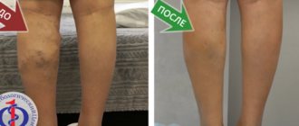
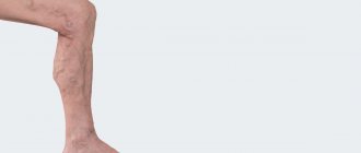
Varicose veins
Varicose veins, which stand out sharply above the surface of the skin, appear at this stage of the disease. From this moment on, the process acquires all the signs of not only a cosmetic, but also a medical problem. Varicose veins are enlarged, bulging and often tortuous. It is at this stage that most people begin to worry.
Stage II of varicose veins: varicose veins
And this is much better than perceiving the problem as purely cosmetic. The very presence of varicose veins already poses a threat of developing complications of varicose veins. In some patients, varicose veins are not accompanied by swelling of the legs or pain, while others experience aching pain along the venous vessels. Also, a number of patients experience a feeling of heaviness, nagging pain in the legs and feet.
Leading symptoms associated with varicose veins:
- Convex dark purple veins.
- Veins that appear twisted or tangled.
- Pain and heaviness in the legs.
- Burning or itching in the legs.
- Increased pain after standing for a long time.
- Bleeding from varicose veins.
- Inflammation and swelling of the legs around varicose veins.
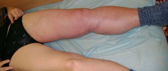
If you experience thickening and pain in the area of a varicose vein, you should immediately contact a good professional phlebologist. These are manifestations of a complication such as thrombophlebitis.
Stage II varicose veins, complicated by thrombophlebitis on the thigh
The lack of timely medical care in this situation can end badly.
Possible causes of varicose veins include:
- Age - As we age, our veins lose their ability to expand and contract well, so they stretch without returning to their original shape. As a result, the valves in the veins weaken, then collapse, and the blood in these veins changes the direction of blood flow. Thus, blood flow in varicose veins takes on a pathological character.
- Pregnancy. When you are pregnant, the amount of blood in the body increases, the enlarged uterus puts pressure on the venous vessels. As a result, we get varicose veins in the legs.
- Rapid weight gain and hormonal imbalance also play a major role in varicose veins. An increased load on the venous system, coupled with a weakening of the venous wall, does not have the best effect on the condition of the veins.
- Increased static load is one of the leading causes of the development of varicose veins of the lower extremities.
Preparing for an ultrasound
Ultrasound examination does not require special preparation. Before the procedure, you do not need to limit yourself in food. Canceling medications that a person is taking also does not make sense. Medicines do not have a negative effect on ultrasound results.
Step by step procedure
The study of the vessels of the lower extremities takes place in several stages:
- the patient is freed from clothing below the waist (pants, shorts, tights, skirt)
- The doctor applies a small amount of gel to the legs, which increases the throughput of ultrasound.
- the person lies down on the couch with his stomach down and then turns over. The specialist examines the surface of the thighs and lower legs. At the final stage, the doctor examines the vessels in a vertical position. In this way, the blood flow of the vascular system is assessed
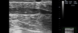
An ultrasound examination lasts more than half an hour. The procedure is absolutely painless. It does not cause any discomfort. During the diagnosis, the phlebologist examines the condition of the deep veins, arteries, venous valves, and junctions of large vessels. During the examination, information is displayed on the monitor. The specialist selects the frequency of ultrasonic waves, depending on the depth of the vessels. The doctor examines the clinical picture and makes the necessary conclusions.
Ultrasound of the veins of the lower extremities
Ultrasound detection of deep vein thrombosis
Edema of the lower extremities
Along with varicose veins, swelling of the legs, ankles and feet often occurs. Edema leads to the development of chronic inflammation in the distal parts of the lower extremities.
Stage III of varicose veins: swelling in the distal parts
When you experience pain, discomfort and heaviness, these are often signs of an inflammatory response. The sensation can be relieved by elevating your legs above your heart, this helps circulate blood to your heart from your legs after long periods of sitting or standing. This problem will not be solved by itself; you need to contact a phlebologist in a good specialized medical center. The best solution would be to choose one of the best Moscow city phlebological centers, where leading specialists in this field work.
Here are the leading symptoms that should prompt you to seek medical help.
Symptoms accompanying inflammation in the legs with stage III varicose veins:
- Feeling of heaviness, fullness, swelling in the calves or ankles.
- Itching in the lower extremities.
- Numbness and pastiness in the distal parts of the lower extremities.
- Aching pain when walking and standing.
- Restless legs syndrome, when it becomes difficult to maintain a static body position.
- Muscle spasms, cramps in the calf muscles (usually at night).
Many of these symptoms are among the first serious signs of venous pathology, so if you have varicose veins and swelling, it is very important to get a diagnosis so as not to make the problem worse.
Surgery of venous and lymphatic pathology
Vein diseases
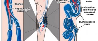
The body's venous system consists of an extensive network of veins - vessels through which blood moves from organs and tissues to the heart and lungs. When moving from the lower extremities to the heart, the blood flow overcomes the force of gravity. The function of the pump that pushes blood up through the veins is performed by the muscles that contract when walking, and the valves on the inner lining of the veins prevent the return flow of blood. Defects of the venous valves play a significant role in the development of venous diseases: varicose veins and its consequences - chronic venous insufficiency, trophic ulcers, thrombophlebitis.
Phlebology is a specialized branch of cardiovascular surgery that studies the anatomy, physiology of blood circulation, as well as diseases of the venous vessels, issues of their prevention, diagnosis and treatment. Treatment of vein diseases is carried out in departments of vascular pathology or phlebology centers. Vein diseases today are one of the most common diseases of the vascular system; these are diseases of young people of working age, mainly from 25 to 45 years. Untimely treatment of venous pathology is fraught with dire consequences, including disability and death.
Symptoms of venous disease that require immediate medical attention include fatigue, heaviness, cramps and pain in the legs, swelling, vascular dilation in the legs, the appearance of areas of discoloration and thickening of the skin and subcutaneous tissues.
To assess the degree of venous circulation disorder and select further treatment tactics, diagnostics are carried out using duplex ultrasound scanning and Doppler ultrasound - modern high-precision methods for studying the condition of blood vessels.
In the treatment of venous pathology, conservative methods (drug and compression therapy), non-surgical endovasal methods (laser coagulation, sclerosing therapy of veins) and surgical treatment methods are used.
In modern phlebology, low-traumatic methods for the treatment of vein diseases, based on the use of endoscopic, laser, and radiofrequency technologies, have become widespread, allowing to minimize complications and the rehabilitation period after the intervention. Timely treatment can achieve a good cosmetic result, which is especially important for women suffering from diseases of the veins of the lower extremities.
To prevent the development of diseases of the veins of the lower extremities, phlebologists advise following simple but very effective rules.
- Avoid prolonged standing or sitting in a cross-legged position.
- Avoid compressing the blood vessels of the legs with tight clothing and elastic bands, wearing high heels
- To improve venous outflow from the lower extremities during rest, it is necessary to give the legs an elevated position relative to the body
- To strengthen the leg muscles, you should perform self-massage, engage in physical therapy, and take a contrast shower.
- Avoid hot baths, frequent visits to baths, saunas, and excessive tanning.
- To reduce blood viscosity and the tendency to form blood clots, increase fluid intake to 2 liters per day
- For the initial manifestations of varicose veins, as well as for the purpose of its prevention during pregnancy, it is recommended to use medical compression hosiery
Varicose veins of the lower extremities (International Classification of Diseases, 10th revision (ICD-10) / IX Diseases of the circulatory system (I00-I99) / I80-I89 Diseases of the veins, lymphatic vessels and lymph nodes, not classified elsewhere / I83 Varicose veins lower extremities) is an important medical and social problem due to the tendency towards an increase in morbidity in people of working age, an increase in the number of complicated forms and, as a consequence, the formation of permanent disability. Varicose veins are characterized by high prevalence. So in Russia, according to the most rough estimates, it is diagnosed in 30 million people. In the USA and Western European countries, about 25% of the population suffers from various forms of varicose veins. After 70 years, the disease occurs in more than 70% of people. Mostly women over the age of 20 are affected (5 times more often than men). Clinical picture The main clinical symptoms of varicose veins are as follows:
- visible dilated veins;
- fatigue of the lower extremities;
- a feeling of fullness in the calf muscles, burning, soreness in the lower extremities;
- periodic and later permanent swelling of the feet and legs;
- night cramps in the calf muscles;
- general bluishness of the skin of the lower extremities;
- age spots, etc. (see pictures).
The severity of the symptom complex is greater in the evening, after physical exertion, and in hot weather. Stages of development In the development of varicose veins of the lower extremities, 4 stages are distinguished: 1st stage (compensation). There are minor cosmetic defects (spider veins), no complaints. 2nd stage. Twisted dilated veins appear, slight swelling of the ankles, mild night pain. Stage 3 (subcompensation). Reporting, night cramps in the calves, rapid fatigue of the legs, a feeling of muscle swelling, and skin pigmentation are observed. Stage 4 (decompensation). Severe swelling of the feet and ankles, a sharp increase in the width of the veins, acute pain, itching, severe cramps. Signs of thrombophlebitis and venous ulcers often appear.
Varicose veins of the lower extremities are one of the most common diseases on earth. The only radical, pathogenetically substantiated method of treatment is surgery. Conservative therapy is an addition to the main treatment.
Drawing. Physiology of venous outflow and pathophysiology of varicose veins n/c Acute venous thrombosis (ICD - I80 Phlebitis and thrombophlebitis; I82 Embolism and thrombosis of other veins) is the formation of a “blood clot”, a thrombus, in the lumen of a vein (more often observed in the deep venous system of the lower limbs, much less often - upper).
Drawing. Venous thrombosis.
Risk factors for venous thrombosis are, for example, young women taking oral contraceptives, fractures of limb bones, mass formations of the pelvis and retroperitoneal tissue, prolonged bed rest, paraplegia, the postpartum period, cancer, however, it is difficult to identify specific causes in a significant number of cases. The pathogenesis of venous thrombosis consists in the acute occurrence of an obstacle to venous outflow and redistribution of blood along collaterals against the background of inflammation of the venous wall; after which the process (period) of recanalization of thrombosed veins and restoration of pathological blood flow through them begins, which (period) ends by the sixth month. But even in recanalized veins, the blood flow does not become normal because the lumen of the vein does not reach its original diameter, and the destruction of the valves after thrombosis leads to retrograde blood flow. The consequence of venous thrombosis is Postthrombophlebitis syndrome (PTPS) or postthrombotic disease. Diagnosis of acute venous thrombosis in outpatient and inpatient settings is based on an assessment of clinical symptoms and the results of instrumental examination methods and should be carried out as soon as possible since the clinical prognosis depends on the speed of determining the fact of thrombosis, its localization, and the nature of the proximal part of the thrombus. In all cases of acute venous thrombosis, it is preferable to begin the examination with ultrasound angioscanning.
Drawing. Ultrasound angioscanning (ultrasound of n/c veins).
Treatment Goals
venous thrombosis: stop the spread of thrombosis, prevent thromboembolism of the pulmonary arteries, prevent the progression of edema and thereby prevent possible venous gangrene and limb loss, restore the patency of the veins (prevention of post-thrombophlebitis disease, prevent relapse of thrombosis. Suspicion of deep vein thrombosis of the lower extremities, especially if established diagnosis are an indication for emergency hospitalization of the patient (if possible, in a specialized angiosurgical hospital, in extreme cases in a general surgery department). The main objective of surgical treatment is to prevent pulmonary embolism. For this purpose, when an embolic (floating) thrombus is identified, depending on the specific clinical situation, direct or catheter thrombectomy, percutaneous implantation of vena cava filters of various designs, ligation or plication of the great veins/inferior vena cava.In all other cases, treatment problems are solved with the help of conservative therapy. Also, conservative therapy must be carried out after any of the listed surgical interventions.
Drawing. Scheme: a) completed operation of thrombectomy from the femoral veins supplemented by plication of the SMV according to the original method of the Bakulev Center; b) front view; c) side view; d) in cross section. Anticoagulant therapy is indicated for all patients with clinical and laboratory signs of active thrombus formation, which usually corresponds to the first three weeks of the disease. This is the most effective means of stopping the progression of thrombosis with a proven therapeutic effect. Anticoagulant therapy should be carried out with mandatory consideration of contraindications to these drugs. Anti-inflammatory drugs (NSAIDs) are used due to the fact that there is inflammation of the venous wall and perivasal tissues, as well as pain syndrome, which makes it difficult for the patient to activate. Local treatment includes local hypothermia in the projection of vascular bundles, as well as the use of ointments (gels are preferable), the main active principles of which are heparin and NSAIDs. You should not use warming alcohol and ointment compresses, which can support the phenomena of phlebitis and contribute to the progression of thrombosis.
Non-drug treatment methods: adherence to a certain motor regimen and elastic compression. The more carefully the patient follows the motor regimen and compression therapy regimen in the acute stage of the disease and during the rehabilitation period, and the longer the time it is carried out, the better the results of treatment of venous thrombosis, and the less pronounced the phenomena of chronic venous insufficiency in the long-term postthrombotic period.
Postthrombophlebitic syndrome (PTFS) or postthrombotic disease (ICD - I87 Other venous lesions / I87.0 Postthrombotic syndrome) is a chronic obstruction of venous outflow from the lower extremities, developing after deep vein thrombosis. Clinically, PTFS can manifest itself several years after acute thrombosis. Patients experience a bursting feeling in the affected limb and painful night cramps, ring-shaped pigmentation and swelling are formed, which over time acquires fibrous density. Diagnosis of PTFS is based on anamnestic data and ultrasound results of the veins of the lower extremities. Increasing decompensation of the venous circulation is an indication for surgical treatment.
With thrombosis, a blood clot forms in the lumen of the vein. After the acute process subsides, the thrombotic masses are partially lysed and partially replaced by connective tissue. If lysis predominates, recanalization occurs (restoration of the vein lumen). When replaced by connective tissue elements, occlusion occurs (disappearance of the lumen of the vessel). Restoration of the vein lumen is always accompanied by destruction of the valve apparatus at the location of the thrombus. Therefore, regardless of the predominance of one or another process, the outcome of phlebothrombosis is a persistent disturbance of blood flow in the deep vein system. Increased pressure in the deep veins leads to dilation (ectasia) and incompetence of the perforating veins. Blood from the deep vein system is discharged into the superficial vessels. The saphenous veins dilate and also become incompetent. As a result, all veins of the lower extremities are involved in the process. Blood deposition in the lower extremities causes microcirculatory disorders. Impaired skin nutrition leads to the formation of trophic ulcers.
Classification There are two variants of the course (edematous and edematous-varicose forms) and three stages of postthrombophlebitic disease.
- transient swelling, “heavy leg syndrome”;
- persistent swelling, trophic disorders (skin pigmentation disorders, eczema, lipodermatosclerosis);
- trophic ulcers.
Symptoms The first signs of PTFS may appear several months or even years after acute thrombosis. In the early stages, patients complain of pain, a feeling of fullness, heaviness in the affected leg when walking or standing. When lying down and placing the limb in an elevated position, the symptoms quickly decrease. Modern research in the field of clinical phlebology shows that in 25% of cases, PTPS is accompanied by varicose veins of the superficial veins of the affected limb. Edema of varying severity is observed in all patients. A few months after the development of persistent edema, inductive changes in soft tissues appear. One of the characteristic signs of PTFS is ring-shaped pigmentation, which begins above the ankles and covers the lower third of the lower leg. Subsequently, dermatitis, dry or weeping eczema often develop in this area, and in the later stages of the disease, poorly healing trophic ulcers appear. The course of PTFS can be different. In some patients, the disease manifests itself with mild or moderately severe symptoms for a long time, while in others it rapidly progresses, leading to the development of trophic disorders and permanent disability. Diagnostics. If post-thrombophlebitis disease is suspected, the doctor finds out whether the patient suffered from thrombophlebitis. When collecting anamnesis, it is necessary to pay attention to episodes of severe prolonged swelling and a feeling of fullness of the affected limb. To confirm the diagnosis, an ultrasound scan of the veins of the lower extremities is performed. To determine the shape, location of the lesion and the degree of hemodynamic disturbances, direct or computer radiopaque venography is used.
Treatment of PTFS. Conservative therapy During the adaptation period (the first year after thrombophlebitis), patients are prescribed conservative therapy. The indication for surgical intervention is early progressive decompensation of blood circulation in the affected limb. At the end of the adaptation period, treatment tactics depend on the form and stage of PTFS. In the stage of compensation and subcompensation of circulatory disorders, constant wearing of elastic compression devices and physiotherapy are recommended. Even in the absence of signs of circulatory disorders, patients are contraindicated in hard work, work in hot shops and in the cold, and work associated with prolonged standing on their feet.
In case of circulatory decompensation, the patient is prescribed antiplatelet agents, fibrinolytics, and drugs that reduce inflammation of the vein wall. For trophic disorders, pyridoxine, multivitamins, and desensitizing agents are indicated.
Surgery. Surgery cannot completely cure a patient with PTFS . The operation only helps to delay the development of pathological changes in the venous system. Therefore, surgical treatment is performed only if conservative therapy is ineffective.
The following types of operations for PTFS are distinguished:
- reconstructive interventions (vein resection and plastic surgery, bypass surgery);
- corrective operations (phlebectomy and miniphlebectomy - removal of dilated saphenous veins, ligation of communicating veins).
Forecast. To date, no type of treatment, including surgical interventions, can stop the further development of the disease if its course is unfavorable. Within 10 years from the moment of diagnosis of postthrombophlebitic disease, disability occurs in 38% of patients. I89 Other non-infectious diseases of the lymphatic vessels and lymph nodes/ I89.0 Lymphoedema, not elsewhere classified
Lymphatic pathology Lymphedema (elephantiasis) (ICD-10: I89.0; I97.2; Q82.0) is a polyetiological disease associated with impaired lymphatic drainage, which causes disruption of protein metabolism in tissues, progressive and irreversible formation of fibrous tissue, leading to significant enlargement and deformation of a limb or any part of the body. The reasons that lead to these changes may be associated with a congenital anomaly of the lymphatic vessels or be a consequence of their damage of various types.
Drawing. Patient M., 23 years old, with stage III congenital lymphedema of the left leg. A) before treatment and B) 4 months after resection, skin-plastic surgery.
Diseases of the venous system of the extremity, in particular postthrombotic disease, are not the cause of the development of lymphedema, but if the patency of the deep veins is impaired, secondary persistent disorders of lymph circulation in the extremity of the “pseudoelephantiasis” type are often observed.
Lymphedema is a common socially significant disease. According to WHO, about 10% of the population suffers from this pathology. More than 10 million people worldwide suffer from lymphedema caused by chronic infection. Patients with lymphedema account for 2.5-7% of all patients with peripheral vascular disease. The main group of patients are young and middle-aged women. The number of newly identified patients increases every year, and there is no downward trend in the incidence rate. The social significance of the topic under discussion is great and is due to the fact that the majority of patients are people of working age (96%), for whom rehabilitation is extremely important.
How does lymphedema develop? Lymphedema can be congenital or acquired. In the first case, it can appear in early childhood or during puberty, when hormonal levels change. But congenital pathologies are less common. Lymphedema often occurs as a complication after skin diseases, oncology and gynecological problems.
Drawing. Patient B., 62 years old, with stage III postmastectomy lymphedema of the left upper limb. Lymphedema tends to constantly progress. In areas of edema, skin changes, trophic disorders (ulcers, dermatitis, eczema), recurrent erysipelas, and fungal skin lesions (mycosis) begin to occur. The last stage of the disease is called “elephantiasis” due to the strong increase in size of the limb. It is important to stop the development of the disease, to eliminate existing problems at the initial stages of the process, when irreversible changes have not occurred in the skin and subcutaneous tissue.
Lymphedema is a chronic, progressive disease; it cannot be cured forever , but lymphedema can be significantly reduced or completely eliminated with the help of comprehensive treatment.
Clinical manifestations of this disease are: swelling of the upper or lower extremities, impaired nutrition of the skin, the appearance of trophic changes in the skin and subcutaneous fat. The most common complications of lymphedema are erysipelas, hyperkeratosis, papillomatosis, and lymphorrhea.
Diagnosis of lymphedema is based on the patient’s complaints, anamnesis of the disease and life, and features of the clinical picture. To confirm the diagnosis, special instrumental research methods are used:
- lymphoscintigraphy (radioisotope study of lymphatic vessels);
- magnetic resonance and computed tomography (allows visualization of tissues and organs layer by layer if an oncological etiology of lymphedema is suspected);
- lymphography.
- imfovenous anastomoses
- Vascularized lymph node transplantation
The choice of a rational method of treatment for lymphedema of the extremities is determined by the clinical course, stage of the disease and extent of the lesion. Treatment should be comprehensive, individual, with constant preventive measures. Currently, conservative and surgical methods are used, each of which has its own indications. The main goal in the treatment of lymphedema is to restore the outflow of lymph from tissues and reduce lymphedema. Conservative therapy as the main method of treatment is indicated for patients with stage I lymphedema. For more severe lymph circulation disorders, the method is auxiliary and is carried out when preparing patients for surgery and as a preventive course after surgical treatment.
Drawing. Patient S., 47 years old, with stage IIb-III lymphedema of the lower extremities before (photo on the left) and after (photo on the right) a course of inpatient complex anti-edematous therapy (20 bed days according to compulsory medical insurance standards). Reduction in the circumference of the left shin by 25 cm and hip circumference by 7 cm.
Among the surgical methods used:
Drawing. Scheme and intraoperative photo of lymphovenous anastomosis. The operation is performed under a microscope using microsurgical techniques and instruments.
Lymphorrhea (lympha - moisture, rhoe - flow), or lymphorrhagia, is a fairly rare pathological condition. May be traumatic or non-traumatic in nature. The traumatic form of lymphorrhea is more often diagnosed in young and middle-aged people. The non-traumatic variant of the pathology is detected mainly in patients of the middle and older age groups. Minor losses of lymph do not pose a danger to the body. With massive single lymphorrhagia or chronic leakage, exhaustion develops and death is possible.
Causes of lymphorrhea. Lymphorrhagia is a polyetiological condition that occurs during injuries, operations, and some diseases. The main causes of pathology are considered:
- open and closed injuries to lymphatic vessels of significant diameter, most often the thoracic duct;
- accidental injury to vessels or manipulation of such vessels during operations in certain anatomical areas;
- rupture of altered vessels with lymphangiectasias, vascular tumors, blockage by cancer emboli, especially against the background of lymphostasis and lymphangitis.
Classification Along with the outflow of chylous fluid, the following variants of lymphorrhea are distinguished: lymphocele - lymph accumulates in the tissues; chylothorax - fluid is detected in the pleural cavity; chylopericardium – lymph flows into the pericardial cavity; chyloperitoneum - fluid is found in the abdominal cavity. The release of lymph with urine due to its leakage into the urinary tract is called chyluria; leakage through the intestinal wall leads to the development of exudative enteropathy.
Diagnostics Depending on the cause of the development of lymphorrhea, thoracic surgeons, oncologists, cardiologists and other specialists can diagnose this pathology. The diagnosis of external lymphorrhagia is made on the basis of anamnesis, complaints and the results of a physical examination. In case of internal loss of lymph, additional research is necessary. Diagnosing external lymphorrhea is usually not difficult. Lymphorrhagia in the body cavity must be differentiated from the accumulation of other fluids. Chylothorax is distinguished from hydrothorax, pleurisy, hemothorax. Chyloperitoneum is differentiated from various variants of ascites, chylopericardium is differentiated from hemopericardium and exudative pericarditis.
Treatment of lymphorrhea The patient is hospitalized in a hospital according to the profile of the underlying disease. Treatment tactics are determined by the localization of lymphorrhagia.
NMITs SSH im. A.N. Bakuleva of the Ministry of Health of the Russian Federation has unique experience in performing all kinds of operations on the main veins and lymphatic system. We operate on patients with varicose veins of the lower extremities, perform reconstructive interventions for PTF, venous aneurysms and compression venous stenoses. To date, at the National Medical Research Center of Sports Sciences named after. A.N. Bakulev Ministry of Health of the Russian Federation performs about 100 operations on the lymphatic system per year (the largest number of operations among clinics in the Russian Federation). The Center performs almost all types of surgical interventions existing in the world for congenital and acquired pathologies of the great veins and lymphatic system, the results of which correspond to the world indicators of the world's leading clinics specializing in such operations.
Venous trophic ulcer
A venous ulcer is classified as a chronic wound that occurs with a long history of varicose veins and has no tendency to heal. This occurs due to severe structural tissue damage due to impaired venous outflow.
Venous ulcers are caused by long-term chronic venous insufficiency.
V stage of varicose veins: trophic ulcer
Blood pools and stagnates in your legs because defective valves in the veins prevent adequate blood return to the heart.
Symptoms of leg ulcers:
- Painful open wounds on the legs or ankles.
- Itching sensations in the lower extremities.
- Discharge of yellow or green fluid from the ulcer.
- Swelling of the legs.
- Change in skin color around the ulcer.
- Pain in the area of the ulcer.
- Feeling of heaviness in the lower extremities.
Venous trophic ulcers account for up to 80% of all ulcers of the lower extremities. In the vast majority of cases, patients can be helped using modern technologies for treating varicose veins. Yes, most of them are treated. It is enough to contact a good phlebological center in Moscow and a competent specialist.
Many of the listed symptoms and complications of varicose veins are hereditary, so if you have family members with chronic vein disease and you experience any of these symptoms, you are definitely at risk. Factors such as obesity, diet, smoking and alcohol consumption can also have a detrimental effect on the health of your feet.
Veins perforating the legs
Straight veins perforators move in the intermuscular septa together with the artery and nerve.
The anterior tibial group of perforators in the anterior muscle bed connects the SAV to the PBBV.
The posterior tibial group lies along Linton's line (Cockett's area) and connects the PAV with the PTA.
The paratibial perforator on the verge of the c/3 and c/3 tibia (Sherman) bridges the gap from the PAV to the PCA.
The paratibial vein perforator immediately below the knee (Boyd's) regulates flow from the GSV and SVV.
Direct veins perforate the adductor (Dodd) and adductor canals (Hunter) in the n/3 and s/3 thighs.
The GSV drains into the SPV through the SPS; in rare cases, you can find a perforator 5 cm below the SPS.
The anterior femoral perforators pierce through the quadriceps muscle and merge into the SMV and GBV.
The sciatic vein perforators are located longitudinally along the midline of the posterior thigh.
Four permanent connections of the SVC with the deep veins:
- paraachilles perforators (Bassi) behind the lateral malleolus;
- a perforator vein at a height of 12 cm from the ground connects the SVC with the MBV;
- the peroneal veins that perforate outside the leg are drained into the MBV;
- indirect perforator of the soleus muscle (May) in the s/3 tibia.