01.11.2017
Interesting
Bogacheva Sharofat Bairovna
The brain is a “mystical” organ that can fill us with incredible sensations, show us our own “movie”, dreams, accumulate experience and wisdom that allows us to think. This is an organ that controls and regulates the functioning of the entire organism as a whole and each organ and system separately; providing the balance, protection, and compensatory reactions to disturbances necessary for our body. This small organ, weighing about 1400–1500 g (2% of body weight), has incredible abilities that have not yet been fully studied.
What does the brain need? Working without rest day and night, he is in dire need of oxygen (the brain consumes 20% of all oxygen entering the body) and nutrients, without which he cannot live even for a few minutes. It is a known fact that oxygen reserves are not created in the brain, and there are no substances that can nourish it under anaerobic (in the absence of oxygen) conditions. That is, the nerve cells of the brain constantly need oxygen, glucose and “cleaning” (cleansing of cell waste products).
Excursion into physiology
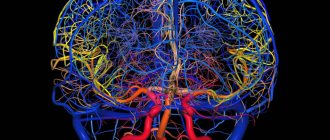
The uninterrupted supply of substances necessary for the nerve cells of the brain and the cleansing of waste are carried out by the cerebral circulatory system, where arterial blood carries oxygen and nutrition to the brain, and venous blood removes toxins and metabolic products.
The vessels of the brain have a unique, perfect structure that ideally regulates blood flow, ensuring its stability. They are designed in such a way that with an increased flow of blood into large vessels, the strong pulse impulse of the blood coming from the heart is weakened due to numerous bends (siphons) of the vessels along the vascular bed, which contribute to the pressure drop and smoothing of the pulsating blood flow. Due to complex regulatory mechanisms, when total blood pressure increases, the pressure in the brain remains stable for a long time. Regulatory systems allow blood flow to be redistributed from parts of the brain with less load to areas with increased brain activity.
The brain has an autonomous regulatory system, which allows it to be in a healthy functional state and control the processes of continuous adaptation of the body to constantly changing conditions of the external and internal environment. In a state of functional rest, the brain receives 750 ml of blood per minute, which is 15% of cardiac output. In children, blood flow activity is 50–55% higher, and in elderly people it is 20% lower than in adults.
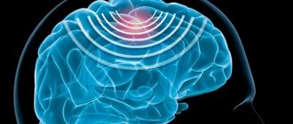
It should be noted that the gray matter of the brain (cell bodies of neurons) is supplied with blood more intensively than the white matter (conducting pathways), which is due to greater cell activity. Thus, during intense mental work, local blood flow in the cerebral cortex can increase 2–3 times compared to the resting state.
The brain has the richest capillary network. Nerve cells are not only intertwined, but also penetrated by capillaries. The vessels of the brain are connected to each other by collaterals (“bridges”). Arterial collateral circulation of the brain, important for maintaining normal blood flow, plays a particularly significant role in compensating for circulatory disorders when one of the cerebral arteries is blocked.
With a high intensity of blood flow in the vessels of the brain, the blood pressure in them is maintained relatively constant. A complex chain of regulatory mechanisms protects the brain from a drop in blood pressure and hypoxia (decreased oxygen). Along the path of blood flow to the brain, there are many sensitive cells (pressoreceptors, chemoreceptors) that can respond to blood pressure and regulate heart rhythm and vascular tone.
The activity of the vasomotor centers of the brain is associated not only with nervous and humoral regulation mechanisms, but also with the autonomic regulation system, which allows, despite significant fluctuations in total blood pressure, to maintain cerebral blood flow at a constant level.
Thus, cerebral circulation is provided with complex regulatory mechanisms that make it possible to maintain a constant supply of the substances it needs.
With excessive blood supply to the brain, excessive hydration (fluid accumulation) may occur, followed by the development of edema and damage to vital centers that are incompatible with life. The cause of excess blood supply can be, for example, an increase in systemic blood pressure to 160–170 mm Hg. Art. and higher.
In the problem of impaired blood supply to the brain, much attention is paid to arteries. But venous circulation is no less important. The veins carry out the removal of waste substances (toxins) with the blood - that is, cleansing the brain. Thanks to these vessels, constant intracranial pressure is maintained.
Violation of the venous outflow leads to stagnation of blood and accumulation of fluid in the brain, causes hydrocephalus with compression of the brain centers, and contributes to the occurrence of phlebitis and thrombophlebitis.
There is one more feature of the cerebral veins that must be taken into account. The wall of a venous vessel in the brain does not have a valve apparatus, unlike, for example, the veins of the extremities (valves help withstand loads by moving blood upward and preventing it from moving in the opposite direction). Therefore, venous blood in the vessels of the brain passes freely in both directions, depending on the pressure that arises. This creates a danger of rapid spread of infection from the sinuses and eye sockets, which is facilitated by the atomic structure of the nose and its paranasal sinuses, located in close proximity to the brain. When coughing, venous pressure increases, reverse venous flow, congestion, and brain hypoxia become possible. There are known cases of loss of consciousness during a coughing attack in the presence of a chronic respiratory tract disease and in young children when they “go into a fit” of coughing during illness and crying and screaming until they cough.
It becomes clear why long-term respiratory problems, accompanied by constant swelling and coughing, can cause cerebrovascular accidents. Because they not only cause brain hypoxia, but also disrupt venous outflow and, being a constant source of infection, contribute to its penetration into the brain.
An ophthalmologist, for example, can observe manifestations of congestion in the brain (dilated, blood-filled vessels of the fundus). But this is also visible to the naked eye: red, puffy eyes after sleep (due to drinking alcohol the night before, overeating at night, lack of sleep) are a symptom of congestion in the brain.
After a brief excursion into physiology, it becomes clear that the reasons for the deterioration of cerebral circulation may be associated with disturbances in the flow of blood to the brain and the outflow of blood from the brain.
Peripheral circulation disorders
The main peripheral circulatory disorders are:
- hyperemia (arterial and venous) – increased blood supply to the tissue;
- ischemia – a decrease in blood supply to an organ or tissue;
- stasis – cessation of blood flow in organs and tissues.
Arterial hyperemia is an increase in the blood supply to an organ due to an increase in the amount of blood flowing through its dilated vessels.
There are physiological hyperemia, which occurs normally with increased organ function, as well as reflexively under the influence of ultraviolet rays, cold, heat, etc., as well as pathological hyperemia, which occurs in the following cases:
- with inflammation;
- with rapid decompression of compressed vessels (for example, when emptying the abdominal cavity from the accumulation of ascitic fluid);
- when creating a rarefied space (vacuum hyperemia) - for example, when using medical cups;
- with overload or drug blockade of the sympathetic nerves that constrict blood vessels (neuroparalytic hyperemia).
Clinically, arterial hyperemia is manifested by redness of tissues and a local increase in their temperature.
Venous (congestive) hyperemia is an increase in blood supply to a tissue area with a decrease in the amount of flowing blood.
Causes of venous hyperemia:
- thrombosis or compression of veins from the outside (tumor, scars, pregnant uterus, surgical ligation of a vessel);
- stagnation and slowing of blood flow in the veins of the lower body with a decrease in the pumping function of the heart (right ventricular heart failure);
- blood stagnation in the lower extremities in people who work for a long time while standing.
Clinically, venous hyperemia is manifested by a bluish color of tissue or cyanosis, and may also be accompanied by edema.
Stasis is a local stop of blood flow in small vessels, mainly capillaries. Stasis occurs due to a complete cessation of blood flow, due to a sharp disruption of blood outflow, as well as due to various diseases of an inflammatory and non-inflammatory nature (true capillary stasis), leading to intracapillary crowding (aggregation) of red blood cells and stopping capillary blood flow.
Stasis can be reversible or irreversible (in this case, blood flow is not restored, and necrosis occurs in the corresponding area of tissue). Externally, when stasis occurs, a “marbled” color may appear on the skin.
Sludge (sludge syndrome) is a blood condition based on the aggregation of red blood cells. The development of sludge represents an extreme degree of expression of the aggregation of blood cells. The main features of blood sludge are: adhesion of formed elements to each other and an increase in plasma viscosity, which leads to a state of blood that makes it difficult for blood to flow through small-caliber vessels.
Ischemia is the reduced blood supply to any area of tissue due to weakening or cessation of blood flow to it through the arteries.
Causes of ischemia:
- compression of the artery (with a tourniquet, tumor, scar, foreign body, surgical ligation of the vessel);
- blockage of an artery (thrombus, embolus, narrowing of the lumen of the artery due to vascular diseases);
- reflex ischemia (when exposed to painful, visual, sound, chemical, emotional stimuli, etc.).
Clinical manifestations of ischemia depend on the location of the ischemic area. With ischemia of the limbs, their pallor, a feeling of numbness, “pins and needles”, pain occur, and the function of the limb is impaired. With ischemia of the heart muscle, pain occurs in the heart, and with ischemia of the brain, one or another neurological symptoms occur.
The outcomes of ischemia depend not only on the location, but also on the diameter of the switched off vessel and on the degree of development of collateral (roundabout) circulation in this area. With a favorable outcome, the blood supply to the ischemic area is restored; with an unfavorable outcome, an area of tissue necrosis occurs - an infarction.
The usefulness of collateral (roundabout) blood circulation depends on the anatomical features of the blood supply to the ischemic area (mainline or branched type of blood supply), the state of the vascular wall, the state of cardiac activity and the nervous regulators of blood circulation. There are functionally absolutely sufficient and functionally insufficient (absolutely and relatively) collaterals. This accordingly affects the nature of the outcome of ischemia.
Thrombosis is the intravital coagulation of blood or lymph in the lumen of a vessel with partial or complete blockage, leading to disruption of blood flow.
The mechanism of thrombus formation consists of a combination of three factors (Virchow’s triad): – slowing down of blood flow; – damage to the vascular wall; – increased blood clotting.
Vein thrombosis is also called phlebothrombosis. If thrombosis is combined with inflammation of the vein wall, then they speak of thrombophlebitis. If there is a combination of thrombosis of an artery with inflammation of its wall, this is called thromboarteritis.
Embolism is the blockage of blood and lymphatic vessels by particles carried by the blood or lymph flow. These particles are called emboli.
The following types of embolism are distinguished:
- thromboembolism - embolism by a migrated fragment of a thrombus;
- tissue and cellular embolism - embolism of a tissue area due to organ injury, tumor cells, etc.;
- fat embolism - blockage of blood vessels with drops of fat, most often due to fractures of long tubular bones;
- gas embolism (an option is air embolism) - blockage of blood vessels by gas bubbles, for example, bubbles of nitrogen dissolved in the blood during decompression sickness in divers;
- bacterial embolism – blockage of blood vessels by bacterial conglomerates in various diseases (acute hematogenous osteomyelitis);
- embolism by a foreign body (bullet, shell fragment).
If the embolus, due to gravity, falls from top to bottom against the direction of blood flow, then they speak of a retrograde embolism. If an embolus from the venous system enters the arterial system through an unprotected septum between the left and right atria, then this embolism is called paradoxical.
Thrombosis and embolism of arterial vessels leads to ischemia of the blood supply areas of these vessels. Venous thrombosis leads to the occurrence of venous stagnation in the areas of venous outflow of a given vessel.
The fate of the blood clot can be different. Over time, the thrombus can grow with connective tissue (organization of a blood clot), partially or completely dissolve (recanalization of a blood clot), or undergo purulent melting.
Bleeding is the outpouring of blood from the lumen of a vessel into surrounding tissues, natural cavities of the body, or into the external environment.
There are arterial, venous, capillary and mixed bleeding. bleeding most often occurs when the vascular wall is damaged, but it can also occur through an intact vessel wall (diapedetic bleeding). Damage to the vessel wall occurs as a result of injury, but can also be the result of a pathological process - corrosion of the vessel wall due to purulent inflammation or a tumor process (arrosive bleeding). An accumulation of blood in soft tissue is called a hematoma. When blood flows into soft tissues and natural cavities of the body, we speak of internal bleeding, and blood flow into the external environment (including into the lumen of the digestive tract) is called external bleeding.
With intense (profuse) bleeding, activation of the blood coagulation system occurs, which leads to multiple thrombus formation in small vessels. This leads to a deficiency of fibrinogen in the blood plasma, a decrease in the blood's ability to clot and increased bleeding. This condition is called disseminated intravascular coagulation (DIC).
What happens when blood pressure rises?
At first, vascular tone is slowly disrupted. Over time, if elevated blood pressure (BP) persists, minor cerebral hemorrhages and strokes may occur.
As a result of a constant increase in blood pressure during hypertension, plasma (part of the blood without formed elements) is released, which ultimately leads to the destruction of the walls of blood vessels.
How does this happen? A specific protein (hyaline-like substance, similar in structure to cartilage) is deposited on the walls of blood vessels, which leads to the development of hyalinosis. The vessels become like glass tubes, lose their elasticity and ability to hold blood pressure. In addition, the permeability of the vascular wall increases, and blood can freely pass through it, soaking the nerve fibers (diapedetic bleeding). The result of such transformations can be the formation of microaneurysms and rupture of the vessel with hemorrhage and blood entering the white medulla. The resulting swelling and hematomas lead to further hemorrhages (hemorrhagic stroke).
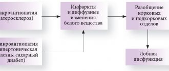
Atherosclerosis that accompanies hypertension, or exists without it (which is rare), contributes to cerebral ischemia - insufficient supply of nutrients and oxygen to the tissues (except for atherosclerotic plaques that narrow the lumen of the arteries, the blood itself can be thick and viscous).
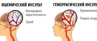
Acute circulatory disorders are strokes (hemorrhagic and ischemic). But it all starts with transient cerebrovascular accidents against the background of hypertension and atherosclerosis, as well as obesity, diabetes mellitus, and respiratory diseases that often accompany them.
Exercises to improve blood circulation in the legs
To minimize the risk of developing hemodynamic disorders, it is important to move as much as possible. Daily walks in the fresh air not only strengthen the muscles and blood vessels of the lower extremities, but also contribute to better oxygen saturation of the blood.
To prevent varicose veins, it is recommended to exercise regularly
If you don’t have the opportunity to walk every day, doctors advise doing simple exercises: heel-to-toe rolls, “scissors”, “birch”, as well as some asanas (the list of recommendations may include a sitting fold, a downward-facing dog with a rise on the toes, reverse flow pose, etc.)
Symptoms of cerebrovascular accident
When a lesion forms in the brain with impaired blood supply, the patient may experience numbness in half of the body (on the side opposite to the lesion) and part of the face around the lips; short-term paresis of the limbs or other parts of the body and face is possible. Speech is impaired and an epileptic seizure may occur.
If there is a circulatory disorder, depending on the location of the lesion, the legs and arms may become weak, the head may become dizzy, the patient may have difficulty swallowing and pronouncing sounds, photopsia (appearance of luminous spots, sparks, etc. in the eyes) or diplopia (doubling of visible objects) may occur. . The person loses orientation and has memory lapses.
Signs of impaired cerebral circulation due to hypertension are manifested in the following: the head and eyeballs begin to hurt very much, the person experiences drowsiness, he experiences stuffiness in the ears (like on an airplane during takeoff or landing) and attacks of nausea. The face turns red and sweating increases.
Unlike strokes, all these symptoms, which are called “transient attacks,” disappear within 24 hours.
Chronic cerebrovascular accident (CVA), unlike acute forms, develops gradually. There are three stages of the disease:
- At the first stage, the symptoms are vague. They are more like chronic fatigue syndrome. A person quickly gets tired, becomes hot-tempered and absent-minded, and forgets some minor points. His sleep is disturbed, his mood often changes, his head hurts and he feels dizzy.
- At the second stage, chronic cerebrovascular accident is accompanied by significant memory deterioration, and minor motor dysfunctions develop, causing unsteadiness in gait. There is a constant noise in my head. A person perceives information poorly, having difficulty concentrating his attention on it. Becomes irritable and unconfident, loses intelligence, reacts inadequately to criticism, and often becomes depressed. He gradually degrades as a person and adapts poorly socially. He constantly feels dizzy and has a headache. He always wants to sleep. Performance is significantly reduced.
- In the third stage, all symptoms intensify. Personality degradation turns into dementia, memory suffers. Having left home alone, such a person will never find his way back. Motor functions are impaired, which manifests itself in hand tremors and stiffness of movements. Speech impairment and uncoordinated movements are noticeable.
Consequences of cerebrovascular accidents
Disability is a sad result of acute and in many cases chronic cerebrovascular accident.
Acute cerebrovascular accident has serious consequences. In most cases, a person who has suffered a stroke becomes completely helpless. He cannot eat, perform hygiene procedures, dress, etc. on his own. Such people have a completely impaired ability to think. They lose track of time and have absolutely no orientation in space.
Some people retain the ability to move. But many people, after a cerebrovascular accident, remain bedridden forever. Many of them maintain a clear mind, understand what is happening around them, but are speechless and cannot express their desires and feelings in words.