Heart
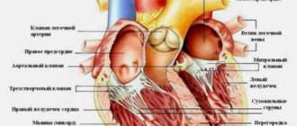
Having determined which sections the circulatory system consists of, they pay special attention to the study of the heart. It is a hollow muscular organ shaped like a cone. The heart is located in the left half of the chest at the level of the second to fifth ribs. The organ is located in the pericardial sac, divided by longitudinal and transverse septa into two ventricles and two atria.
Features :
- The walls have three layers: the outer layer is the epicardium, the middle layer is the myocardium, formed by a special striated muscle, and the inner layer is the endocardium.
- The thickness of the muscle layer of the left ventricle reaches 10-15 mm, the right - from 5 to 8 mm, the atria - from 2 to 3 mm.
- The one-way flow of blood from the atria to the ventricles, to the aorta and pulmonary artery is ensured by the heart valves - folds of the inner membrane.
The work of the heart consists of contraction (systole) and relaxation (diastole). Myocardial contractions occur rhythmically: 70-75 times per minute at rest. Electrical impulses originate in the muscle itself. This ability is called “automaticity of the heart.” The high efficiency of the organ is due to increased blood supply and active metabolism in the heart muscle.
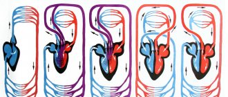
Circulation circles
What is the circulatory system in the body? The largest artery is the aorta, into which blood is pushed under high pressure from the left ventricle. Further movement occurs through arteries, capillaries and veins. Large vessels in organs branch into smaller and microscopically thin ones. The capillaries then enlarge and gather into veins.
Table "Blood circulation"
| Circles | Big | Small |
| Where does it begin | In the left ventricle | In the right ventricle |
| Where does it end? | In the right atrium | In the left atrium |
| Oxygen content | Decreases as oxyhemoglobin releases O2 and binds CO2 | It increases because in the alveoli of the lungs carboxyhemoglobin releases CO2 and binds O2 |
| Importance for the body | Delivers O2, nutritional and mineral elements, hormones, vitamins to cells | Removal of CO2 for removal from the body into the environment, enrichment of blood with O2 |
Small veins enlarge and join the vena cava, which flows into the right atrium. As a result of its contraction, blood enters the right ventricle. The pulmonary artery emerges from it, carrying venous blood. The pulmonary veins carry arterial blood into the left atrium.
Functions of the circulatory organs:
- Providing cells with nutrition, hormones, vitamins, oxygen, water, organic and inorganic ions.
- Maintaining a constant temperature and other important parameters of the body.
- Removing CO2 and unnecessary metabolic products from cells.
- Participation in humoral regulation.
- Carrying out gas exchange.
The average volume of blood in the human body is from 4 to 6 liters. Arterial blood makes up 20%, venous blood - up to 80% of the total volume. Half does not participate in the bloodstream, but is located in the blood depot: liver, spleen, abdominal veins. The supply quickly replenishes and increases the mass of circulating blood in critical situations.
See also: What are the causes of cardiovascular diseases and what are the prerequisites?
Algorithm for diagnosis and treatment of chronic forms of venous circulation disorders
In the Russian Federation, 35–38 million people suffer from chronic venous insufficiency. Unfortunately, the stereotype according to which venous pathology is considered only a surgical pathology has led to the fact that a huge number of patients do not receive adequate medical care [1]. At the same time, changes in venous circulation are one of the important pathogenetic mechanisms for the development of vascular diseases of the brain.
Regional changes in the tone of intracranial veins lead to venous congestion and impaired cerebral circulation in atherosclerotic lesions of cerebral vessels, arterial hypertension (AH) and hypotension, chronic lung diseases, and cardiac pathology. It has been recorded that in 15% of patients with hypertension, compression of the jugular, brachiocephalic and vertebral veins is recorded, signs of impaired venous outflow of the brain occur in 91% of cases of hypertension, and in patients with stage 1-2 hypertension - in 55% of cases [2, 3] . At the same time, the compensatory capabilities of the brain and its circulatory system are so great that even serious difficulties in the outflow of venous blood may not cause clinical manifestations of increased intracranial pressure and impaired brain function for a long time [4], so early diagnosis of this pathology causes certain difficulties.
To simplify the doctor’s work, the following algorithm for diagnosing and treating chronic forms of venous circulation disorders can be used.
Algorithm for diagnosis and treatment of chronic forms of venous circulation disorders
Step 1. Identify risk factors
The doctor should always remember that venous congestion in the vast majority of cases is secondary in nature, that is, it occurs as a symptom of an underlying disease that impedes the outflow of venous blood from the cranial cavity. Therefore, diagnosis first of all involves identifying the underlying disease (Table 1).
Obstruction of venous outflow from the cranial cavity is observed in a number of diseases [5]:
- cardiac and cardiopulmonary failure;
- common pulmonary tuberculosis, pulmonary emphysema, bronchiectasis, bronchial asthma, pneumothorax;
- compression of extracranial veins - internal jugular, innominate, superior cava - by a neoplasm in the neck, aneurysm; hypertrophied neck muscles in reflex-muscular-tonic syndromes of cervical osteochondrosis;
- tumors of the brain, its membranes, and skull;
- thrombosis of veins and sinuses, infectious-toxic lesions of veins, cerebral thrombophlebitis;
- compression of the veins during craniostenosis (premature fusion of the sutures between the bones of the skull with compression, in particular, of the jugular veins), in these conditions the venous collectors dilate compensatoryly;
- asphyxia of newborns and adults;
- venous and arteriovenous hypertension;
- when nasal breathing stops;
- infectious and toxic lesions of the brain;
- with the consequences of traumatic brain injuries;
- epilepsy.
Diseases that cause disruption of venous outflow are given in Table. 1.
In addition, the development of venous encephalopathy may also be due to the classical causes of cerebrovascular pathology: hypertension, atherosclerosis, smoking, diabetes, use of hormonal drugs (estrogens), alcohol and drug abuse, use of nitrates and some vasodilators (nicotinic acid, papaverine). Venous outflow can also be impaired under physiological conditions, for example, when straining, during a prolonged cough, during physical stress, singing, playing wind instruments, childbirth, screaming, bending the head (for example, during physical exercise), in a lying position without pillows under the head, when the neck is compressed by a tight collar.
Step 2. Analysis of complaints and medical history
Disturbances of venous circulation, as a rule, are genetically determined. Currently, the role of the initial tone of the veins in the formation of venous discirculation is undeniable. Constitutional and hereditary factors are key for the development of venous dysgemia [6]. Patients with a family “venous” history usually have several typical manifestations of constitutional venous insufficiency - varicose veins or phlebothrombosis of the lower extremities, hemorrhoids, varicocele, impaired venous outflow from the cranial cavity, esophageal varicose veins. Pregnancy is often the trigger.
Typical complaints:
- morning or afternoon headache of varying intensity;
- dizziness, depending on changes in body position;
- noise in the head or ears;
- visual disturbances (decreased visual acuity, photopsia);
- “tight collar” symptom - increased symptoms when wearing tight collars or ties;
- “high pillow” symptom - increased symptoms when sleeping with a low headboard;
- sleep disorders;
- feeling of discomfort, “fatigue” in the eyes in the morning (symptom of “sand in the eyes”);
- pastiness of the face and eyelids in the morning (with a pale, purple-cyanotic tint);
- mild nasal congestion (outside the symptoms of acute respiratory infections);
- darkening of the eyes, fainting;
- numbness of the limbs.
The course of the disease has chronic, episodic and remitting variants.
Step 3. Examination of the patient
When examining the patient, a “venous triad” is detected:
1) swelling of the face in the morning after a night's sleep, which decreases significantly in the evening with sufficient physical activity; 2) cyanosis of the facial skin; 3) expansion of the saphenous veins of the neck and face.
With severe venous stagnation, patients are unable to lower their heads and remain in a horizontal position for a long time. Blood pressure (BP) in such patients is usually within normal limits, venous pressure ranges from 55 to 80 mmH2O. Art. A low difference between systolic and diastolic pressure is characteristic, in contrast to hypertension. In severe cases, epileptic seizures and mental disorders are possible [7]. Venous discirculation is characterized by a decrease in corneal reflexes. On palpation, pain is detected at the exit points of the first, less often the second, branches of the trigeminal nerve (“transverse sinus syndrome”) with the formation of hypoesthesia in the innervation zone of the first branch of the trigeminal nerve, which is probably associated with the development of neuropathy caused by venous stagnation and impaired microcirculation in the vaza system nervorum [8].
According to the type of prevailing symptom, the following variants of chronic venous insufficiency (encephalopathy) are distinguished: cephalgic, hypertensive (pseudotumorous), bettolepsy, polymorphic (scattered small-focal brain damage), sleep apnea syndrome, psychopathological/asthenovegetative [9].
Cephalgic syndrome is the most common clinical manifestation of the pathology of the venous system. As a rule, headaches increase when moving the head to the sides, changes in atmospheric pressure, changes in ambient temperature, after excitement, drinking alcohol, etc. This syndrome has a number of characteristic signs (Table 2).
Hypertension (pseudotumor syndrome) is characterized by clinical signs of increased intracranial pressure (ICP) in the absence of focal neurological symptoms and the presence of congestive optic discs [10]. Develops acutely. Patients, as a rule, complain of intense paroxysmal headaches, are euphoric, irritable, and often angry. Bradypsychism appears with slowness of movements. When examining the cerebrospinal fluid, increased pressure attracts attention. The protein content is slightly increased or normal, cytosis is not increased, serological reactions are negative. Pseudotumor syndrome in chronic venous pathology must be carefully differentiated from brain tumors.
Bettolepsy (cough syncope) is the development of short-term fainting with convulsive twitching during a coughing attack. Cases of “cough” fainting (bettolepsy) are quite rare and account for no more than 2% of patients with venous pathology. This form of venous blood flow disorder develops when:
- chronic bronchitis;
- emphysema;
- pneumosclerosis;
- bronchial asthma;
- cardiopulmonary failure.
In the pathogenesis, the main role is played by brain hypoxia, which occurs during a prolonged cough, caused by an increase in intrapleural pressure, disruption of venous blood flow in the superior vena cava system, a slowdown in pulmonary blood flow with an increase in intrapleural pressure, with a decrease in the filling of the left ventricle with blood, a slowdown in cardiac activity, and a decrease in cardiac output. . In most cases, paroxysms during coughing are not related to epilepsy, since they develop according to pathogenetic mechanisms characteristic of fainting conditions. Coughing attacks occur in patients while sitting or standing, often during or shortly after eating. Provoking factors: cold air, pungent odor, tobacco smoke, excessive laughter, etc. Simultaneously with the cough, facial hyperemia develops, followed by cyanosis with pronounced swelling of the neck veins. Usually there are no warning signs, there may only be slight dizziness. Loss of consciousness occurs within the first minute from the onset of coughing. The duration of syncope varies from several seconds to a minute. Cyanosis appears, patients often fall, often hurt themselves, the cough stops, the color of the face changes from cyanotic to marble-pale. Seizures are usually not observed (sometimes tonic seizures are possible). There is no tongue biting or involuntary urination.
Bettolepsy is observed mainly in older people with chronic diseases of the respiratory tract and lungs (pharyngitis, laryngitis, emphysema, bronchial asthma, etc.). At a younger age, the appearance of fainting when coughing is observed quite rarely, mainly in individuals with increased sensitivity of the carotid sinus, or with functional insufficiency of the mechanisms that support postural tone.
The syndrome of scattered small-focal brain lesions is clinically manifested by individual symptoms, such as asymmetry of the nasolabial folds, mild nystagmus, and slight staggering when walking. Motor, sensory, and coordination disorders are less common. Parkinson-like syndrome may develop [11].
Psychopathological and asthenovegetative syndromes are the earliest signs of venous insufficiency. They are characterized by increased fatigue, irritability, unstable or bad mood, sleep disorders in the form of constant drowsiness or persistent insomnia, autonomic disorders (unpleasant sensations from the heart, shortness of breath, hyperhidrosis of the extremities). It is possible to develop hyperesthesia (intolerance to bright light, loud sounds, strong odors), intellectual disorders (disorders of attention and memory, ability to concentrate). Headaches are often observed. Patients experience a change in mental state depending on atmospheric pressure: when it falls, fatigue increases, irritable weakness, and hyperesthesia (Pirogov's symptom) increase. In rare cases, psychosis develops with delusions and visual and auditory hallucinations [12]. Determinants of asthenia are constant complaints of increased fatigue, weakness, exhaustion after minimal effort in combination with at least two of the following complaints:
- muscle pain;
- dizziness;
- tension headache;
- sleep disorders;
- inability to relax;
- irritability;
- dyspepsia.
The most characteristic signs of asthenic disorders can be divided into several groups depending on the dominant complaints [13].
1. Physical disorders:
- muscle weakness;
- decreased endurance.
2. Intellectual disorders:
- disorders of attention, ability to concentrate;
- Impaired memory and vigilance.
3. Psychological disorders:
- lack of self-confidence;
- decreased motivation.
4. Sexual disorders:
- lack of libido;
- decreased erection.
Psychopathological and asthenovegetative syndromes predominantly develop in young and middle-aged patients.
Sleep apnea syndrome. In patients with sleep apnea, the absence of a physiological nocturnal decrease in blood pressure and impaired cerebral venous hemodynamics have been established.
Step 4. Additional research methods
For more accurate diagnosis, instrumental research methods are used: ophthalmoscopy, skull radiography (craniography), ultrasound (US) methods for studying the venous system of the brain, computed tomography or magnetic resonance imaging, cerebral angiography. When conducting any diagnostic study, it is necessary to take into account that venous circulation is extremely labile, and this is associated with the state of central hemodynamics, the respiratory cycle, muscle activity, and posture. It is advisable to conduct the examination on days with a favorable geomagnetic situation, provided that the patient does not have an increase in blood pressure at the time of examination, or complaints of headache or a feeling of “heaviness” in the head during the last week. Patients should not drink alcohol for several days. In women of reproductive age, it is advisable to assess cerebral hemodynamics in the first half of the menstrual period.
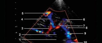
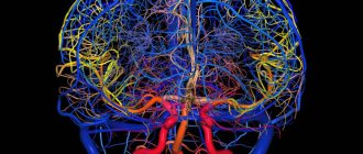
Craniograms can reveal an increase in the vascular pattern, expansion of diploic veins, and venous outlets. Ophthalmological methods allow already in the early stages of vascular diseases of the brain, along with changes in the arteries, to detect the dilation of veins, their tortuosity, uneven caliber, and with a pronounced increase in intracranial pressure - congestion in the fundus. The methods of biomicroscopy of the conjunctiva of the eyeball and venous ophthalmodynamometry are quite informative. To clarify the causes and extent of impairment of venous outflow at the neck level, duplex ultrasound scanning (USDS), selective contrast venography, scintigraphy and computed tomography are used. Each of these methods has advantages and disadvantages. Ultrasound scanning makes it possible to reliably assess the speed of blood flow and the relationship of blood vessels with surrounding tissues, but has limitations since a relatively small area of the brachiocephalic veins is available for study. Selective contrast venography is associated with a certain risk when administering a contrast agent, which is often unjustified for this pathology [15]. Scintigraphy does not provide information about the structures surrounding the veins. Standard computed tomography allows assessing the diameter of the veins and their relationship with surrounding structures only in cross sections, but does not display the characteristics of blood flow, and in addition, is accompanied by radiation exposure. Magnetic resonance venography of the brain is characterized by a decrease in the intensity of the blood flow signal, up to its loss, in the superior sagittal sinus, great cerebral vein and straight sinus. It is also possible that there is a decrease in size or a complete absence of the signal from the blood flow along the transverse and sigmoid sinuses, the internal jugular vein of one of the hemispheres of the brain, combined with the expansion of these venous structures on the opposite side; expansion of emissary and superficial cerebral veins [16].
Step 5. Choice of therapy
Unfortunately, the issues of pharmacotherapy of cerebral venous circulation disorders still remain controversial and insufficiently studied; there is no doubt that, first of all, it is necessary to treat the underlying disease. The earliest possible energy correction can additionally affect the survival of neurons, reduce damage to brain tissue caused by chronic ischemia and hypoxia, and primarily affect the core of the asthenic syndrome - hypoergosis with increased exhaustion of mental functions [17]. Taking into account modern ideas about the pathogenesis of venous encephalopathy, the main efforts should be aimed at eliminating the following pathological factors:
1) normalization of the tone of the venous bed; 2) leukocyte aggression and inflammation; 3) correction of microcirculatory disorders; 4) increasing the capacity of the venous bed.
In the treatment of chronic disorders of venous blood flow at various stages, pharmaceutical drugs belonging to various groups (anticoagulants, agents that improve microcirculation, venotonics) are most often used. The spectrum of action of most drugs is quite narrow (dextrans affect blood rheology, antiplatelet agents reduce platelet aggregation activity, venotonics improve the tone of the venous wall, vasodilators enhance the hypotonic effect, etc.), therefore, to achieve an optimal therapeutic effect, it is necessary to use several drugs of different groups [ 2]. In recent years, there has been a search for an ideal drug for the treatment of disorders of cerebral venous circulation, which should affect as many pathogenetic links as possible, have a minimum number of side effects and high bioavailability. Naturally, the greatest interest is in drugs that have energy-correcting and microcirculatory mechanisms of action in the spectrum of their pharmacological activity with the maximum possibility of combination with venotonic drugs.
Treatment of venous circulation disorders
Clinical symptoms of brain damage in the initial stages of venous circulation disorders are minimal, but the microvasculature is already damaged, which leads to further progression of the pathological process, thus, the basic therapy is the prescription of drugs that have an angioprotective effect.
Angioprotectors
The first group of basic therapy is angioprotectors - drugs whose main effect is to restore vascular tone and their permeability. As a rule, they also have a multimodal mechanism of action.
One of these pharmacological agents is Actovegin, a drug that activates metabolism in tissues, improves trophism and stimulates regeneration processes. Of particular importance in its mechanism of action is the activating effect on the energy metabolism of cells of various organs [18]. This is due primarily to the ability to increase the uptake and utilization of glucose and oxygen, leading to improved aerobic energy production in the cell and oxygenation in the microcirculatory system. At the same time, anaerobic energy exchange in the vascular endothelium improves, accompanied by the release of endogenous substances with powerful vasodilating properties - prostacyclin and nitric oxide. As a result, organ perfusion improves and peripheral resistance decreases [19]. This mechanism ensures stabilization of the functional metabolism of tissues under conditions of temporarily induced stress and hypoxia in peripheral arterial disorders. Improvement in the processes of tissue utilization of oxygen and glucose is not isolated, but is associated with changes in the functional state of both the blood inflow pathways to the capillaries (arterioles) and the blood outflow pathways (postcapillary venules), as well as with changes in hemodynamic parameters at the capillary level [20].
A structural feature of precapillary arterioles is that there are no elastic elements in their wall, the number of smooth muscle elements is minimal, and neighboring muscle cells spiraling around the endothelial tube are located at a considerable distance from each other [21]. As a result, along the precapillary arterioles there are areas in which the vascular wall consists only of endothelial cells, outside of which there is a basement membrane, which allows them to be compared with venous vessels. Changes in the functional state of the microvascular bed, as an integral part of the cardiovascular system, affect the parameters of central hemodynamics, and, in particular, the venous system. There are probably also correlations between the functional state of the tone-forming links of microblood flow modulation and the level of blood pressure; a slight but significant decrease in diastolic (p < 0.03) and mean blood pressure (p < 0.04) is associated with a decrease in the tone of precapillary arterioles [22]. Considering the close relationship of metabolic and hemodynamic processes at the level of the microcirculatory bed, changes in the functional activity of all three tone-forming mechanisms of blood flow modulation, recent studies have proven the role of Actovegin as a corrector of microcirculation disorders [18–22]. The use of the drug resulted in a unidirectional change in the functional activity of all three tone-forming mechanisms of blood flow modulation, a significant increase in the amplitude (decrease in tone) of the myogenic rhythm on average for the group by 54% (p < 0.03) and the amplitude of the neurogenic rhythm by 50% (p < 0.003) ), and therefore Actovegin has a pronounced vasomotor component. An indicator of the metabolic activity of the microvascular endothelium is a decrease in the size of the pericapillary zone, which reflects the degree of hydration of the interstitial space. A significant decrease in the size of this zone against the background of the drug’s action confirmed that reabsorption processes predominate in the filtration-reabsorption metabolic mechanism. The filtration-reabsorption mechanism of exchange is directly related to the hemodynamic parameters of the blood flow, since it is based on the difference between the hydrostatic and colloid-osmotic pressure of the blood. Filtration processes are associated directly with the amplitude of pulse oscillations, which reflects the hemodynamic parameters of arterial blood flowing into the microcirculatory bed, and reabsorption processes with the amplitude of the venular rhythm. The results obtained during the study demonstrated the close relationship between metabolic processes and microhemodynamics and, as a final result, the possibility of using Actovegin as a means of basic therapy in patients with chronic forms of venous circulation disorders. The advantages of Actovegin include its low toxicity and good tolerability. The only contraindication to the use of the drug is hypersensitivity. Recommended regimen: 200 mg 2-3 times a day orally for a long period of 3-6 months. Courses can be held twice a year, preferably in spring and autumn.
In hospital settings, it is possible to prescribe the drug parenterally. The clinical effect is usually achieved gradually (within 3–4 weeks), and therefore the average duration of use is 1 to 3 months. Repeated courses of treatment are recommended.
Venotonics
The second group of basic therapy drugs are venotonics. Venotonics can be divided by origin into preparations from herbal raw materials and synthetic preparations.
Derivatives of ergot alkaloids (dihydroergotamine mesylate, ditamine, clavigrenin, ergotamine) are preparations from plant materials that have pronounced α1- and α2-adrenergic blocking activity, dilate peripheral vessels, increase venous tone.
Venoruton stimulates the release of endothelin from endothelial cells, which, by activating the endothelium-A receptors of the myocyte membrane, stimulates the contractile apparatus of the smooth muscles of the veins and increases the tone of the vascular wall.
Troxevasin increases the production of endothelin by endothelial cells, which, by activating endothelium-A receptors of the SMC membrane, stimulates the contractile reactions of venous myocytes and causes an increase in the tension of the vascular wall.
Detralex is a synthetic drug that reduces the distensibility of veins and venous stasis and reduces capillary permeability and increases resistance. In the presence of chronic venous circulation disorders, the maximum effect of treatment is ensured in combination with a certain, well-balanced lifestyle, in which it is recommended to avoid long exposure to the sun, reduce body weight, go for walks and, in some cases, wear special stockings that improve blood circulation.
Unfractionated and low molecular weight heparins increase the production of nitric oxide by endothelial cells, which leads to dilatation of the saphenous vein. In the portal vein, which has spontaneous phasic activity, heparins stimulate endothelial nitric oxide synthase, as a result of which the tone decreases and the frequency of phasic contractions decreases and, according to the mechanism of chronoinotropic dependence, the amplitude of phasic contractions of smooth muscles increases.
However, these drugs are used primarily for the treatment of varicose veins of the extremities or as a complement to the main treatment for venous encephalopathy.
Drugs for symptomatic treatment
Therapy for cephalgic syndrome, which often occurs in the form of tension headaches, consists of normalizing venous outflow by eliminating the increased tone of the pericranial muscles and benign intracranial hypertension. The drugs of choice in this case are muscle relaxants and diuretics.
The main diuretic drug is acetazolamide (Diacarb), used in a dosage of 250 mg 3 times a day. According to recent studies, the use of Diacarb can reduce the frequency and duration of sleep apnea by 6 months after taking it for 1 month [23]. Precautions must be taken when using acetazolamide.
- Use with caution in patients with a history of thromboembolic syndrome and in persons with pulmonary emphysema.
- The simultaneous use of Diacarb and acetisalicylic acid is not recommended.
- With long-term use, it is necessary to monitor the level of blood electrolytes, the number of platelets and leukocytes, as well as the acid-base status.
In conclusion, I would like to emphasize that chronic forms of venous circulation disorders are a common pathology in clinical practice today. It should be noted that making a diagnosis at the initial stages may not even require expensive diagnostic methods; it is sufficient to carry out a thorough analysis of complaints and the clinical picture at the stage of the patient’s first treatment. Identification of characteristic “venous complaints” allows for timely complex therapy including drugs that have a venotonic effect with an adequate treatment period (at least three months), which will minimize pathological changes in the venous bed and eliminate the phenomena of cerebral ischemia and hypoxia.
Literature
- Belova L. A. Venous cerebral discirculation in chronic cerebral ischemia: clinical picture, diagnosis, treatment // Neurological Bulletin. 2010. T. XLII, issue. 2, p. 62–67.
- Mishchenko T. S., Zdesenko I. V., Mishchenko V. N. Therapeutic possibilities for the treatment of cerebral venous disorders // International Neurological Journal. 2011, 1, p. 39.
- Savelyeva L. A., Tulupov A. A. Features of venous outflow from the brain, according to magnetic resonance angiography // Bulletin of the Novosibirsk State University. Series: Biology, clinical medicine. 2009. T. 7, issue. 1, p. 36–40.
- Svistov D.V. Pathology of the sinuses and veins of the dura mater // Health of Ukraine. K., 2004, No. 9, p. 3.
- Manvelov L. S., Kadykov A. V. Venous insufficiency of cerebral circulation // Atmosphere. Nervous diseases. 2007, no. 2, p. 18–21.
- Putilina M.V., Ermoshkina N.Yu. Venous encephalopathy // Journal of Neurology and Psychiatry named after. S. S. Korsakova. 2013, v. 113, no. 4, p. 26–34.
- Chukanova E. I., Chukanova A. S., Daniyalova N. D. Cerebral venous disorders: diagnosis, clinical features // Neurology. Neuropsychiatry. Neurosomatics. 2014, no. 1, p. 26–34.
- Berdichevsky M. Ya. Venous discirculatory pathology of the brain. M.: Medicine. 1989. 224 p.
- Caso V., Agnelli G., Paciaroni M. Frontiers of Neurology and Neuroscience. Handbook on Cerebral Venous Thrombosis. 2008. V. 23.
- Kholodenko M.I. Disorders of venous circulation in the brain. M.: Publishing house of medical literature, 1963. 226 p.
- Neimark E. Z. Thrombosis of intracranial sinuses and veins. M.: Medicine, 1975.
- Shemagonov A.V. Chronic cerebral venous discirculation syndrome. www.medicusamicus.com.
- Skorobogatykh K.V. State of the intracranial venous system in patients with chronic tension-type headache. Author's abstract. ...cand. honey. Sci. M., 2009. 27 p.
- Putilina M.V. Asthenic disorders in general medical practice // Nervous diseases. 2014, no. 4, p. 26–34.
- Savelyeva L. A., Tulupov A. A. Features of venous outflow from the brain, according to magnetic resonance angiography // Bulletin of the Novosibirsk State University. Series: Biology, clinical medicine. 2009, vol. 7, issue. 1, p. 36–40.
- Skorobogatykh K.V. State of the intracranial venous system in patients with chronic tension-type headache. Author's abstract. ...cand. honey. Sci. M., 2009. 27 p.
- Putilina M.V. The role of arterial hypertension in the development of chronic cerebrovascular accident // Journal. neurology and psychiatry named after. S. S. Korsakova. 2014, no. 9, p. 119–123.
- Nordvik B. Mechanism of action and clinical use of the drug Actovegin. Actovegin. New aspects of clinical application. M., 2002. pp. 18–24.
- Fedorovich A. A., Rogoza A. N., Kanishcheva E. M., Boytsov S. A. The effect of the drug Actovegin on the metabolic and vasomotor functions of the microvascular endothelium in human skin // Rational pharmacotherapy in cardiology. 2010, No. 1, vol. 6, p. 119–123.
- Fedorovich A. A. Non-invasive assessment of vasomotor and metabolic function of microvascular endothelium in human skin // Regional blood circulation and microcirculation. 2013, No. 2 (46), p. 15–25.
- Hayward CS et al. Assessment of endothelial function using peripheral waveform analysis // J. Am. Coll. Cardiol. 2002, No. 40, p. 521–528.
- Fedorovich A. A. Disturbances of microcirculation processes in the skin in diseases of the peripheral vascular bed // Farmateka. 2013, No. 12.
- DeBacker WA et al. // Am J Respir Crit Care Med. 1995; 151(1).
M. V. Putilina, Doctor of Medical Sciences, Professor
GBOU VPO RNIMU im. N. I. Pirogova Ministry of Health of the Russian Federation, Moscow
Contact Information