Almost all of the images presented here were taken using a scanning electron microscope (SEM). The electron beam emitted by such a device interacts with the atoms of the desired object, resulting in 3D images of the highest resolution. Magnification of 250,000 times allows you to see details measuring 1-5 nanometers (that is, billionths of a meter).
The first SEM image was obtained in 1935 by Max Knoll, and already in 1965 the Cambridge Instrument Company offered its Stereoscan. Now such devices are widely used in research centers.
Looking at the pictures below, you will take a journey through your body, starting from your head and ending with your intestines and pelvic organs. You'll see what normal cells look like and what happens to them when they are attacked by cancer, and you'll also get a visual understanding of how, say, the first meeting of an egg and sperm occurs.
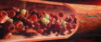
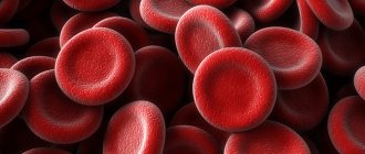
Red blood cells
This is what you might call the heart of your blood, the red blood cells (RBCs). These cute biconcave cells have the responsible task of carrying oxygen throughout the body. Typically, in one cubic millimeter of blood there are 4-5 million such cells in women and 5-6 million in men. People living at high altitudes, where there is a lack of oxygen, have even more red cells.
Cells that are found in blood
Substances necessary for the full functioning of all our organs constantly circulate through the bloodstream. There are also elements in the blood that protect the human body from diseases and the effects of other negative factors.
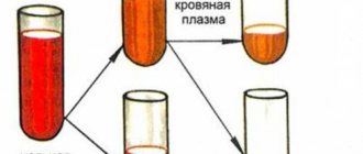
Attention! Blood is divided into two components. These are the cellular part and plasma.
Plasma
In its pure form, plasma is a yellowish liquid. It makes up about 60% of the total blood mass. Plasma contains hundreds of chemicals that belong to different groups:
- protein molecules;
- ion-containing elements (chlorine, calcium, potassium, iron, iodine, etc.);
- all types of saccharides;
- hormones secreted by the endocrine system;
- all types of enzymes and vitamins.
All types of proteins that are in our body are also in plasma. For example, from blood test results we can remember immunoglobulins and albumins. These plasma proteins are responsible for defense mechanisms. There are about 500 of them. All other elements enter the blood due to its constant circulating movement. Enzymes are natural catalysts for many processes, and three types of blood cells make up the main part of plasma.
This is interesting! Blood plasma contains almost all the elements of D.I. Mendeleev’s periodic table.
About red blood cells and hemoglobin
Red blood cells are very small. Their maximum size is 8 microns, and their number is large - about 26 trillion. The following structural features are distinguished:
- absence of nuclei;
- lack of chromosomes and DNA;
- they do not have an endoplasmic reticulum.
Under a microscope, a red blood cell looks like a porous disk. The disc is slightly concave on both sides. He looks like a little sponge. Each pore of such a sponge contains a hemoglobin molecule. Hemoglobin is a unique protein. Its basis is iron. It actively contacts the oxygen and carbon environment, transporting valuable elements.
At the beginning of maturation, the red blood cell has a nucleus. Later it disappears. The unique shape of this cell allows it to participate in the exchange of gases, including the transport of oxygen. The red blood cell has amazing plasticity and mobility. While traveling through the vessels, it undergoes deformation, but this does not affect its operation. It moves freely even through small capillaries.
In simple school tests on medical topics, you can come across the question: “What are the names of the cells that transport oxygen to the tissues?” These are red blood cells. It’s easy to remember them if you imagine the characteristic shape of their disk with a hemoglobin molecule inside. And they are called red because iron gives our blood a bright color. When the blood binds with oxygen in the lungs, it turns bright scarlet.
On a note! Few people know that the precursors of red blood cells are stem cells.
The name of the hemoglobin protein reflects the essence of its structure. The large protein molecule that is part of it is called “globin”. The structure that does not contain protein is called heme. In its middle there is an iron ion.
The process of producing red blood cells is called erythropoiesis. Red blood cells are formed in flat bones:
- cranial;
- pelvic;
- sternum;
- intervertebral discs.
Until the age of 30, red blood cells are produced in the bones of the shoulders and hips.
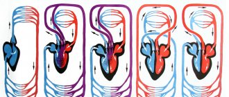
Collecting oxygen in the alveoli of the lungs, red blood cells deliver it to all organs and systems. The process of gas exchange takes place. Red cells give oxygen to cells. In return, they collect carbon dioxide and carry it back to the lungs. The lungs remove carbon dioxide from the body, and everything repeats all over again.
At different ages, a person experiences different degrees of red blood cell activity. The fetus in the womb produces hemoglobin, which is called fetal hemoglobin. Fetal hemoglobin transports gases much faster than in adults.
If the bone marrow produces few red blood cells, a person becomes anemic or anemic. Oxygen starvation occurs throughout the body. It is accompanied by severe weakness and fatigue.
The lifespan of one red blood cell can be from 90 to 100 days.
There are also red blood cells in the blood that have not had time to mature. They are called reticulocytes. With large blood loss, the bone marrow releases immature cells into the blood, since there are not enough “adult” red blood cells. Despite the immaturity of reticulocytes, they can already be carriers of oxygen and carbon dioxide. In many cases this saves human life.
Antigens, blood groups and Rh factor
In addition to hemoglobin, red blood cells contain another special antigen protein. There are several antigens. For this reason, the composition of blood in different people cannot be the same.
Important! Blood group and Rh factor depend on the type of antigen.
If there is an antigen on the surface of the red blood cell, the Rh factor of the blood will be positive. If there is no antigen, then the rhesus is negative. These indicators are critical if blood transfusion is necessary. The donor's group and Rh must match those of the recipient (the person receiving the blood transfusion).
Leukocytes and their varieties
If red blood cells are carrier cells, then leukocytes are called protectors. They contain enzymes that fight foreign protein structures, destroying them. White blood cells detect harmful viruses and bacteria and begin to attack them. By destroying harmful substances, they cleanse the blood of harmful decay products.
Leukocytes provide the production of antibodies. Antibodies are responsible for the body's immune resistance to a number of diseases. White blood cells take part in metabolic processes. They provide tissues and organs with the necessary composition of hormones and enzymes. Based on their structure, they are divided into two groups:
- granulocytes (granular);
- agranulocytes (non-granular).
Among granular leukocytes, neutrophils, basophils and eosinophils are distinguished.
Leukocytes are divided into 2 groups: granular (granulocytes) and non-granular (agranulocytes). Non-granular bodies include monocytes and lymphocytes.
Neutrophils
They make up about 70% of all white blood cells. The prefix “neutro” means that a neutrophil has a special property. Due to its grainy structure, it can only be painted with neutral paint. Based on the shape of the nucleus, neutrophils are:
- young;
- stab;
- segmented.
Young neutrophils do not have nuclei. In rod cells, the nucleus looks rod-shaped under a microscope. In segmented neutrophils, the nuclei consist of several segments. There can be from 4 to 5. When conducting a blood test, the laboratory technician calculates the number of these cells as a percentage. Normally, young neutrophils should be no more than 1%. The norm for the content of rod cells is up to 5%. The permissible number of segmented neutrophils should not exceed 70%.
Neutrophils carry out phagocytosis - they detect, capture and neutralize harmful viruses and microorganisms.
This is interesting! One neutrophil can destroy about 7 microorganisms.
Eosinophils
This is a type of leukocyte, the granules of which are stained with dyes that have an acidic reaction. Basically, eosinophils stain with eosin. The number of these cells in the blood ranges from 1 to 5% of the total number of leukocytes. Their main task is to neutralize and destroy foreign protein structures and toxins. They also take part in the mechanisms of self-regulation and purification of the bloodstream from harmful substances.
Basophils
Few cells among leukocytes. Their percentage of the total is less than 1%. Cells can only be stained with alkali-based dyes (“bases”).
Basophils are producers of heparin. It slows down blood clotting in areas of inflammation. They also produce histamine, a substance that expands the capillary network. The expansion of capillaries ensures resorption and healing of wounds.
Monocytes
Monocytes are the largest human blood cells. They look like triangles. This is a type of immature white blood cell. Their kernels are large and of different shapes. The cells are formed in the bone marrow and mature through several stages.
The lifespan of a monocyte is from 2 to 5 days. After this time, the cells partially die. Those that survive continue to mature into macrophages.
Fun fact! A macrophage can live in the human bloodstream for about 3 months.
The role of monocytes in our body is as follows:
- participation in the process of phagocytosis;
- restoration of damaged tissues;
- regeneration of nervous tissue;
- bone growth.
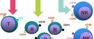
Lymphocytes
They are responsible for the body’s immune response, protecting it from foreign invasions. The place of their formation and development is the bone marrow. Lymphocytes that have matured to a certain stage are sent through the bloodstream to the lymph nodes, thymus and spleen. There they ripen to the end. Cells that mature in the thymus are called T lymphocytes. B lymphocytes mature in the lymph nodes and spleen.
T lymphocytes protect the body by participating in immune responses. They destroy harmful microorganisms and viruses. With such a reaction, doctors talk about nonspecific resistance - that is, resistance to pathogenic factors.
The main task of B lymphocytes is to produce antibodies. Antibodies are special proteins. They prevent the spread of antigens and neutralize toxins.
Important! B lymphocytes produce antibodies to each type of harmful virus or microbe.
In medicine, antibodies are called immunoglobulins. There are several types:
- M-immunoglobulins are large proteins. Their formation occurs immediately after antigens enter the blood;
- G-immunoglobulins are responsible for the formation of the fetal immune system. Their small size makes it easy to cross the placental barrier. The cells transmit immunity from mother to child;
- A-immunoglobulins - include defense mechanisms in the event of a harmful substance entering from the outside. Type A immunoglobulins are synthesized by B lymphocytes. They enter the blood in small quantities. These proteins accumulate on mucous membranes and in women's breast milk. They are also contained in saliva, urine and bile;
- E-immunoglobulins - released during allergies.
In the human bloodstream, a microorganism or virus can encounter a B lymphocyte on its way. The response of the B lymphocyte is to create so-called “memory cells”. “Memory cells” determine a person’s resistance (persistence) to diseases caused by specific bacteria or viruses.
We can obtain “memory cells” artificially. Vaccines have been developed for this purpose. They provide reliable immune protection against those diseases that are considered especially dangerous.
Platelets
Their main function is to protect the body from critical blood loss. Platelets ensure stable hemostasis. Hemostasis is the optimal state of the blood, which allows it to fully supply the body with the elements necessary for life. Under a microscope, platelets appear as cells that are convex on both sides. They lack nuclei and can range from 2 to 10 microns in diameter.
Platelets can take on a round or oval shape. When they are activated, growths appear on them. Because of the growths, the cells look like small stars. The formation of platelets occurs in the bone marrow and has its own characteristics. First, megakaryocytes arise from megakaryoblasts. These are cells with huge cytoplasm. Several separation membranes are formed inside the cytoplasm and division occurs. After division, parts of magekaryocytes “bud off” from the mother cell. These are already full-fledged platelets that are released into the blood. Their lifespan ranges from 8 to 11 days.
Platelets are divided by their diameter (in microns):
- microforms – up to 1.5;
- standard forms – from 2 to 4;
- macroforms – 5;
- megaloforms – 6-10.
The site of platelet formation is the red bone marrow. They mature in six cycles.
This is interesting! The growths that appear on platelets during the period of their activity are called pseudopodia. This is how cells stick together. They close the damaged vessel and stop bleeding.
Stem cells and their features
Stem cells are called immature structures. Many living beings have them and are capable of self-renewal. They serve as the initial material for the formation of organs and tissues. Blood cells also appear from them. There are more than 200 types of stem cells in the human body. They have the ability to renew (regenerate), but the older a person gets, the fewer stem cells his bone marrow produces.
Medicine has long practiced successful transplantation of certain types of stem cells. Among them, hematopoietic structures are distinguished. As already mentioned, hematopoiesis is a complete process of hematopoiesis. If it is normal, the composition of a person’s blood does not cause concern among doctors.
When treating leukemia or lymphoma, donor stem cells responsible for hematopoietic functions are transplanted. In systemic blood diseases, hematopoiesis is impaired, and bone marrow transplantation helps restore it.
Fun fact! Stem structures can turn into any type of cell, including blood cells.
Purkinje cells
Of the 100 billion neurons in your brain, Purkinje cells are some of the largest. Among other things, they are responsible in the cerebellar cortex for motor coordination. They are adversely affected by alcohol or lithium poisoning, as well as autoimmune diseases, genetic disorders (including autism), as well as neurodegenerative diseases (Alzheimer's, Parkinson's, multiple sclerosis, etc.).
Types of human tissues and their functions
Tissues are an association of groups of cells according to their purpose (function). Organs are formed from tissues. Some organs are made up of the same type of cells (for example, heart muscle and skeletal muscle). Some cells have a high degree of proliferation (that is, multiplication by division), under the influence of hormones, for example.
Others, when mature, lose the ability to divide or mutate - nerve cells, blood cells.
Types of organic fabric:
- Epithelium - performs the integumentary function; the outer covering is formed from it - skin, mucous membranes, soft tissues. Forms the outer capsule of some organs.
- Connective – cartilaginous, fatty, bone.
- Muscular – it forms all types of muscles, performs a motor and contractile function.
- Nervous – consists of nerve cells (neurons). Provides communication between organs and tissues and the brain through electrical impulses.
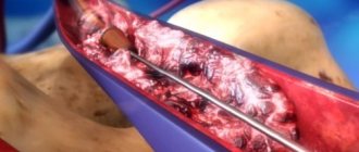
Villi of the small intestine
The villi of the small intestine increase its area, which promotes better absorption of food. These are irregularly cylindrical outgrowths up to 1.2 millimeters high. The basis of the villi is loose connective tissue. In the center, like a rod, runs a wide lymphatic capillary, or lacteal sinus, and on the sides of it there are blood vessels and capillaries. Fats pass through the milky sinus into the lymph and then into the blood, and proteins and carbohydrates enter the bloodstream through the blood capillaries of the villi. Upon careful examination, you can notice food debris in the grooves.
Distinctive features of plant and animal cells in the table
Plant and animal cells have both similarities and differences, which are briefly described in the table:
| Sign | Vegetable | Animal |
| Getting food | Autotrophic. Photosynthesizes nutrients | Heterotrophic. Does not produce organic matter. |
| Power storage | In vacuole | In the cytoplasm |
| Storage carbohydrate | starch | glycogen |
| Reproductive system | Formation of a septum in the maternal unit | Formation of constriction in the maternal unit |
| Cell center and centrioles | In lower plants | All types |
| Cell wall | Dense, retains its shape | Flexible, allows change |
The main components are similar for both plant and animal particles.
Human embryo and sperm
It looks like a war of the worlds, but in fact, you have an egg in front of you 5 days after fertilization. Some sperm are still retained on its surface. The image was taken using a confocal microscope. The egg and sperm nuclei are purple, while the sperm flagella are green. The blue areas are nexuses, intercellular gap junctions that communicate between cells.
How is the cell membrane structured?
The cytoplasmic membrane is the membrane that separates each cell from the extracellular space and you, of course, are wondering what the plasma membrane of a cell consists of? It consists of two layers of phospholipids and glycolipids.
These molecules have a polar end (which attaches to other molecules and atoms) and are a hydrophilic end. This part comes into contact with the intercellular environment and the cell plasma. The ends of non-polar lipids (hydrophobic) form the double inner layer of the cell membrane.
Also built into the shell are large protein molecules that act as opening pores, electrolyte channels and connecting mechanisms. Such a protein molecule passes through the bi-lipid layer. There are also glycocalyx threads on the surface. The shell may have folds, roughness and bulges, and processes like flagella.