What's happened
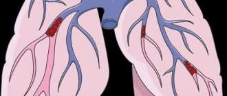
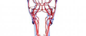
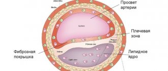
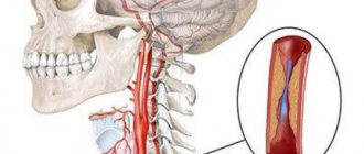
First, it’s worth understanding what a carotid artery is: in the human body there are two carotid arteries, running from the chest through the sides of the neck to the head. Their main task is to nourish the brain. Like any artery, they are confirmed by atherosclerosis - a serious disease, the nature of which is not fully understood, as well as the causes. This disease manifests itself in the deformation of the walls of blood vessels, when the tissues lining the vessel grow, cholesterol deposits appear between them, and the lumen of the vessel becomes smaller and smaller until it becomes completely blocked (obstruction). Oxygen-enriched blood flows through the carotid arteries for brain cells, so narrowing the lumen of the vessel is very dangerous - it can cause a stroke.
4.Treatment
With confirmed dissection of the carotid artery, long-term treatment with anticoagulants and antiplatelet agents is prescribed as a measure to prevent thrombus formation. In most cases, it is also indicated and advisable to prescribe drugs that stimulate trophism and strengthen vascular walls.
Precautions and necessary restrictions in terms of physical activity must be explained: in approximately 10% of cases, a relapse of RSA occurs within a year.
Despite this, the prognosis is generally favorable. Timely request for help and qualified provision of it allows you to completely restore the patency of the artery and the associated brain functions in approximately half of the patients; in other cases, as a rule, it is possible to achieve significant and lasting improvement.
Causes and prevention
The causes of vasoconstriction are being actively studied, but there is still no clear understanding. A hereditary cause is identified, and everything else can be attributed to risk factors. Among them:
- arterial injuries,
- obesity,
- smoking,
- diabetes,
- sedentary lifestyle,
- older age,
- high level of “bad cholesterol” in the blood (low-density lipoproteins are a combination of cholesterol and protein that do not take excess cholesterol from the vessels, but, on the contrary, attach them to the walls, forming cholesterol plaques).
Prevention of carotid artery stenosis, like any other arteries, consists of maintaining a healthy active lifestyle and a balanced diet to prevent an imbalance in the ratio of low- and high-density lipoproteins, and therefore the development of atherosclerosis.
Vascular stenosis
Atherosclerosis
Diabetes
Ulcer
3197 12 August
IMPORTANT!
The information in this section cannot be used for self-diagnosis and self-treatment.
In case of pain or other exacerbation of the disease, diagnostic tests should be prescribed only by the attending physician. To make a diagnosis and properly prescribe treatment, you should contact your doctor. Vascular stenosis: causes, symptoms, diagnosis and treatment methods.
Definition
Vascular stenosis (Greek στενός - “narrow, cramped”) is a partial or complete persistent narrowing of the lumen of blood vessels with limitation or complete cessation of blood flow.
Causes of vascular stenosis
Depending on which vessels are affected, a distinction is made between stenosis of arterial vessels (aorta, arteries, arterioles) and stenosis of venous vessels (superior vena cava, inferior vena cava, veins, venules).
Vascular stenosis can be either congenital or acquired.
The main cause of acquired stenosis of the aorta, arteries of the lower extremities, and coronary arteries of the heart is atherosclerosis, a systemic metabolic disease with predominant damage to the vascular wall. The degree of narrowing of the artery and its length may vary. When blood pressure increases, the sclerotic inner layer of the vessel (endothelium) is easily damaged, as a result, the blood clotting process is activated and a blood clot is formed.
Blockage of the vessel can lead to ischemia or necrosis of the tissue or organ.
Risk factors for the development of atherosclerosis include:
- male gender;
- elderly age;
- smoking;
- dyslipidemia (violation of the normal ratio of blood lipids);
- diabetes,
- arterial hypertension,
- increased blood homocysteine;
- elevated levels of C-reactive protein (CRP);
- increased blood viscosity and hypercoagulable states;
- chronic renal failure.
Another disease leading to arterial stenosis is obliterating endarteritis (spontaneous gangrene) - a chronic disease of peripheral blood vessels (mainly affecting the arteries of the feet and legs).
Mostly men under the age of 25-40 are affected. Those at risk include smokers, as well as people with frostbite on their feet. Diabetic angiopathy, characterized by damage to both small vessels and large and medium-sized arteries, develops in patients with diabetes mellitus. In diabetic macroangiopathy, when large blood vessels are affected, changes characteristic of obliterating atherosclerosis are found in the wall of the great vessels. With microangiopathies, when small blood vessels are affected, thickening of the walls of microvasculature vessels (arterioles, capillaries, venules) occurs, which leads to a narrowing of the lumen and deterioration of the blood supply to organs and tissues.
Coarctation of the aorta (congenital segmental narrowing of part of the aorta that obstructs blood flow) occurs as a result of improper fusion of the aortic arches in the embryonic period. The length of the narrowing is usually 1-2 cm. The ascending aorta and branches of the aortic arch expand, their diameter increases significantly, and the walls of the arteries participating in the collateral circulation become thinner. Two modes of blood circulation are formed in the systemic circle: up to the point of obstruction to blood flow there is arterial hypertension, and distal (or below) there is hypotension.
Venous stenosis most often occurs as a result of direct damage to the vascular wall during catheter insertion and is then aggravated by the constant presence of a foreign body and mechanical irritation. Inflammation and activation of the blood coagulation system are observed in the vessel wall. These changes lead to proliferation (multiplication) of smooth muscle cells, thickening of the vein wall, and the formation of microthrombi.
Thus, risk factors for the development of central venous stenosis are: the use of a central venous catheter, infections associated with the installation of a catheter, and concomitant diseases.
Systemic vasculitis, tumor diseases and other causes of vascular stenosis are detected much less frequently.
Classification of the disease
According to the type of blood vessels:
- arterial stenosis;
- venous stenosis
Due to the occurrence:
- congenital;
- acquired.
By localization:
- Stenosis of the arteries of the lower extremities.
- Stenosis of the carotid (carotid) and cerebral arteries.
- Stenoses of the arteries of internal organs:
- renal arteries,
- mesenteric arteries etc.
- Aortic stenosis.
- Stenosis of coronary vessels.
By caliber of damage:
- stenosis of large vessels (aorta and its branches);
- stenosis of medium-diameter vessels;
- stenosis of small vessels (arterioles and capillaries).
Symptoms of vascular stenosis
Damage to the blood vessels of the brain is one of the main causes of mortality and disability in the population. 2/3 of ischemic strokes are associated with narrowing and deformation of the carotid arteries.
The risk of developing ischemic stroke is directly related to the degree of narrowing of the artery lumen.
Occlusion (closure) of the internal carotid artery leads to the development of stroke in 40% of cases.
Damages to the blood vessels of the brain can occur in several forms:
- The asymptomatic form is characterized by the absence of focal and cerebral neurological symptoms (impaired consciousness, headache, vomiting, slow pulse).
- Discirculatory encephalopathy is characterized by a predominance of general cerebral symptoms; focal neurological symptoms are absent or appear in a very mild form.
- Transient ischemic attacks manifest themselves in the form of transient disorders of cerebral circulation of the ischemic type and are accompanied by the appearance of focal neurological symptoms that resolve within 24 hours.
- The consequences of a minor stroke are an acute ischemic cerebrovascular accident with the development of neurological symptoms, which almost completely regress within a month as a result of conservative therapy.
- The consequences of a completed stroke are an acute ischemic disorder of cerebral circulation, accompanied by the development of persistent focal neurological and cerebral symptoms.
- Ischemic stroke is damage to brain tissue with disruption of its functions due to obstruction or cessation of blood flow.
Atheroslerotic lesion of the coronary arteries of the heart is manifested by angina pain, but can sometimes be perceived by the patient as discomfort, a feeling of heaviness, compression, tightness, distension, burning or lack of air.
Most often, the pain is localized behind the sternum or along the left edge of the sternum; it can radiate (give) to the neck, lower jaw, teeth, interscapular space, and less often to the elbow or wrist joints, mastoid processes. Pain with angina pectoris usually lasts from 1 to 15 minutes. Occurs during significant physical or emotional stress. After taking nitroglycerin or stopping the exercise, the pain stops. As angina progresses, an attack may occur with minimal exertion and then at rest.
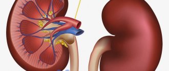
The main symptom of renal artery stenosis is a persistent increase in blood pressure, which is difficult to respond to drug therapy. Approximately 90% of cases of renal artery stenosis are caused by atherosclerosis; in 10% of cases, stenosis occurs due to fibromuscular dysplasia, a group of diseases that affect the walls of the arterial vessel.
With renal artery stenosis, the blood supply to the kidney tissue is reduced, hormonal factors (renin-angiotensin-aldosterone system) that regulate blood volume and blood pressure are activated, and the development of chronic kidney disease is accelerated.
Stenosing damage to the vessels supplying blood to the abdominal organs (mesenteric arteries) is more often observed in middle-aged and elderly people and manifests itself as chronic abdominal ischemia syndrome, so the main complaint of patients is pain, which appears after 20-25 minutes. after eating, lasts 1-2 hours and usually subsides on its own. The pain can be localized in the epigastric region, directly under the xiphoid process, and radiate to the right hypochondrium or spread from the periumbilical region throughout the abdomen. Some patients note a feeling of constant heaviness in the abdomen, and vomiting is rarely observed.
Other symptoms of chronic abdominal ischemia are intestinal dysfunction, expressed by disturbances in its motor, secretory, absorption functions, and progressive weight loss.
Obliterating atherosclerosis of the aorta and main arteries of the lower extremities is more common in men over 40 years of age and deprives them of their ability to work. The process can be localized in large vessels (aorta, iliac arteries) or medium-sized arteries (femoral, popliteal).
Small atherosclerotic lesions of the arteries of the lower extremities may not be clinically manifest. With continued vasoconstriction, intermittent claudication occurs, which is manifested by discomfort or pain in the muscles of the lower limb during physical activity. Damage to the terminal aorta and iliac arteries can cause pain in the buttocks, thigh, and calf. Impaired patency of the femoral-popliteal segment is characterized by pain in the calf. Occlusion of the arteries of the leg usually causes pain in the calf, foot, absence or decrease in skin sensitivity in them.
With obliterating endarteritis, trophic disorders are observed (cracks, dry skin, brittle nails, ulcers), intermittent claudication, leg pain, necrosis and gangrene of the limb.
In the generalized form of obliterating endarteritis or atherosclerosis, not only the vessels of the extremities are affected, but the visceral branches of the abdominal aorta, branches of the aortic arch, cerebral and coronary arteries.
The clinical picture of diabetic macroangiopathy consists of the clinical picture of microangiopathy and atherosclerosis of the great vessels, but is characterized by a more severe and progressive course, often ending in gangrene.
The clinical picture of diabetic microangiopathy of the lower extremities is similar to that of obliterating endarteritis.
With coarctation of the aorta, symptoms depend on the severity of the disease. In the case of significant narrowing of the aorta, the parents of the newborn pay attention to the pale skin, sweating, and difficulty breathing of the child. In older children and adults, the symptoms are usually mild: high blood pressure, headache, cold extremities, nosebleeds.
Stenosis of the central veins is clinically manifested by swelling of the extremities, pain in them and trophic changes (cyanosis, thinning of the skin, cracks, ulcers, etc.).
Diagnosis of vascular stenosis
Diagnosis of the disease is based on the analysis of patient complaints, medical history data, clinical picture, data from laboratory and instrumental research methods.
To clarify the cause of vascular stenosis, the following may be recommended:
- clinical blood test: general analysis, leukoformula, ESR (with microscopy of a blood smear in the presence of pathological changes);
Symptoms of carotid artery stenosis
The disease is dangerous because the symptoms are practically invisible, and only when stenosis becomes the cause of other pathological conditions does a person consult a doctor. Among such conditions, ischemic attacks (mini-stroke, small stroke) are primarily distinguished, when the blood supply to the brain is disrupted for a short period of time. This condition manifests itself as follows:
- numbness of hands/arms,
- speech distortion,
- temporary loss of vision, visual acuity,
- temporary disorientation,
- numbness of the face.
And more general symptoms
- headache,
- nausea,
- weakness.
Carotid artery stenosis develops slowly, often asymptomatically, and people attribute general fatigue and increased fatigue to excessive stress or the weather. Therefore, doctors recommend carefully monitoring your condition, and, if you experience prolonged general weakness without obvious reasons, contact a specialist. Some diseases are so dangerous if not diagnosed promptly that the risk of their presence justifies an unplanned trip to the doctor, who will reassure you if there is no cause for concern, or carry out the necessary diagnostics if suspicions arise.
How to diagnose pathology?
To diagnose abnormal clamping of the CC pathways, a comprehensive neurological examination is performed. The most accessible technique is ultrasound screening of head and neck vascular tissues. The most informative method confirming the initial diagnosis is angiography of cerebral vessels. Angiography is used in difficult cases, as well as before instrumental intervention (for planned surgery).
Magnetic resonance angiography is as safe and informative as possible due to the inaccessibility of other diagnostic methods. Additionally, a referral is made for a nuclear resonance scan of the brain. With joint hardware diagnostics, damage to the carotid “traces” is 100% detected.
Computer scanning helps to identify areas affected by oxygen deprivation. Color differences in degenerative areas in the resulting images help differentiate different types of ischemia. This type of disease can be determined only after 2-3 days. In the first hours of ischemia development, changes in brain tissue are not visible. Destruction can be determined only by indirect signs.
Diagnostics
Diagnosing a narrowed blood vessel may require one or more types of tests because the location of the narrowing is not always easy to find. For this purpose, our center uses:
- ultrasound - as the main method - since modern equipment provides maximum information not only about the presence of stenosis, but also the degree of deformation of the vessel, while being a non-invasive safe diagnostic method;
- MRI or CT;
- angiography - thanks to the introduction of a contrast agent, a complex diagnostic device monitors blood flow throughout the body, noting even minor deviations from the norm).
To diagnose atherosclerosis (which is the cause of narrowing), a laboratory method is also used - a blood test.
In which organs can stenosis occur?
Stenosis can occur in any organs that have a lumen, in the heart and in vessels of different diameters, in various anatomical canals. Symptoms of stenosis are very diverse - they depend on the affected organ and impaired functions.
Common types of stenoses:
- Stenoses of the digestive system organs: esophagus, stomach, intestines, bile ducts, pancreatic duct, papilla of Vater (the place where the bile duct and pancreatic duct enter the duodenum).
- Stenosis of the respiratory tract: larynx, trachea, bronchi.
- Stenoses of organs of the cardiovascular system: heart valves, aorta, arteries and veins of different diameters.
- Stenoses of the urinary system organs: ureters, urethra, bladder neck, renal arteries.
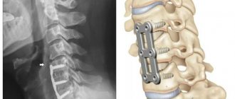
- Spinal stenosis. Frequent causes are previous spinal injuries, osteophytes, tumors, herniated intervertebral discs.
- Stenoses in the nervous system: the most striking example is non-communicating hydrocephalus; Among the causes may be tumor processes in the brain and spinal cord.
Book a consultation around the clock +7+7+78
Treatment of carotid artery stenosis
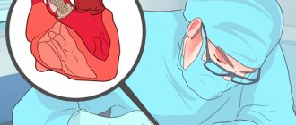
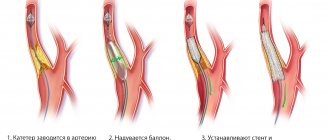
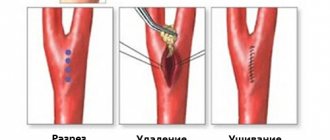
The most common treatment for stenosis is artery stenting. Stenting is a surgical treatment method, therefore, like any other surgical intervention, it requires a highly qualified doctor in diagnosis, deciding on the need/possibility of surgery and direct insertion of the stent. A stent is a metal frame inserted directly into an artery. At the point of narrowing, it unfolds and expands the walls of the vessel as necessary. The stent remains in the artery. Only in the first days its presence can be inconvenient due to changes in blood flow, but over time the patient stops feeling it and returns to normal life without the risk of a stroke. But of course, the doctor will give certain recommendations for further lifestyle in order to prevent re-stenosis.
Our doctors understand the importance of high-quality patient management and postoperative monitoring, so by contacting us, you can count on an integrated approach to diagnosis, treatment and the postoperative period. To implement this approach, the neurology department of the Federal Scientific Research Center has a powerful diagnostic base and a strong team of specialists with extensive experience in the treatment of stenosis, who additionally undergo advanced training courses on a regular basis. The department closely cooperates with the cardiac surgery service and the department of vascular surgery, which is separated into a separate unit.
The advantage of our center is the provision of a full range of services to our patients in one place, starting with an initial consultation, which you can sign up for by phone or through a special form on the website.
Causes of the pathological condition
There are many reasons for the development of arterial occlusion. The most common is atherosclerosis. This disease is characterized by the formation of a plaque-growth on the inner side of the vascular wall. The object consists of a cholesterol substrate, fat cells, blood cells - platelets. Plaque growth occurs at a slow pace. As it grows, the internal vascular lumen decreases. The growth itself becomes the basis for the formation of a blood clot. With the blood flow, the thrombus can move into narrower sections of the canal and block the arterial lumen. If the plaque has resulted in only a partial occlusion of the bloodstream, the flow may cause the plaque itself to rupture. This also results in overlapping of narrower areas.
Other diseases of the vascular system and heart that contribute to thrombosis of the bloodstream also lead to narrowing or blocking of the ducts. The root cause of the abnormal condition can be a bacterial infection that develops in the heart muscle, internal parts, and valves. Often, congenital disorders, acquired defects, and tumor formations of various nature play a decisive role. Anatomical tortuosity and confused configuration of the vascular network become the basis for the risk of formation of wall closure. External injuries to intracranial structures, such as traumatic brain injuries of varying severity, can also lead to abnormal degeneration of the CC network.
Associated unfavorable factors are:
- bad habits (smoking, alcoholism);
- diabetic syndrome;
- constant consumption of “heavy” foods with a high concentration of animal fats;
- excess weight gain.
Forecast
Without treatment, atherosclerotic occlusion of the subclavian artery leads to a gradual decrease in working capacity, and the likelihood of ischemic stroke and gangrene of the hand increases.
After restoring blood circulation by any method, the problem is completely eliminated. Normal blood flow promotes normal hand function and eliminates brain steal syndrome.
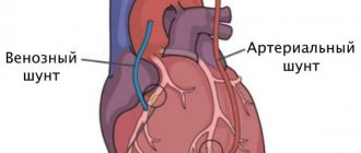
Relapses after stenting of the subclavian artery occur in approximately 10% of cases, due to the development of new plaques inside the stent (restenosis). If a carotid-subclavian bypass operation was performed, then the probability of relapse is no more than 2% of cases.
Complications
The most common complication is the phenomenon of vertebral-subclavian steal (steal syndrome). To compensate for blood circulation in the arm, cerebral blood flow is used, which moves in the opposite direction from the brain to the arm through the vertebral artery. In this case, physical work with the hand can cause disruption of cerebral circulation, including loss of consciousness.
Occasionally, atheroembolic complications may occur. Pieces of atherosclerotic plaque may be carried downstream into the arm. This is manifested by a sharp deterioration in blood circulation in the hand, blueness and pain in the fingers. If help is not given in a timely manner, necrosis of the finger may develop, which will require its amputation.