Pulmonary embolism (PE) is a serious, potentially life-threatening condition. Pulmonary embolism occurs due to blockage of blood vessels by thrombotic masses in the lungs. Pulmonary embolism (PE) can cause symptoms such as chest pain and/or shortness of breath. Also, in some cases, PE may have no symptoms at all, and it is difficult to detect without specialized examination. Massive pulmonary embolism can cause collapse and death. PE usually occurs due to the formation of a thrombosis in the lower extremity - deep vein thrombosis (DVT).
Pulmonary embolism (PE)
Prompt treatment is important and can save lives. Pregnancy, various medical conditions and medications, immobility, and major surgery all increase the risk of PE. Anticoagulant therapy, including heparin drugs, direct oral anticoagulants, or warfarin, is the usual treatment for PE.
Venous thromboembolic complications
Pulmonary embolism (PE) is part of a group of clinical problems collectively known as venous thromboembolic events (VTE).
Thrombosis is the blockage of a blood vessel by a blood clot (thrombus). Embolisms occur when part or all of a blood clot displaces from where it formed. The thrombotic masses then move with the bloodstream until they get stuck in narrower blood vessels in other parts of the body. In this case, the blood clot is called an embolus. Deep vein thrombosis (DVT) is the cause of venous thromboembolism, including pulmonary embolism (PE). Thrombosis most often occurs in the veins of the lower extremities.
Prognosis and prevention
Source: Mark Stebnicki: Pexels If pathology is detected in a timely manner and complex therapy is prescribed, the prognosis is favorable. However, there are several risk factors:
- advanced age;
- bed rest;
- the presence of tumors;
- history of thrombosis.
All these factors worsen the survival prognosis. In diseases of the cardiovascular and respiratory systems, approximately one third of all cases of pulmonary embolism are fatal.
Preventive actions:
- regular ultrasound examination of the veins of the lower extremities;
- giving up bad habits (it is especially important to give up smoking);
- maximum reduction of forced bed rest for various diseases and the postoperative period;
- wearing compression garments.
Separately, we should touch upon nutrition issues. It is important to minimize your intake of foods rich in vitamin K as it increases blood clotting. This includes milk and cottage cheese, broccoli, cauliflower and cabbage, bran, olive oil. You also need to reduce the amount of fatty and spicy foods in your diet. It is also recommended to avoid foods with a high glycemic index - in other words, sweets should also be significantly limited.
A sedentary lifestyle should be avoided. Any type of physical activity is suitable: walking (at least half an hour a day), running, swimming, moderate strength training (dumbbells, exercise machines). During long flights or transfers, it is important to consume at least 1.5 liters of fluid per day and ensure a minimum level of physical activity: it will be enough to just get up and walk for at least a few minutes about once every hour and a half.
What causes pulmonary embolism?
The usual cause of pulmonary embolism is deep vein thrombosis (DVT).
In almost all cases, the cause is a blood clot (thrombosis) that originally formed in a deep vein of the lower limb (known as deep vein thrombosis). This clot travels through the circulation and eventually becomes lodged in one of the lung's blood vessels. The broken thrombus is now called an embolus (and therefore can cause an embolism). Most deep vein thromboses arise from the veins of the legs or pelvis. Sometimes PE can occur from a blood clot in a vein in the arm or from a blood clot that forms in the heart. However, these situations are quite rare.
Other causes of pulmonary embolism
In rare cases, a blockage of a blood vessel in the lung may be caused by an embolus, which is not a blood clot. It could be:
- Fatty tissue from the bone marrow of a broken bone (if a large long bone such as a femur is broken).
- Foreign material from unsterile injection - for example, due to drug abuse.
- Amniotic fluid during pregnancy or childbirth (rare).
- Entry of a significant volume of air into the vein (rare).
- A small fragment of cancerous material (tumor) that has broken off from a larger tumor in a different location.
- Mycotic emboli are material from the focus of a fungal infection.
Treatment of pulmonary embolism
Typically, treatment is carried out in the intensive care unit. Moreover, not only patients with an acute form of pulmonary embolism are sent there, but also those whose diagnosis has not yet been definitively confirmed: this is done taking into account the high risk of sudden onset of the disease and the rapid development of life-threatening complications.
Treatment of high-risk patients
To prevent the continuation of blood clot formation (in different patients this process takes from several hours to several days), it is necessary to start using fast-acting direct anticoagulants (sodium heparin, calcium nadroparin) as soon as possible.
Since systemic hypotension aggravates right ventricular failure, it is necessary to use cardiotonic drugs with a vasopressor effect (dopamine, dobutamine) to increase blood pressure due to the vasoconstrictor effect.
Thrombolytic therapy (streptokinase, urokinase, alteplase) is also carried out. If there are contraindications to thrombolysis (ischemic and hemorrhagic stroke, bleeding, brain tumor), percutaneous catheter embolectomy is recommended. This is a minimally invasive surgical procedure during which the blood clot is removed from the vessels using a catheter.
Treatment of low-risk patients
For this group of patients, thrombolysis is not recommended; anticoagulant therapy (fondaparinux sodium or heparin sodium) is the preferred option.
The average duration of a course of anticoagulant therapy is 2-3 months. The exact timing is determined by the attending physician, based on the general condition of the patient and hemodynamic parameters.
Patients are also recommended to wear compression orthopedic underwear and take oral vitamin K antagonists (warfarin, acenocoumarol).
During pregnancy, low molecular weight heparin is used, which does not cross the placental barrier, does not harm the fetus and is practically not excreted in breast milk. As for warfarin, experts agree that this drug is contraindicated for pregnant women, since it has a negative effect on the development of the central nervous system of the unborn child.
Who develops pulmonary embolism?
Almost all cases of pulmonary embolism are caused by deep vein thrombosis. Thus, people who are more likely to get pulmonary embolism are people who are prone to deep vein thrombosis. Let's look at some risk factors for pulmonary embolism. Important factors are immobility, severe somatic pathologies and major surgical intervention (especially gynecological operations, operations on the pelvis and lower extremities). The risk of developing deep vein thrombosis or PE in hospital can be significantly reduced by activating the patient to walk as soon as possible. Medicines to help prevent deep vein thrombosis and PE are also prescribed to those patients who are at high risk.
Symptoms of pulmonary embolism
Symptoms of pulmonary embolism will depend on how large or small the clot is and how well a person's lungs can handle the clot. Frail or already ill people will have more severe symptoms than relatively healthy people. Symptoms of pulmonary embolism often occur suddenly.
A small pulmonary embolism can cause the following symptoms:
- Be asymptomatic (often).
- Shortness of breath - The degree of shortness of breath can range from very mild to severe.
- Chest pain, which is pleuritic in nature, that is, acute pain is felt when inhaling. Often you feel like you can't breathe deeply because it forces you to catch your breath. This happens because the blood clot can irritate the lining (pleura) around the lung. Shallow breathing is more comfortable for such patients.
- Coughing up blood (hemoptysis).
- Increased body temperature (from low-grade fever to fever).
- Increased heart rate (tachycardia).
A large pulmonary embolism or large number of clots (multiple embolism) may cause the following symptoms:
- Severe shortness of breath.
- Chest pain – With a massive pulmonary embolism, pain may be felt in the center of the chest behind the sternum.
- Confusion, feeling unwell, or fainting. This is because a large blood clot interferes with the heart and circulation, causing a sharp drop in blood pressure.
- Rarely and in extreme cases, a massive pulmonary embolism can cause cardiac arrest, when the heart stops pumping due to a blood clot. This may result in death even if resuscitation is attempted.
- Symptoms of deep vein thrombosis may occur, such as pain in the back of the leg, tenderness of the calf muscles, or swelling of the leg or foot.
Massive pulmonary embolism is so called not because of the actual large size of the blood clot (embolus), but because of the magnitude of its effect. PE is high risk if it causes serious problems, such as collapse or a drop in blood pressure. Major thromboembolism is by definition associated with a high risk.
Massive pulmonary embolism kills approximately 1 in 7 people.
How is pulmonary embolism diagnosed?
The diagnosis is based on clinical symptoms and medical history. For example, if a bedridden patient undergoing major surgery in a hospital develops shortness of breath, it is likely to be a pulmonary embolism.
Pregnant women who experience symptoms and/or signs suggestive of PE should be hospitalized immediately, as pulmonary embolism during pregnancy is a very serious condition and requires prompt diagnosis and treatment. For other patients, the Wells score is used to decide whether hospitalization is necessary. This allows you to assess the risk of developing pulmonary embolism, taking into account the following clinical signs:
- Clinical features of deep vein thrombosis.
- Heart rate more than 100 beats per minute.
- Immobilization for more than three days or surgery in the previous four weeks.
- Previous deep vein thrombosis and pulmonary embolism.
- Coughing up blood (hemoptysis).
- Treatment of cancer over the past six months.
- The alternative diagnosis is less likely than PE. Example: pneumothorax, pneumonia, myocardial infarction or gastroesophageal reflux.
If the likelihood of pulmonary embolism is high, emergency hospitalization of the patient is necessary. Various tests will be used to confirm the diagnosis. These may include the following activities:
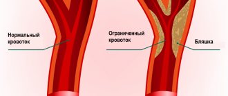
Duplex ultrasound scanning of the veins of the lower extremities
This study is used to determine blood flow disorders in the veins of the lower extremities. Lower extremity ultrasound is a simple, non-invasive test that can detect deep vein thrombosis. If DVT is detected, it can be assumed that the cause of other symptoms (such as shortness of breath or chest pain) was a pulmonary embolism. It is better to start treatment with anticoagulants immediately both in cases of deep vein thrombosis and in cases of suspected pulmonary embolism. If thrombotic masses are detected in the veins of the lower extremities, further studies may not be required. However, if the ultrasound results are negative, both deep vein thrombosis and pulmonary embolism cannot always be excluded. Further clinical studies will be required.
D-dimer blood test
This test detects fragments of the breakdown products of a thrombotic blood clot. The higher the level, the greater the likelihood of a blood clot forming in the vein. Unfortunately, the test can be positive in a number of other situations, such as if you have recently had surgery or are pregnant. Therefore, a positive result does not diagnose DVT or PE. However, in a complex of studies, this test can show how likely the presence of thrombosis is. To some extent, this may help decide whether further research is needed. A negative D-dimer result with a low risk of thrombosis means that the likelihood of a blood clot is extremely low. However, if your risk of venous thromboembolism is high, the D-dimer test cannot rule out the possibility of a blood clot and you will need further testing.
Ultrasound scan of the heart (echocardiography)
Echocardiography is useful for patients who may have massive PE, as it can show the effect on the heart. If there is massive PE, this puts stress on the right side of the heart. This test can be performed at the patient's bedside.
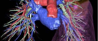
Computed tomography of the lungs with blood flow contrast
This is a specialized test that examines blood circulation in the lungs. This technique is the most accurate for detecting pulmonary embolism.
General studies
These studies are focused on diagnosing the condition of the heart, lungs and blood. This may help with diagnosis or identify other conditions:
- An electrocardiogram (ECG) is performed on all patients. This is necessary to identify any signs of cardiac strain that may occur with PE. Any heart rhythm abnormalities, such as atrial fibrillation, that may result from thromboembolism are identified.
- Blood tests are used to look for signs of heart attack, infection, or inflammation. An arterial blood gas test may also be done, which involves taking a blood sample from an artery rather than a vein. This is necessary to check the oxygen level in the blood.
- A chest x-ray is used to detect pneumonia or other chest conditions.
- Treatment of pulmonary embolism
Possible CT errors in the diagnosis of a body
As for pulmonary embolism of the large (central) branches of the pulmonary arteries, the sensitivity of spiral CT in detecting thromboembolism approaches 100%. For subsegmental and small branches, sensitivity ranges from 5% (according to the results of the PIOPED study) to 36% in other studies. The true significance of small emboli is not clearly established, however, thromboembolism of small branches of the pulmonary arteries may have clinical significance in patients with limited cardiopulmonary reserve.
Conventional pulmonary angiography allows for more detailed assessment of subsegmental vessels compared to CT, however, overlaps encountered when visualizing small vessels remain a limiting factor. As a result, the agreement between studies for isolated subsegmental pulmonary embolism is only 45%.
Studies have shown that favorable clinical outcome was achieved in patients (with a negative predictive value of 99%) whose CT findings were interpreted as negative for PE and who did not receive anticoagulation therapy or catheter-based thrombolysis. The outcome was similar to that of patients in whom there was clinical suspicion but no emboli were detected on pulmonary angiography. Therefore, although some small emboli may be missed on CT, the morbidity associated with PE is not high.
Modern multidetector computed tomographs are characterized by a significantly higher scanning speed, allowing thin-slice (1.25 mm) spiral CT angiopulmonography to be performed during a short breath hold (10-15 seconds). At the same time, segmental and subsegmental vessels become better distinguishable, changes are easier to interpret; The consistency of results across studies is also improved.
And, although the use of MSCT increases diagnostic capabilities, a large amount of data (for example, with CT angiopulmonography using thin slices, performed on a tomograph with 16 rows of detectors, 500–600 slices are obtained) leads to an increase in the load on any system intended for analysis and archiving information. In the future, the advent of automated detection algorithms and increased use of maximum intensity projection (MIP) reconstructions will be useful for identifying pulmonary emboli based on the large volume of MDCT data.
Possible errors in the interpretation of CT results are due to the partial volume effect: the overlap of perivascular soft tissues, bronchial branching zones, and blood vessels that do not run in a vertical direction. Thus, lymphoid and connective tissue, located mainly near large vessels, between the artery and the bronchial wall, can be mistaken for a blood clot.
Artifacts caused by blood flow and movement can lead to false filling defects: the likelihood of their presence must be taken into account when assessing the quality of the study and analyzing the data obtained. False filling defects due to blood flow can also occur with CT venography, causing a false-positive result.
Basic methods of treating pulmonary embolism
Anticoagulant therapy
Anticoagulant therapy is often referred to as blood thinning. However, it does not actually thin the blood. Anticoagulant therapy changes certain chemicals in the blood to prevent thrombotic clots from forming. This therapy does not dissolve the clot (as some people mistakenly think). Anticoagulation prevents the formation of new clots, and therefore thromboembolism. The body’s own mechanisms in the blood destroy the clot. Anticoagulant treatment is usually started immediately (if PE is suspected) to prevent worsening of the thrombosis while awaiting test results. Anticoagulation medications come in two forms: injections and tablets (or syrup for those who cannot swallow tablets). The first include heparin and fondaparinux. Tablet forms include warfarin and direct oral anticoagulants.
Supportive treatment is also used in the treatment of pulmonary embolism.
This therapy helps the body cope with the consequences of pulmonary embolism.
- Oxygen to reduce shortness of breath.
- In some cases, intravenous fluids are given to support circulation.
- If the patient is unwell or has a massive pulmonary embolism, careful monitoring and possibly intensive care are necessary.
- Additional procedures
They are usually used to treat severe or massive PE when the patient is very unwell or when treatment with anticoagulants is not possible.
Thrombolysis is an injection of drugs that is used to dissolve a blood clot. The drugs commonly used are alteplase, streptokinase, or urokinase. They are more powerful than the anticoagulants heparin and warfarin described above. However, there is a greater risk of side effects such as unwanted bleeding. Unwanted bleeding may even include bleeding into the brain (intracerebral hemorrhage).
Kava filters . They can be used to prevent blood clots from reaching the lungs. The filter is placed in a large vein called the inferior vena cava. The filter is inserted through a thin tube, which is inserted into a large vein and then inserted along the vein into the correct position. This procedure does not require anesthesia and can be performed at the patient's bedside. Filters may be useful when anticoagulant treatment alone is not sufficient or for patients who for some reason cannot undergo anticoagulant treatment. Currently, the indications for the use of vena cava filters are narrowing.
Surgical embolectomy. In some cases, it is possible to remove the embolus surgically. This is called an embolectomy. This is a major operation because it involves surgery on the organs of the chest, near the heart. A specialized medical facility and a highly specialized surgical team are required. This is usually considered a last resort for seriously ill patients. The operation carries a significant risk of death. This intervention will only be considered as an option if there is a massive PE, limiting other treatment options.
Catheter thrombolysis . This type of treatment is called catheter embolectomy or catheter clot fragmentation. It involves advancing a catheter through the blood vessels until it reaches the clot in the lung. Once the clot is reached, it can be removed or broken up (fragmented) with treatment given through the tube. This is a highly specialized treatment, so it is only available in certain treatment facilities.
Diagnosis of thromboembolism
It is often very difficult to diagnose PE due to the fact that its symptoms are similar to many other CVDs. Therefore, for the correct differentiation of the disease, the experience of the doctor and decent technical equipment of the clinic are very important.
After talking with the patient and analyzing his complaints, the doctor conducts a physical examination, measures blood pressure and pulse, and listens to the heart and lungs with a stethoscope. Next, laboratory and instrumental diagnostics are prescribed:
- clinical blood test, biochemical blood test, blood test for D-dimer;
- electrocardiography (ECG);
- echocardiography (ultrasound of the heart);
- chest x-ray;
- duplex/triplex scanning of the veins of the lower extremities;
- MSCT of the chest organs;
- ventilation-perfusion lung scintigraphy.
1 ECG for thromboembolism
2 Diagnosis of pulmonary embolism in MedikCity
3 ECHO-CG for thromboembolism
Complications of pulmonary embolism
Most people with PE are treated successfully and have no serious complications. However, serious complications are possible, including:
- Collapse - due to the effect of a blood clot on the heart and circulation. This can cause cardiac arrest and can be fatal. PE can cause heart strain. This can lead to a condition called heart failure, where the heart pumps less than normal.
- Recurrence of pulmonary embolism. Blood clots may occur again later (called recurrent PE). Treatment with anticoagulants helps prevent this.
- Bleeding. Complications due to treatment. Treatment with anticoagulants may have side effects. The main one is bleeding in various locations, for example, from a stomach ulcer. Approximately 3 in 100 patients will have significant bleeding due to treatment of PE with anticoagulants. This type of bleeding can usually be treated successfully. However, it is almost always safer to take anticoagulants to prevent recurrent PE, which can be even more severe.
- Chronic pulmonary hypertension. If small PEs recur, they may contribute to the development of a condition in which there is increased pressure in the blood vessels of the lungs (pulmonary hypertension).
3. Factors that increase the risk of developing pulmonary embolism
All factors that increase the likelihood of blood clots and blood clots also increase the risk of developing a pulmonary embolism. Some people have an innate tendency to form blood clots. In other cases, the following factors may influence the formation of blood clots:
- Prolonged physical inactivity. This can happen when a person remains in bed for a long time after surgery or a serious illness, or, for example, during long trips by car;
- Previous surgery that affects the legs, hips, abdomen, or brain;
- Certain diseases such as cancer, heart failure, stroke or severe infectious diseases;
- Pregnancy and childbirth, especially by caesarean section;
- Taking birth control pills or hormone therapy;
- Smoking.
The risk of blood clots increases in older people (especially those over 70) and those who are overweight or obese.
About our clinic Chistye Prudy metro station Medintercom page!
What are the prospects for pulmonary embolism?
This depends on the type of pulmonary embolism and the presence of concomitant diseases.
If PE is treated correctly and promptly, the prognosis is good and most patients can make a full recovery.
The prognosis is worse if there is a serious disease that contributed to the embolism, such as advanced cancer. Massive PE is more difficult to treat and is life-threatening. PE is a serious condition and can have a high risk of death, but this is greatly reduced with early treatment.
The most dangerous time for complications or death is the first few hours after an embolism. In addition, there is a high risk of developing a new pulmonary embolism within six weeks of the first one. Therefore, treatment must be started immediately and continued for at least six months.
How to prevent pulmonary embolism?
To avoid pulmonary embolism, it is necessary to prevent and promptly treat chronic diseases. One of the causes of venous thrombosis is varicose veins. Any person who suspects he has this disease should visit a phlebologist and have the venous system of the lower extremities diagnosed. After diagnosis, you need to follow the doctor's recommendations. Also, patients planning major surgery should be assessed for the risk of deep vein thrombosis, and people at high risk of DVT may require prophylactic doses of anticoagulants before and after surgery.
Frequently asked patient questions about pulmonary embolism (PE)
What are the symptoms of pulmonary embolism?
The main symptoms to suspect pulmonary embolism are cough, shortness of breath, chest pain and hemoptysis.
How to avoid pulmonary embolism?
In order to prevent the development of pulmonary embolism, it is necessary to prevent venous thrombosis: promptly treat varicose veins and other vein pathologies, avoid prolonged immobilization for various diseases and surgical treatment, use anticoagulants in case of increased risks of thrombosis.
Who is more likely to develop pulmonary embolism?
Pulmonary embolism, as a rule, affects patients with severe cardiac and pulmonary pathology, cancer patients, and patients in various surgical hospitals after long-term surgical interventions.
What should you do if you suspect pulmonary embolism?
At the slightest suspicion of pulmonary embolism, you must call an ambulance. Before the arrival of medical personnel, the patient must be laid on a flat surface and ensure free access of air.