General information
What is vertebral artery hypoplasia?
Hypoplasia of the right vertebral artery and hypoplasia of the left vertebral artery are an abnormal decrease in the lumen of these arteries (approximately to a diameter of less than 2 mm).
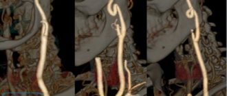
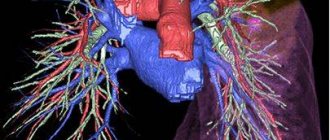
But there is no single criterion for narrowing the diameter of the vessel, and a number of authors include a decrease in the diameter of the vessel of less than 3 mm as signs of VA hypoplasia. The right vertebral artery is a branch of the right subclavian artery, which arises from the brachiocephalic trunk, and the left one arises from the left subclavian artery, which originates from the aortic arch. Next, both vertebral arteries (VA) rise in the bone canal to the brain and merge in the cranial cavity, forming the basilar artery. The vertebral arteries supply 15–30% of blood to the structures of the brain stem, the temporal/occipital lobes, the cerebellum, the hypothalamic region, and segments of the spinal cord, that is, they play a significant role in ensuring blood flow to the brain structures. Below is a picture of hypoplasia of the right/left VA.
Hypoplasia is one of the most common pathologies of the vertebral arteries. Its occurrence in the population, according to various sources, varies widely between 2.34% and 26.5% and is predominantly congenital in nature. Hypoplasia of one of the arteries occurs predominantly; cases of bilateral hypoplasia of the VA are relatively rare. The main place of narrowing of the lumen of the vertebral arteries is the place of entry into the bone canal (in the cranial cavity), which leads to a significant decrease in blood flow mainly to the posterior parts of the brain with the gradual development of non-vertebrogenic PA syndrome .
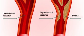
Hypoplasia of the vertebral artery is a factor in a significant reduction in blood circulation in the vertebrobasilar region, which disrupts the hemodynamics of the brain as a whole, increasing the risk of developing ischemic stroke (acute cerebrovascular accident). The situation is sharply worsening against the backdrop of the development of cerebral atherosclerosis , which is extremely common in the population due to poor lifestyle and especially dietary habits.
Treatment
Therapy depends on the phase of the pathological process. If we are talking about a compensated violation, there is no need for medical correction.
Dynamic observation under the supervision of a vascular surgeon and neurologist (possibly one or the other) is indicated.
When blood flow abnormalities develop, medications are used. Several groups of drugs are prescribed:
- Protective agents, nootropics. To protect nerve tissue and, on the other hand, restore normal metabolic processes in the brain. Glycine, Piracetam, Mexidol, Mildronate.
- Antihypoxic agents with the ability to dilate blood vessels. Actovegin, Cavinton and others.
- Antiplatelet agents. At risk of developing blood clots. Aspirin in modern modifications is suitable for long-term use.
- Antihypertensive. In some cases. On the recommendation of the attending physician.
- Dizziness is relieved with specialized drugs: Tagista, Vestibo, Betagistin and others like that.
In case of decompensation or if conservative methods are useless, surgical intervention is indicated.
There are three treatment options:
- Stenting. Mechanical expansion using a special flexible device. It prevents the artery from narrowing and forcibly normalizes blood flow.
- Angioplasty. Another name for this is ballooning. The essence is approximately the same, only the mechanism of action is different.
- Reconstruction and prosthetics. Replacement of the narrowed fragment with a natural analogue, which is created from the patient’s own vein.
The effectiveness of the described methods is sufficient to achieve high-quality results.
In the future, it is recommended to undergo a course of physiotherapy. UHF, magnet, currents. Here are the basic techniques.
Physical therapy is optional, at the discretion of the specialist.
It is mandatory to give up smoking, alcohol, and excess amounts of animal fat. Salt no more than 7 grams per day.
Adequate sleep of 8 hours per night and adequate physical activity (at least an hour, two walks in the fresh air) are also indicated.
It is recommended to change your professional activity if it is physical in nature. When working sedentarily, every 30-60 minutes you need to do a little exercise for the neck, but without fanaticism, so as not to provoke compression of the vertebral artery.
It is important to avoid stress. In the modern world, this is almost impossible to do. Therefore, there is an alternative - mastering the simplest relaxation techniques in order to reduce the harmful effects of negative emotions on the body.
Attention:
All recommendations should be clarified with your treating specialist, because there can be no universal recipes. The role of the individual factor is too great.
Treatment of hypoplasia of the right vertebral artery is carried out in a system; the method is chosen depending on the severity of the pathological process.
Pathogenesis
Currently, hypoplasia of the vertebral arteries is considered as one of the manifestations (during the formation of the fetal vascular system) of undifferentiated connective tissue dysplasia. Also in the pathogenesis of clinical manifestations, the development of vasospastic reflex reactions caused by irritation of the sympathetic plexus of the vertebral arteries is of significant importance. The resulting flow of afferent impulses irritates the overlying centers of vascular-motor regulation, which is manifested by local/diffuse reactions that predominantly affect the vessels of the vertebrobasilar system.
Diagnostics
Diagnosis of SPA is carried out by a neurologist. The specialist needs to be able to identify the patient’s main complaints and compare them with the characteristics of each form of the disease. If the existing symptoms correspond to the clinical form of SPA, it is important to diagnose the causes of its development, i.e., to detect which vascular or spinal pathologies led to impaired blood flow through the vertebral arteries.
To this end, the doctor initially conducts a thorough examination and neurological tests, which allow one to suspect what could have caused the SPA. To confirm guesses, obtain more accurate information and detect possible concomitant diseases, the patient is prescribed:
- X-ray or CT scan of the cervical spine;
- MRI of the cervical spine;
- Ultrasound of neck vessels with Doppler sonography;
- stabilometry;
- general and biochemical blood test;
- audiometry, etc.
In our clinic, you can also learn in more detail about the composition of your body and the state of the vascular system, which is involved in the blood supply to internal organs, skeletal muscles, and the brain. Our experienced doctors will explain the data obtained to you in detail. Bioimpendansometry calculates the ratio of fat, muscle, bone and skeletal mass, total fluid in the body, and basal metabolic rate. The intensity of recommended physical activity depends on the state of muscle mass. Metabolic processes, in turn, affect the body's ability to recover. Based on the indicators of active cell mass, one can judge the level of physical activity and nutritional balance. This simple and quick test helps us identify disturbances in the endocrine system and take the necessary measures. In addition, it is also very important for us to know the condition of blood vessels for the prevention of diseases such as heart attacks, hypertension, heart failure, diabetes and much more. Angioscan allows you to determine such important indicators as the biological age of blood vessels, their stiffness, stress index (which indicates heart rate), and blood oxygen saturation. Such screening will be useful for men and women over 30, athletes, those undergoing long-term and severe treatment, as well as everyone who monitors their health.
Often, for the correct diagnosis of vertebral artery syndrome and the causes of its development, consultations with other specialized specialists, including cardiologists, ENT specialists, ophthalmologists, etc. are required.
Today, SPA is often falsely diagnosed, which is due to the insufficient completeness of the examination.
Causes
Hypoplasia is predominantly congenital in nature, the development of which can be caused by both chromosomal mutations and factors with an adverse effect on the mother’s body during pregnancy. These factors include:
- Bruises/injuries during pregnancy .
- Infectious diseases that have a negative impact on the fetus, especially in the early stages of development ( influenza , rubella ).
- Bad habits (smoking, alcoholism, drug addiction).
- Prolonged contact with harmful substances (work in hazardous industries, toxic chemicals, radiation).
- Drug abuse.
Hypoplasia of the posterior communicating arteries of the brain
Hypoplasia is a fundamental term in pathological anatomy, denoting underdevelopment of the tissues of a particular organ or the entire organism, which is determined by defects during embryonic maturation. Any organ can succumb to hypoplasia: arteries, heart, brain, kidneys, testicles or knee joint.
Intrauterine underdevelopment of an organ refers to disorders of adaptation and adaptation of the body. This disease is an irreversible process. Related concepts:
- Aplasia is an extreme degree of underdevelopment of an organ that appears in a newborn in its rudimentary form.
- Dysplasia is the abnormal formation of an organ.
The disease does not always manifest itself from the moment the child is born. Underdevelopment of an organ, if it is paired, can be compensated by another organ. For example, each kidney is 10% loaded. If one organ is hypoplastic, the other kidney will be 30-50% loaded. Often, a malformation is discovered by chance during a routine examination.
Symptoms
The initial signs of VA hypoplasia are nonspecific, and they can be mistaken for manifestations of encephalopathy , cervical osteochondrosis or vegetative-vascular dystonia . PA hypoplasia syndrome is clinically manifested by three groups of symptoms:
- A group of vertebral symptoms (pain in the cervical spine/occipital region).
- Local symptoms - pain in the vertebral artery/structures of the corresponding vertebral segment (during palpation) with pronounced irradiation to the head.
- Symptoms at a distance from the vascularization zone of the VA, as well as due to irritation of the sympathetic plexus of the vertebral artery (increased blood pressure, migraine -type pain , body instability when walking).
The specificity of the clinical manifestations of VA hypoplasia is determined by a number of factors: the localization/extension of the lesion, the functional state of the arterial-venous system of the brain - the presence of anastomoses, collaterals, the state of the vascular wall.
Standard patient complaints include frequent, severe headaches ; dizziness ; surges in blood pressure ; inability to concentrate; memory loss; increased weather sensitivity ; deterioration of vision/appearance of “spots” in the field of vision; unsteady gait, decreased performance; difficulty making sudden movements/turns.
The presence of neurological complaints is traditionally considered to be indirect signs of stenosis . The symptoms of left-sided and right-sided VA hypoplasia are generally similar, however, the pathology of the vertebral artery on the right is often localized higher (in the intracranial area), which causes a more rapid development of symptoms and their more pronounced manifestation (a sharp drop in vision, loss of consciousness, severe coordination disorder).
The left vertebral artery initially has a larger working diameter, so symptoms do not appear so sharply and later. With left-sided hypoplasia, the characteristic symptoms are sharp, throbbing occipital pain; shots to the head; pain on palpation of the 1st and 2nd vertebrae of the neck; feeling of sand/double vision; tinnitus/hearing impairment. With bilateral VA hypoplasia, due to the lack of a compensatory effect, symptoms develop quickly, and there is a high risk of developing serious complications.
Types of hypoplasia
In a broad sense, hypoplasia is an anomaly in the formation of any organ, in which it is completely or partially reduced. One of the most severe forms of this type of anomaly is considered to be hypoplasia of the corpus callosum of the brain in a child. With it, there is no corpus callosum, a structure of the central nervous system that connects the hemispheres. In 70-75% of clinical cases, the child becomes disabled.
The brain is most often affected by vascular diseases of varying complexity. Moreover, any pathology immediately affects the human condition, since this organ is sensitive to a lack of microelements.
Hypoplasia of cerebral arteries can be acquired or congenital. The first develops against the background of exposure to unfavorable factors, and the second type of pathology is a consequence of improper development of tissues of arteries and veins during the period of intrauterine development of the child. In medical practice, congenital hypoplasia is most often diagnosed.
The blood supply to the head of the central nervous system occurs through two internal carotid arteries and two vertebral arteries - left and right. In a healthy person they are developed evenly throughout. If a malfunction occurs during the formation of the circulatory system of the fetal brain, this leads to underdevelopment of some blood vessels. Experts distinguish right-sided, left-sided and bilateral hypoplasia.
When the main vessels are blocked, the blood supply to the brain is carried out through the structures of the circle of Willis, which form the main main arteries near the base of the skull.
A normally developed circle of Willis occurs only in 25-40% of the population; in other cases, its components have an abnormal structure. Often this is hypoplasia of the anterior or posterior communicating arteries, absence and deformation of the first segments of the anterior and posterior cerebral arteries.
Pathology can also affect the venous sinuses - collectors into which waste blood is collected from the surface of the brain. From them it enters the two jugular veins - paired blood vessels located in the neck. With hypoplasia of the venous sinuses, a decrease in venous outflow is observed.
Consequences and complications
In general, VA hypoplasia significantly impairs quality of life. The most serious complications of VA hypoplasia primarily include a high risk of cerebrovascular accident/ stroke , which is caused by a decrease in blood supply to brain structures. In addition, frequent complications of the pathology of the vertebral arteries include neurological symptoms: headaches ( migraine ), irritability, depression, severe fatigue, disorders of the autonomic nervous system, impaired auditory/visual function, weakened cognitive abilities, dementia, decreased ability to work, arterial thrombosis
Syndrome of blood flow insufficiency in the arteries of the vertebrobasilar system
Maksimova M.Yu., Piradov M.A. RMJ. 2021. No. 7. pp. 4-8 The article is devoted to the problem of blood flow insufficiency syndrome in the arteries of the vertebrobasilar system. Methods for diagnosing and treating vertebrobasilar insufficiency are presented, which should be aimed at preventing its progression, improving blood supply to the brain, and correcting certain syndromes and symptoms.
The independent clinical concept of “blood flow insufficiency syndrome in the arteries of the vertebrobasilar system” was formed in the 1950s, during the period of revision of views on the pathogenesis of ischemic cerebrovascular accidents (CVA) and the emergence of the concept of the leading role of cerebrovascular insufficiency in this case [1]. The peculiarities of the structure and functions of this arterial system, which provides nutrition to vital structures of the brain, and the uniqueness of clinical symptoms in case of disturbances of blood flow in it led to its identification in the latest version of the international classification into an independent symptom complex - “vertebrobasilar arterial system syndrome” within the framework of “transient transient cerebral ischemic attacks (attacks) and related syndromes” (International Classification of Diseases, 10th Revision, G45.0). Even earlier, a group of experts from the World Health Organization defined “vertebrobasilar insufficiency” as “a reversible impairment of brain function caused by a decrease in the blood supply to the area supplied by the vertebral and basilar arteries.” The ischemic nature and reversible nature of the disorders were emphasized, but the duration of neurological symptoms was not indicated, which previously did not allow them to be classified as transient ischemic attacks (TIA) and which has now become possible. Blood flow disturbances in the arteries of the vertebrobasilar system account for about 70% of all TIAs. Stroke with localization of focal changes in areas of the brain that receive blood through the arteries of this system develops 2.5 times less frequently than in regions belonging to the arterial basins of the carotid system [1].
Causes of blood flow insufficiency syndrome in the arteries of the vertebrobasilar system
The main causes of blood flow insufficiency syndrome in the arteries of the vertebrobasilar system, caused by arterial hypertension (AH) and atherosclerosis (AS), include [1, 2]:
- atherostenosis or atherobliteration of one of the vertebral arteries;
- characteristic arterial tortuosity, which in some cases can lead to kinking of the vertebral artery with the formation of septal stenosis and disruption of blood flow in it;
- congenital anomalies of the vertebral arteries (hypoplasia of one of the vertebral arteries, lateral displacement of the mouth of the vertebral artery), in which the insufficiency of blood flow in one of the vertebral arteries is compensated by another vertebral artery, but decompensation occurs against the background of AS and hypertension;
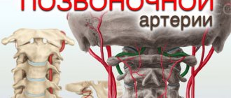
- compression of the vertebral artery by an osteophyte in the bone canal of the cervical spine, an articular process with instability of the cervical spine, an accessory cervical rib, a spasmodic neck muscle (posterior scalene muscle, longus colli muscle, inferior oblique muscle of the capitis), which is most often observed with congenital abnormally high entry vertebral artery into the spinal canal - at the level of 3–5 cervical vertebrae.
The syndrome of blood flow insufficiency in the arteries of the vertebrobasilar system can also be observed:
- with subclavian “steal syndrome”, in which, as a result of occlusion of the subclavian artery, blood flows not only to the entire vertebrobasilar system, but also to the arm through only one vertebral artery;
- with occlusion or severe atherostenosis of both internal carotid arteries
(ICA), since the vertebrobasilar system plays a significant role in the blood supply to the cerebral hemispheres and, under certain conditions, “steal syndrome” may occur; - with disturbances of general hemodynamics.
Subclavian “steal syndrome” is characterized by a phenomenon when a patient, during intensive work of the arm (retrogradely supplied with blood from the contralateral vertebral artery), experiences brainstem symptoms - most often dizziness. A certain contribution to the development of blood flow deficiency syndrome in the vertebrobasilar system can be made by changes in the rheological properties of blood (increased levels of fibrinogen, blood viscosity, platelet aggregation and hematocrit, increased rigidity of erythrocytes), leading to deterioration of microcirculation.
Diagnosis of blood flow insufficiency in the arteries of the vertebrobasilar system
Subjective data
The diagnosis of insufficiency of blood flow in the arteries of the vertebrobasilar system is based on a characteristic symptom complex that combines several groups of clinical symptoms found in patients with AS and hypertension.
These are visual and oculomotor disorders, disorders of statics and coordination of movements, vestibular disorders. In this case, the presumptive diagnosis is determined on the basis of at least two of these symptoms. They are short-lived and often go away on their own, although they are a sign of impaired blood flow in the arteries of this system, which requires clinical and instrumental examination. A thorough medical history is especially necessary to clarify the circumstances of the occurrence of certain symptoms [1, 2]. Visual disturbances
include a feeling of blurred vision, photopsia, scotoma, changes in visual fields, decreased visual acuity and are associated with transient ischemia of the occipital lobes of the brain.
Blurred vision in the form of a veil before the eyes and blurred vision often occurs at the height of a headache. Photopsia appear in the form of flashes of colored dots, most often red or green, black, with a light halo, as well as spots, fiery lightning, lines, rings, zigzags. Photopsia differ from the rainbow circles characteristic of glaucoma in that their appearance is not associated with an external light source; they also occur with closed eyes. Changes in visual fields are usually observed in the form of their concentric narrowing. Decreased visual acuity often develops after the onset of headache and progresses; vision deteriorates noticeably during headache attacks and after them. Oculomotor disorders
manifest themselves in the form of transient diplopia with mild paresis of the eye muscles and impaired convergence.
In most patients, these disorders are among the initial manifestations of the disease, and in a quarter of them they serve as one of the main complaints with vertebrobasilar insufficiency. Static and dynamic ataxia are also among the permanent symptoms that are manifested by patient complaints of instability and staggering when walking and standing. Coordination of movements is significantly less impaired; a persistent change occurs, as a rule, with cerebellar infarctions. Vestibular disorders
manifest themselves in the form of sudden dizziness - systemic, which is characterized by a feeling of “rotation of objects”, “an inverted room”, and non-systemic with a feeling of “motion sickness”, nausea, and less often vomiting.
Spontaneous nystagmus is also detected, sometimes only after special tests with turning the head to the side and fixing it in these positions (De Klein test). The development of dizziness is associated with ischemia of either the vestibulocochlear organ or the vestibular nuclei and their connections. The vestibular nuclei are most sensitive to ischemia and hypoxia. In this case, dizziness as a monosymptom can be regarded as a sign of impaired blood flow in the arteries of the vertebrobasilar system only in combination with other signs of its impairment in patients with a relatively persistent otoneurological symptom complex. Less known, although not uncommon, are optic-vestibular disorders. These include symptoms of “shading shadow” and “convergent vertigo,” in which patients experience dizziness or unsteadiness when flashing light and shadow or when looking downward. Characteristic symptoms are attacks of sudden falling
without loss of consciousness (“drop attacks”), usually occurring during sudden turns or throwing back the head.
Syncopal vertebral Unterharnscheidt syndrome has been described, in which loss of consciousness and muscle hypotonia are observed in the absence of evidence of epilepsy and other paroxysmal conditions. Manifestations of diencephalic
disorders include severe general weakness, irresistible drowsiness, disturbances in the rhythm of sleep and wakefulness, as well as various autonomic-visceral disorders, a sudden increase in blood pressure (BP), and heart rhythm disturbances.
These disorders are associated with ischemia of the structures of the reticular formation of the brain stem. The described symptom complex has now been supplemented by other signs, which, in combination with them, also make it possible to judge the insufficiency of blood flow in the arteries of the vertebrobasilar system. At various stages of vertebrobasilar insufficiency, patients often complain of decreased memory
(“forgetfulness”), concentration disorders and instability of active attention. Most often, memory for names, numbers, and recently occurring events decreases. The ability to memorize new material decreases, it becomes more difficult to retain what has been read in memory, what is planned for implementation is forgotten, and the need to write it down arises. It becomes difficult for patients to comprehend a large amount of information, which leads to a certain decrease in performance and limitation of creative possibilities in people engaged in mental work. At the same time, professional memory and memory of past events are preserved. This applies more to RAM than to logical memory. Often, a decrease in memory and performance is regarded by others as a result of overwork, and not as a manifestation of cerebral vascular insufficiency. During neuropsychological research, the preservation of the level of generalization, the correspondence of judgments to the general educational and cultural level, and the preservation of the stock of ideas and skills are noted. Impaired cognitive functions significantly reduce the quality of life and also affect the progression of cerebrovascular insufficiency. Decreased memory for current events in patients with vertebrobasilar insufficiency is associated with chronic ischemia of the medial parts of the temporal lobes, primarily the hippocampus and mammillary bodies. With vertebrobasilar insufficiency, attacks of transient global ischemia are also observed, during which working memory (the ability to remember new information) is impaired for several hours. The patient looks absent-minded, he is disoriented in space and time, sometimes excited, persistently tries to find out from those around him where he is, how he got here, but being unable to remember the answers, he constantly asks the same questions. With the return of the ability to remember, orientation is also restored, only the episode itself is amnesic. Acute amnesia can also be caused by acute cerebrovascular accident in the basins of both posterior cerebral arteries. In this case, amnesia may be accompanied by limitation of visual fields (unilateral or bilateral hemianopia), visual agnosia, alexia, amnestic aphasia, and sensory impairment. The combination of a number of characteristic symptoms makes it possible to diagnose the syndrome of blood flow insufficiency in the arteries of the vertebrobasilar system, although in this case only the ischemic nature of the cerebrovascular accident and the localization of the source of ischemia are determined, and not the reasons that determined this nature.
Objective data
The most accessible and safest methods for determining insufficiency of blood flow in the arteries of the vertebrobasilar system are neurological examination and ultrasound methods of studying the vascular system of the brain.
Among the objective signs revealed during a neurological examination
, one should first of all mention nystagmus, static and dynamic ataxia.
In the Romberg test, the patient deviates to the side. Walking with eyes closed reveals unsteadiness and persistent deviation to one side in a patient with insufficient blood flow in the vertebrobasilar system. When performing the Unterberger test, the patient is asked to march in one place with his eyes closed for 1–3 minutes. Normally, it remains in place or moves slightly relative to the starting point or rotates slightly around its axis. A forward shift of more than 1 m and a rotation of more than 40–60° (after 50 steps in place) are considered pathological. The results of the Babinski-Weil test (“star test”) are interpreted in a similar way. With eyes closed, the patient is asked to take two steps forward, turn 180° and take two steps back. Any deviations to the side or rotation indicate dysfunction of the vestibular labyrinth. If the patient is asked to walk in the forward and reverse directions several times, then as a result of a deviation to one side, the trajectory of his movement resembles the outline of a star (hence the name of the test). It is also necessary to measure blood pressure in both arms in a sitting and lying position. Objective signs of the syndrome include differences in pulse and blood pressure in the arms and noise in the supraclavicular region. With a significant decrease in systolic blood pressure (more than 20 mm Hg) in an upright position, symptoms reminiscent of insufficiency of blood flow in the vertebrobasilar system should be attributed to orthostatic hypotension. Subclavian “steal syndrome” is characterized by a phenomenon when a patient, against the background of intense hand work, develops brainstem symptoms—usually dizziness. Doppler ultrasound
allows you to obtain data on blood flow in the vertebral arteries, linear speed and direction of blood flow in them.
Compression-functional tests make it possible to assess the condition and resources of collateral circulation, blood flow in the carotid, temporal, supratrochlear and other arteries. Duplex scanning allows you to determine the condition of the artery wall, the structure and surface of atherosclerotic plaques that stenose these arteries. Transcranial Doppler ultrasound with pharmacological tests is important for determining cerebral hemodynamic reserve. Data on the condition of the great arteries of the head (MAG) and intracerebral arteries obtained from CT and MRI angiography are extremely informative. X-rays
of the cervical spine can provide information about the condition of the structures around the vertebral arteries and the effect of these structures on the vertebral arteries and the blood flow in them;
functional tests are used in this case. A special place among instrumental methods is occupied by otoneurological research
, especially if it is supported by electronystagmographic and electrophysiological data on auditory evoked potentials characterizing the state of brain stem structures, as well as MRI of these structures. The algorithm for using the listed instrumental research methods is determined by the logic of constructing a clinical diagnosis.
Treatment of vertebrobasilar insufficiency
Treatment of vertebrobasilar insufficiency is aimed at preventing its progression, improving blood supply to the brain, and correcting individual syndromes and symptoms.
The most effective measures in this direction are the elimination or correction of the main risk factors
for the development of vertebrobasilar insufficiency, which include smoking, hyperlipidemia, AS of the cerebral arteries, hypertension, diabetes mellitus, obesity, heart disease, disorders of the rheological properties of the blood, psycho-emotional stress, alcohol abuse [3 ].
A large place in the prevention of the progression of vertebrobasilar insufficiency is occupied by recreational activities, climate therapy at local resorts, in low-altitude conditions, at sea resorts, balneotherapy (radon, brine, carbon dioxide, sulfide, iodine-bromine baths). Moderate physical activity (therapeutic exercises, walking, swimming) and regular mental exercise are needed. The diet
should not be burdensome for the patient (do not overeat, limit the consumption of animal fats, easily digestible carbohydrates and foods rich in cholesterol, reduce the total calorie content of food, introduce fresh vegetables and fruits, wholemeal products, fish products into the diet).
Smoking is excluded and alcohol consumption is limited. When treating vertebrobasilar insufficiency, the following measures should be taken: early detection; determination of the severity of clinical symptoms; exclusion or correction of the main risk factors for the development of cardiovascular diseases; dynamic observation; timely initiation of treatment; its duration and continuity; treatment of concomitant somatic, neurological and mental disorders; medical, professional and social rehabilitation. Methods of drug treatment
of chronic musculoskeletal disorders include: antihypertensive therapy, the use of lipid-lowering drugs, improving blood supply to the brain using antithrombotic drugs, neuroprotective therapy [3, 4].
One of the most promising neuroprotective drugs from the perspective of evidence-based medicine is citicoline
[4, 5].
Citicoline, a natural endogenous compound also known as cytidine-5'-diphosphocholine (CDP-choline), is a mononucleotide consisting of ribose, cytosine, pyrophosphate and choline. When taken orally, citicoline is rapidly absorbed and hydrolyzed into choline and cytidine in the intestinal wall and liver. These substances enter the systemic circulation, pass through the blood-brain barrier and recombine to form citicoline within the central nervous system [6]. Phosphatidylcholine in brain cell membranes is broken down into fatty acids and free radicals by phospholipases under ischemic conditions. By restoring the activity of Na+/K+-ATPase of the cell membrane, reducing the activity of phospholipase A2 and participating in the synthesis of phosphatidylcholine, the membrane-stabilizing effect of citicoline is realized. In addition, citicoline affects the formation of free fatty acids, the synthesis of acetylcholine and an increase in the content of norepinephrine and dopamine in nervous tissue. Citicoline is also able to inhibit glutamate-induced apoptosis and enhance neuroplasticity mechanisms [7]. The first studies of citicoline, conducted at the end of the twentieth century, concerned patients with vascular dementia. Thus, R. Lozano et al. (1986) observed 2067 elderly patients treated in geriatric psychiatry departments and found a positive effect of a 2-month course of citicoline therapy on the severity of neuropsychological symptoms [8]. In a study by B. Chandra (1992), assessing the effectiveness of the drug in 146 patients with vascular dementia, it was demonstrated that therapy with citicoline at a dose of 750 mg/day IV for 2 months. led to a significant improvement in cognitive function scores (assessed on the MMSE scale) compared to placebo. Moreover, the effect of therapy was maintained after 10 months. after completion of treatment [9]. In 2005, a Cochrane review of the effectiveness of citicoline in the treatment of cognitive and behavioral impairment due to chronic cerebrovascular insufficiency in elderly patients was published [10]. The review included the results of 14 randomized placebo-controlled trials involving 1336 patients. The average dose of citicoline in these studies was 1000 mg/day, the duration of treatment was 3 months. The effectiveness of treatment was assessed using tests for memory, attention, and behavior. The review demonstrated the positive effect of citicoline on behavioral disorders, as well as improving memory. The only significant limitation of this review was the short duration of the included clinical studies. In subsequent years, researchers focused on studying the drug's effectiveness in patients with mild cognitive impairment (MCI) and post-stroke cognitive impairment. Thus, according to M. V. Putilina (2009), already at the initial stages of manifestations of cognitive impairment in patients with chronic cerebrovascular insufficiency, the use of citicoline (at a dosage of 1000 mg IM or IV for 10 days followed by oral administration in the form of a solution for oral administration for 3 months) contributes to the regression of these disorders. In addition, the drug has a positive effect on concomitant emotional, affective and behavioral disorders in this group of patients [11]. In 2013, the results of two controlled studies evaluating the effect of the drug on cognitive function in patients with chronic cerebrovascular diseases were published. In a placebo-controlled study, L. Alvarez-Sabin et al. (2013) took part in 347 elderly patients (mean age 67.2±11.3 years) who had suffered a stroke and had cognitive impairment. In the active treatment group (172 patients), citicoline was prescribed at a dose of 2000 mg/day per os for 6 months, then 1000 mg/day for another 6 months. The criteria for the effectiveness of treatment were the results of a neuropsychological examination (a battery of tests for memory, attention, executive (regulatory) functions, time orientation), as well as an assessment of clinical outcomes using the modified Rankin Scale after 6 and 12 months. after starting treatment. Long-term therapy with citicoline resulted in a slowdown in the progression of cognitive impairment and better functional recovery (compared to placebo) due to improved attention, regulatory functions, and time orientation [12]. The IDEALE study assessed the effectiveness of citicoline in the long-term treatment of vascular MCI in elderly patients. 349 patients with MCI of predominantly vascular origin were prescribed citicoline (265 patients) at a dose of 1000 mg/day per os for 9 months. or placebo (84 patients). Treatment with citicoline had no effect on measures of functional daily activities compared with placebo. At the same time, during treatment with citicoline, there was a positive dynamics of cognitive functions when assessed on the MMSE scale (improvement after 9 months by an average of 0.5 points); in the placebo group, progression of cognitive impairment was observed (after 9 months - worsening by an average of 1.9 points) (p=0.0001). Thus, long-term therapy with citicoline is associated with a decrease in the rate of progression of cognitive impairment in patients with vascular MCI [13]. Recently, new generic dosage forms have been widely introduced into practice. Among them is the domestic drug Neypilept
. The drug is produced from the Japanese substance of the KYOWA company in the form of a solution of 125 and 250 mg/ml for IV and IM administration, as well as two oral forms - an oral solution of 100 mg/ml in bottles of 30 ml and 100 ml. The volume of the 100 ml bottle corresponds to the sachet form of the original drug. As part of post-registration multicenter randomized studies, Neypilept was compared with the original drug in 152 patients in the acute period of ischemic stroke in the carotid system (RCT No. 396 of June 24, 2013) [14] and the effectiveness and safety of its oral form was studied in 128 patients with cognitive impairment (RCT No. 145 dated March 26, 2015) [15]. The results of the studies demonstrated the tolerability and effectiveness of Neypilept in these conditions comparable to the original drug [16, 17].
Conclusion
It should be emphasized that timely and systematic treatment can prevent the progression of cerebrovascular insufficiency and significantly improve the quality of life of patients. The adequacy and effectiveness of taking citicoline (Neipilept) is of particular importance. Adequacy of therapy implies a course of taking the drug, as well as cooperation between the patient and the attending physician in prescribing and carrying out treatment, the goals of which are to preserve the ability to work and maintain the patient’s quality of life. The following areas for assessing the effectiveness of treatment for vertebrobasilar insufficiency can be recommended (as early as 6–12 months from the start of treatment): reduction or disappearance of cerebral complaints, improvement of cognitive functions (primarily memory).
The original article was published on the RMJ website (Russian Medical Journal): https://www.rmj.ru/articles/nevrologiya/Sindrom_nedostatochnosti_krovotoka_varteriyah_vertebrobazilyarnoy_sistemy/#ixzz5NyXkC9vV
Appointment form...
Forecast
The prognosis is determined by many factors, in particular, unilateral/bilateral VA pathology, since with unilateral VA hypoplasia, blood flow compensation mechanisms develop, the functional state of the arterial-venous system of the brain, and the condition of the vascular wall (in particular, the presence of atherosclerotic plaques). After surgical treatment and drug rehabilitation, in most cases the lumen of the vertebral arteries can be normalized, and neurological symptoms regress.
conclusions
TS BCA is the leading available non-invasive method for visualizing VA hypoplasia. The technique makes it possible to assess with great accuracy the diameter, course and condition of the walls of the arteries, and also, unlike other imaging methods, to study the speed indicators of blood flow and objectively judge the signs of hypoperfusion in the vertebrobasilar region.
Among additional examination methods for detailed visualization of intracranial arteries, we consider CT with contrast to be the most appropriate.
- Views: 4366
- Comments:
Did you like the post? Do you find it useful or interesting? Support the author!
List of sources
- Shmidt E.V. Classification of vascular lesions of the brain and spinal cord // Journal. neuropathology and psychiatry named after. S.S. Korsakov. 1985. T. 85, No. 9. p. 1281-1288.
- Kurtusunov B. T. Variant anatomy of the vertebral arteries at the stages of human ontogenesis. Author's abstract. diss. Doctor of Medical Sciences Volgograd, 2011.
- Pizova N.V., Druzhinin D.S., Dmitriev A.N. Hypoplasia of the vertebral arteries and cerebrovascular accidents // Journal of Neurology and Psychiatry. 2010. No. 7. P. 56–58.
- Kometov A.V., Zhukova M.V. Variants of the structure of the vessels of the arterial circle of the cerebrum depending on the type of Kimmerle anomaly. Congress of Manual Therapists of Russia, 1st: Materials. M 1999; 32.
- Odinak M.M., Mikhailenko A.A., Ivanov Yu.S. and others. Vascular diseases of the brain. St. Petersburg: Hippocrates 1997.
How to make a diagnosis
The optimal way to detect hypoplasia of the cerebral vascular system is considered to be classical angiography and its variations, for example selective research. With its help, specialists evaluate the functioning of all arteries and veins of the head, their length, and identify the presence of additional blood flow paths.
Despite the advantages, this research method has a number of contraindications:
- Allergy to iodine (which is a component of the contrast agent);
- Chronic diseases: renal failure, heart failure, pulmonary failure, oncology;
- Inflammatory processes in the body;
- Thrombophlebitis;
- Mental illnesses;
- Pregnancy.
For a pathology such as hypoplasia of the right or left vertebral artery of the brain, a specialist may prescribe Doppler ultrasound (USDG). This non-invasive research method allows you to quickly obtain information about the thickness of the walls of the affected vessel, the nature and phase of blood flow in it, the symmetry of paired structures, blood flow speed and resistive index.