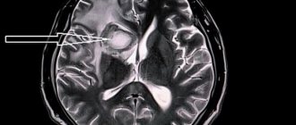
MRI expansion of convexital liquor spaces
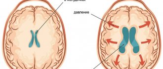
The cerebrospinal fluid spaces are filled with cerebrospinal fluid, which transports nutrients and regulates the level of intracranial pressure.
The distribution of cerebrospinal fluid within the skull is ensured by the subarachnoid space, the third and fourth ventricles.
The movement of cerebrospinal fluid is ensured by cardiac activity, body position, the respiratory cycle, and the movement of epithelial cilia. Normal indicators of intracranial pressure are ensured by the optimal amount of cerebrospinal fluid - 140 ml.
The expansion of convexital spaces is detected by ultrasound and MRI in newborns. Acquired forms of nosology in infants occur after traumatic brain injuries. Doppler ultrasound (USDG) allows for timely diagnosis of pathology before the fontanelles of the skull close. In other cases, magnetic resonance imaging reveals the disease. The procedure is harmless to the baby's health. For high-quality results, a motionless position is required during the scan, so the procedure is performed for the child under anesthesia.
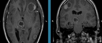
MRI of expanded cerebrospinal fluid spaces
Reasons for the expansion of convexital liquor spaces in children
Etiology of congenital forms:
- Benign and malignant neoplasms;
- Inflammatory processes (meningitis, meningoencephalitis);
- Traumatic brain injuries;
- Cerebral origin when passing through the birth canal;
- Hydrocephalus (dropsy);
- Increased intracranial pressure.
The first signs of pathology can be detected by parents by the protrusion of the skull in the area of the sutures. The second specific manifestation is the divergence of the gap between the hemispheres. The changes lead to limited head elevation. Initially, neuropathologists make a diagnosis of perinatal encephalopathy. Diagnostic methods allow you to more accurately identify cerebral changes.
Secondary symptoms:
- Decrease in reflex activity of the limbs;
- Loss of appetite, weight loss;
- “Moonlight gaze” - lower drooping of the eyelids, visualization of the iris and pupil.
The expansion of the convexital grooves of the brain in the subarachnoid spaces can result in atrophy of the parietal, frontal, temporal, and occipital regions. Against the background of pathology, deformation of the ventricular system occurs.
Atrophic cerebral changes provoke post-ischemic conditions, meningococcal, tuberculous encephalitis, and cancer processes. Expansion of the subarachnoid space is a consequence of the destruction of certain areas of the cerebral parenchyma. A decrease in the amount of white and gray matter leads to the filling of free spaces with cerebrospinal fluid and blood.
Causes and symptoms
Hydrocephalus in adults is an acquired pathology and is divided into forms and degrees. Clinical manifestations depend on the speed of the pathological process.
Root causes of pathology:
- The secretion of cerebrospinal fluid and its absorption by the villi of the arachnoid membrane are impaired. The circulation of secretions is not impaired;
- Violation in the cerebrospinal fluid spaces;
- The disorder is caused by changes in the brain parenchyma (senile age, central nervous system pathologies);
- Hypersecretion of cerebrospinal fluid.
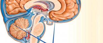
The liquor system in the brain fills all free cavities (ventricles, cisterns, gaps between the membranes). A closed system allows the liquor fluid to circulate, as a continuous cycle of secretion formation and absorption occurs.
Impaired cerebrospinal fluid dynamics may be caused by concomitant diseases.
Secondary causes:
- Malignant or benign neoplasm, cyst;
- Inflammation of the meninges;
- Vascular pathology;
- Traumatic brain injuries;
- Intracranial hemorrhage;
- Intoxication due to poisoning (food, heavy metals, poisons).
Acquired external hydrocephalus is a fairly rare pathology in adults. A small accumulation of cerebrospinal fluid in the subarachnoid space has a blurred clinical picture. The patient experiences only minor headaches and does not seek medical help.
Symptoms increase as the subarachnoid cavity expands (circulation in the ventricles of the brain is not impaired). An increase in intracranial pressure leads to damage or death of nerve cells, which can have serious consequences.
Common clinical manifestations:
- Jet lag;
- Fatigue and drowsiness immediately after rest;
- Continuous headaches;
- Memory disorder;
- Disorientation in space;
- Change in gait (elementary purposeful actions are impossible, movements are uncertain and shaky);
- Development of strabismus;
- Attacks of vomiting, followed by a decrease in the intensity of the headache;
- Meteosensitivity.
The increase in fluid leads to pressure from the membranes on vital centers in the brain. The chronic form is manifested by additional symptoms:
- Disturbance in the respiratory center (from difficulty breathing to complete stop);
- Enuresis (urinary incontinence);
- Convulsive syndrome;
- Paresis or paralysis (lower limbs);
- Development of dementia;
- Heart rhythm failure.
For children, this symptomatology is characteristic of the expansion of the subarachnoid convexital spaces (the area of the central sulcus). This pathology is not typical for adults.
Stages of expansion of subarachnoid spaces
The location of the liquor-containing spaces determines the possibility of free flow from one tank to another. An increase in intracranial pressure is promoted by compression of blood vessels and ventricles of the brain by external formations, accumulation of blood (hematoma), edema, and inflammatory foci.
The main stages of expansion of the subarachnoid spaces:
- Moderate – exceeding the size by up to two millimeters;
- Average - up to four millimeters;
- Heavy – over 4 mm.
In an adult, the expansion of liquor-containing spaces is proportional to the growth of the head and swelling of the fontanelles. Detection of external changes requires timely treatment to prevent irreversible complications. Correct therapy allows you to restore dilated ventricles and liquor spaces to normal.
What it is
The brain contains billions of nerve cells that interact with each other, supporting the functioning of all organs and systems. External protection for the organ is provided by a strong bone frame – the skull. Internal protection is provided by cerebrospinal fluid.
What is cerebrospinal fluid - it is a biological fluid that acts as a shock absorber between the membranes of the brain and spinal cord, protecting the central nervous system from mechanical stress.
Liquor performs many functions:
- Maintains intracranial pressure balance;
- Supports metabolic processes in the central nervous system;
- Maintains oncotic and osmotic pressure at the tissue level;
- Supports cellular immunity;
- Delivers nutrients.
The fluid is formed from glandular cells in the ventricles of the brain and circulates in a closed liquor system, being renewed several times a day (up to 4). The spaces in the central nervous system filled with biological fluid are called liquor spaces.
External expansion of the cerebrospinal fluid spaces of the brain in adults refers to neurological pathology. The accumulation of excess secretion occurs in the ventricles of the organ and in the cavity between the soft membrane and the arachnoid. Obstructed outflow leads to compression of the meninges and increased intracranial pressure.
Clinical symptoms of increased liquor spaces
Signs of dilation of the ventricles and subarachnoid cavities in newborns:
- Constant irritability;
- Negative reaction to strong noises, flashes of light;
- Restless sleep;
- Frequent regurgitation;
- Strabismus;
- Different pupil sizes;
- Moodiness when weather changes;
- Slow overgrowth of the fontanel;
- Twitching of the chin.
Deviations must be detected immediately after their occurrence. Correction of changes should be carried out immediately after detection.
Clinical symptoms of subarachnoid enlargement in adults:
- Dyspeptic disorders (vomiting and nausea);
- Headache;
- Drowsiness;
- Constant dizziness;
- Increased intracranial pressure;
- Dementia;
- Memory problems;
- Disorders of spatial orientation;
- Unsteady gait.
Determining clinical signs in a child and an adult requires contacting a neurologist. The specialist will detect the pathology and give a referral for an MRI of the convexital cerebrospinal fluid cavities.
At the initial stages there are no manifestations of pathology. Symptoms depend mainly on the severity of the deformity and the causes of the pathology. In small children, the disease is provoked by arachnoiditis, birth trauma, and meningitis. Tumors are a common etiological factor in the occurrence of pathology in adults. Compression of the cerebral parenchyma causes defects in the destruction of white and gray matter. The formed cavities are filled with liquor.
Analysis of brain tomograms in people with enlarged cerebrospinal fluid cavities: atrophic changes in the cortex of the frontal, occipital, and temporal regions. The number of convolutions is smoothed out, and blood accumulations are visible.
The presence of concomitant complications in a child is dangerous, so invasive diagnostic measures are postponed to the second year of life. A reliable diagnosis can be made after taking a biopsy of the enlarged cavities. Microscopic assessment of the properties of cerebrospinal fluid makes it possible to differentiate inflammatory, tumor, and ischemic changes.
Magnetic resonance imaging reveals accompanying pathological changes:
- Leukomalacia is a pathology of impulse signal transmission due to softening of the meninges;
- Inflammatory foci;
- Collections of blood.
An MRI picture of a moderate expansion of the external cerebrospinal fluid spaces shows morphological changes, but a whole range of diagnostic methods is required to make a correct diagnosis.
Tomograms of hypertensive hydrocephalus
Treatment
Treatment tactics for neurological disorders caused by expansion of external liquor spaces depend on the form and stage of the disease:
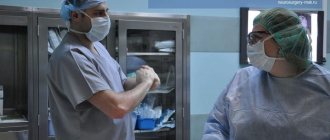
- Acute hydrocephalus of the brain threatens the patient’s life and therefore requires radical measures: neurosurgical surgery. During this procedure, craniotomy is performed, followed by the installation of an external drainage system, which will ensure continuous outflow of excess cerebrospinal fluid. One of the advantages of this method of treatment is the possibility of administering drugs directly to the brain structures, for example, blood thinners and anti-inflammatory drugs. At the moment, this treatment option is considered outdated and is rarely used in practice due to the inconvenience of the drainage system design and the high risk of patient infection.
- Treatment of chronic expansion of external liquor spaces involves performing liquor shunt operations. This type of surgery involves the installation of a complex system of valves and catheters through which excess cerebrospinal fluid is discharged into the natural cavities of the body - the abdominal, pelvic or atrium.
If the patient has been diagnosed with the initial stage of expansion of the external liquor spaces, then conservative treatment is used.
It consists of taking the following medications:
- To reduce intracranial pressure, diuretics (Diacarb, Mannitol) are prescribed - drugs that can remove excess fluid from the cerebrospinal fluid spaces through urine. They are used only when the natural outflow of cerebrospinal fluid is preserved.
- Maintaining the required potassium concentration in the body (Panangin).
- To restore brain activity and improve metabolic processes in brain tissue, nootropic drugs (Cebrolysin, Cavinton, Actovegin) are prescribed.
These measures are aimed at reducing intracranial pressure, normalizing the main functions of the central nervous system, as well as providing brain tissue with the necessary nutrients. If conservative therapy is ineffective and the patient’s condition continues to deteriorate, then surgical treatment is indicated.
Principles for diagnosing convexital dilatation
Let's consider the main diagnostic methods:
- Neurosonography is an ultrasound examination of the brain structure through open slits of the fontanelles. It is carried out for each child after birth;
- Computed tomography (CT) measures the size of the cerebral ventricles, subarachnoid space;
- Comprehensive MRI of the head – monitors soft tissue changes. Layer-by-layer mapping of brain areas followed by three-dimensional remodeling shows the spatial structure of objects;
- Cisternography – determines the direction of movement of the cerebrospinal fluid, the type of dropsy (hydrocephalus).
Additional diagnostic methods are contrast angiography, PET/CT. Positron emission tomography is a radiation examination, therefore it is performed according to strict indications to limit the level of radiation exposure to tissues.