Choroid plexus cyst in fetus and adult: causes, symptoms
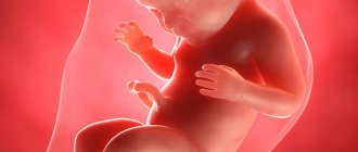
Choroid plexus cyst (CPC) in the fetus and newborn is an ultrasound term formed due to excess accumulations of cerebrospinal fluid along the vessels. In most cases, as the baby grows, the formations go away on their own. If the structures are preserved, they do not pose a danger to the child's health. If the structures are preserved, they do not pose a danger to the child's health. Only in rare cases is surgery required to remove an overgrown cystic cavity.
The optimal period for diagnosing formations is the fetus at 18-22 weeks, when the structure of the brain is formed. Repeated ultrasound at 24-28 weeks notes a decrease in the number of cystic cavities.
Choroid plexus cyst in the fetus - what is it?
CSS is dangerous in children with chromosomal abnormalities such as Edwards disease (trisomy 18). The nosology is characterized by multiple malformations, so intracerebral cavities with cerebrospinal fluid are a minimal problem in treatment.
Down's disease is not characterized by the formation of the cerebral coronary artery system.
Interesting facts about choroid plexus cysts in children:
- Spontaneous disappearance in 90% of fetuses by 28 weeks of development;
- The prevalence rate of nosology among pregnant women is 2%;
- The shape, size, number of cavities varies;
- Found in adults and healthy people;
- The frequency of villous and choroid plexus cysts is about 3%.
It is impossible to determine the initial type of formations in the fetus due to physiological degradation. The absence of pronounced symptoms does not allow doctors to accumulate enough information about the pathology.
Morphological structure of choroid cystic cavities in a child
The accumulation of cerebrospinal fluid inside the choroidal formations does not pose a threat to the health of the baby. Morphologically, the cavity is represented by a thin wall, capable of changing size and shape.
Vascular plexuses are early structures in the formation of the central nervous system of the fetus. Bilateral localization is due to the presence of two hemispheres that require the content of cerebrospinal fluid. Scientists were unable to explain the need for the formation of limited cavities around the plexuses. Presumably, the structures are part of the formation of the central nervous system, and therefore disappear by 28 weeks. If development slows down, CSS can be observed in newborns, infants 3 months and somewhat older.
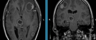
Cystic cavities of the brain are sometimes observed in adults. The pathology does not produce clinical symptoms, so they are detected by chance on an MRI performed due to the verification of another etiology.
Practice shows that there is no health hazard in the persistence of choroid plexus cysts after 30 weeks, with the exception of Edwards disease.
Diagnosis of pathology
Changes are detected by ultrasound or sonography. According to the recommendation of the World Health Society, these studies are performed on all children under one year of age. Neurological disorders are determined using ultrasound.
Sonography is prescribed if there is a history of:
- birth trauma in a baby;
- infectious disease of the mother during pregnancy;
- difficult pregnancy;
- fetal prematurity;
- deviations in size at birth;
- pronounced disturbances in the shape of the head;
- a defect in the structure of the baby’s organs.
There are no signs of a cyst in the infant. This deviation appears and disappears without symptoms. It is not pathological and in most cases does not affect the child’s brain activity.
According to research, such cysts are found not only in children who are developmentally delayed. These formations are equally found in normally developed children.
Causes of CSS
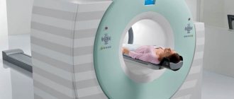
The etiology has not been established. It is considered likely that cystic cavities will persist for a long time against the background of viral infections, herpes, or complicated pregnancy. The absence of clinical manifestations until the end of a person’s life indicates the absence of harm from the pathology. The formations require dynamic monitoring. Blind fontanelles in a child allow an ultrasound of the skull to be performed, and periodic MRI is recommended for adults.
Viral cysts can change: they grow, they degenerate. Dynamic observation determines the prognosis of consequences for a person with CSS.
Two different diagnoses should be distinguished: “fetal choroid plexus cyst” and “cerebral vascular cyst”. The first nosology is harmless, goes away on its own or persists without dynamics for many years. The second type is dangerous, as it sometimes provokes symptoms.
There is a definition of “pseudocyst”. On the screen of an ultrasound monitor, doctors can detect a cavity formed by physiological structures.
The exclusion of pathological conditions after the diagnosis of “choroid plexus cyst” in a newborn and fetus is ensured by an additional comprehensive examination. Genetic counseling rules out Edwards disease. Magnetic resonance imaging shows the structure of the soft tissues of the brain with high resolution, three-dimensional modeling of the area under study.
Detection of a choroid plexus cyst in a child requires verification of provoking factors: viral and bacterial infections, changes in the composition of the cerebrospinal fluid, increased intracranial pressure. If there are no additional changes, it can be argued that there are no conditions for the negative development of a vascular cyst.
Types and times of examinations
To identify dysfunction of brain tissue, all babies undergo neurosonography. Such a study can be performed before the child reaches one year of age. In older children, the large fontanel, as a rule, is already closed, and it is like a window through which you can see the state of the brain. You can also perform an ultrasound when the baby reaches the age of 3, 6 and 12 months. Particular attention should be paid to premature babies, as well as to those born with weight problems. Newborns who were injured during childbirth or suffered from hypoxia in the womb should not be overlooked.
Choroid plexus cysts of a newborn baby
Detection of CSS after birth is not a dangerous condition, but requires examination of the newborn baby. Herpetic infection is activated when the immune system is weakened and can cause changes in the choroid plexus. As the baby grows, the immune system strengthens, so after 3 months, active development of immunity is observed. A decrease in viral activity stabilizes the condition. Cystic cavities can be observed in infants in the absence of clinical pathology. According to the standard scheme, children are examined three times:
- 3 months;
- 6 months;
- 12 months.
The absence of progression indicates the absence of changes that will contribute to the development of dangerous conditions.
Left choroid plexus cyst in a child
The choroid plexus is formed in the fetal body quite early. Education takes part in the formation of cerebrospinal fluid, which provides nutrition to the brain.
The diagnosis of left plexus cyst is common in newborns, although bilateral localization is more common. Pathology develops before the age of 1 year, when intrauterine infections are present. Cystic cavities are equally likely to form in any part of the brain.
Choroid plexus cyst of the right lateral ventricle
The nosology is considered the most benign, since most variants disappear on their own. Found after 22 weeks. An ultrasound scan after 28 weeks shows no pathology. The cystic cavity of the right ventricle can resolve. If there is no associated pathology (with damage to other ventricular spaces), this location of the cyst will not cause complications throughout life, even if the nosology persists in the child after 1 year. In adults, the nosology is rare.
Bilateral choroid plexus cysts
The pathology can persist into adulthood. Some cavities disappear, some remain. Despite the bilateral location, no clinical symptoms occur. Dynamic observation can reveal an increase in size and a change in shape. Only then is conservative treatment carried out. Chromosome mutations are preliminarily excluded. Geneticists can identify a genetic predisposition to Edwards disease. True, pathology can be determined without consulting on the external symptoms of the nosology, characterized by multiple malformations.
Ultrasound examination every 3 months allows you to monitor the condition of the brain parenchyma in infants. The procedure is sufficient to obtain information about the degradation or progression of cystic cavities.
Small cystic cavities in a child's brain
The nature of the CSS has not been reliably established. The pathology is an incidental finding. Small lesions do not affect the human psyche or metabolic reactions. There is no metabolic disorder, so there is no danger.
Small plexus and vascular cysts should be differentiated. The latter option provokes some symptoms. There is no threat to life or health. In adults, minor hemorrhages may occur, soaking into surrounding tissues.
Intrauterine infection provokes inflammatory damage to the walls of the cavity. Detection of infection requires the prescription of antibacterial drugs.
Detection of a plexus cyst at 19 or 20 weeks is not a reason for parents to worry. A repeat examination at week 29 will show no cavities.
Most scientists consider a cystic plexus of intracerebral vessels to be normal. The absence of a clinic suggests that the nosology is a variant of the norm. Improvements in diagnostic methods have made it possible to detect the disease in fetuses and infants more often.
You should pay attention to late cyst formation, which is the result of infection with herpes and a number of other viruses.
Clinical manifestations
Frequent crying and a child’s bad mood is a reason to consult a neurosurgeon
As a rule, if cysts do not disappear on their own, they do not interfere with life and for a long time do not in any way remind of their existence. Brain tissue can be affected in the presence of infectious pathogenesis, trauma, or late appearance of a neoplasm.
The growth of neoplasia provokes the appearance of a certain clinic due to compression of neighboring structures. In addition, increased formation of cerebrospinal fluid is recorded, which causes the appearance of negative manifestations characteristic of hydrocephalus
A typical clinical picture in the presence of large or growing cysts in a newborn:
- attacks of nausea;
- constant regurgitation, vomiting is possible;
- decrease or increase in muscle tone of the arms and legs;
- convulsions;
- coordination disorders;
- strabismus.
The baby becomes lethargic, capricious, sleeps poorly, shows no interest and often cries, which is associated with constant headaches.
Symptoms of choroid plexus cysts
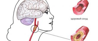
A brain cyst in adults can be a consequence of a hematoma. Limited accumulation of blood (subdural, epidural) with penetration into the ventricles of the brain contributes to ventricular cyst formation.
Pathology contributes to the appearance of clinical symptoms:
- Dizziness, headache;
- Movement coordination disorder;
- Muscle cramps, epilepsy;
- Hypertonicity in newborns.
Localization of manifestations near important nerve centers contributes to the emergence of specific symptoms.
The presence of cytomegalovirus or herpetic infection complicates the course of the disease. Detection of a pathogen and CSS in an adult with a high degree of probability indicates a viral etiology of cystic cavities.
Microcyst of the choroid plexus of the right lateral ventricle rarely causes neurological disorders.
In children, vascular microcysts and larger formations are not accompanied by pathological symptoms. The International Classification of Diseases (ICD 10) does not classify nosology as a number of pathological conditions.
Difference from other types of cysts
This pathology should be distinguished from a cyst that forms in the medulla. The appearance of these formations is often caused by infections suffered by the fetus. To identify a cyst and eliminate it, PCR diagnostics are prescribed. This is done to determine the causative agent of the infection. After the examination, treatment is prescribed, and after its completion, the examination is repeated.
Cystic formations of the brain are divided into subependymal and arachnoid. Formations of the first type appear when there is insufficient nutrition of the brain in the area of the ventricles. This entails tissue death and the appearance of empty cavities.
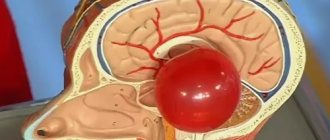
Arachnoid cysts develop inside the meninges. This cavity occurs as a result of infection. This pathology requires medical treatment, and sometimes surgical intervention. If the subcutaneous cyst is not removed, it will lead to disruption of the motor system and damage the visual and other areas of the brain.
Consequences of choroid plexus cysts
Complicated cystic formations cause specific manifestations:
- Hypertonicity of newborns;
- Neurological symptoms due to compression of brain structures;
- Epilepsy (muscle cramps);
- Slight loss of vision and hearing.
Magnetic resonance imaging will need to distinguish a true vascular cyst from a pseudocyst. The latter type is a variant of the physiological development of the brain, but the anatomical structure when examined by ultrasound resembles a cavity. Magnetic resonance imaging is an accurate study (informativeness is about 96%). The three-dimensional modeling mode will allow you to correctly verify the nosology.
Edwards syndrome can be detected by ultrasound by identifying abnormalities of the limbs and changes in internal organs. Additional diagnostic tests:
- Determination of the concentration of human chorionic gonadotropin;
- Blood chemistry;
- Analysis of amniotic fluid.
A special risk group is women aged 32 years and older with hormonal disorders.
Signs of a cyst
Cystic formation can become a provoking factor in disturbances in the functioning of various organs and destruction of their tissues. The volume of the cyst, as well as its predisposition to enlargement, are very important indicators in this case.
A cerebral vascular cyst can grow for several reasons:
- free space with increasing fluid pressure inside the cyst;
- progression of infection or inflammation;
- brain injuries and head contusions with a cyst already present;
- the presence of cysts in brain tissue.
As a child’s body develops, especially during adolescence, cysts that have previously behaved calmly can suddenly increase in size.
Prevention measures
Since the reasons for the formation of CSS are not known for certain, and such a cyst itself is not considered a pathology, there are no specific preventive measures. A woman is recommended to lead a healthy lifestyle, eat properly and nutritiously, walk in the fresh air, do gymnastics for pregnant women and do breathing exercises, and take complex vitamin preparations as prescribed by a doctor.
Particular attention should be paid to protection against all kinds of infections. You should not visit crowded places, especially during the seasonal rise in the incidence of ARVI and influenza. In intimate life, it makes sense to use a condom.