The heart is a four-chamber organ, consisting of two atria and two ventricles. The atria are divided by the interatrial septum into left and right. The left and right ventricles of the heart are separated by the interventricular septum. Blood in the heart normally moves only in one direction, which is ensured by the functioning valvular apparatus of the heart, consisting of four valves: tricuspid, pulmonary, mitral and aortic.
- The tricuspid valve is located between the right atrium and the right ventricle and allows unidirectional blood flow from the right atrium to the right ventricle.
- The pulmonary valve is located at the exit of the right ventricle and allows blood to flow into the pulmonary artery.
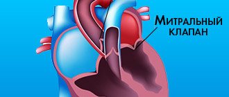
- The mitral valve is located between the left atrium and the left ventricle, through it blood flows from the left atrium to the left ventricle.
- The aortic valve is located at the exit of the left ventricle and allows blood to flow into the aorta and further into the systems and organs of the body.
Causes of heart valve defects
Heart valve defects in adults are diseases that are based on disorders of the valve apparatus (valve leaflets, annulus fibrosus, chordae, papillary muscles), congenital or developed as a result of diseases and injuries, disrupting proper blood flow through the valves and leading to serious complications, such such as severe heart failure, syncope, arrhythmias, sudden cardiac death, etc.
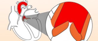
Stenosis is a narrowing of the valve opening for various reasons, as a result of which the heart has to work excessively to overcome this obstacle.
Valve insufficiency is a malfunction of the valve leaflets, leading to their incomplete closure and the return of blood back into the heart chamber, which leads to the heart working with excess blood volume.
Under the lamp light
The biological prosthesis completely replicates the properties of the original valve.
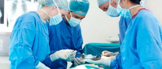
It's stuffy in the operating room. At such moments, I am amazed how heart surgeons, without a hint of fatigue, manage to continuously work their magic on the patient’s main organ. Surgery takes place directly on the open heart, so perfusionists are located next to the main team of doctors. They provide artificial blood circulation and support the vital functions of the body while the patient’s heart is “turned off”. Doctors help doctors see heart defects with special glasses that magnify the image 2-3 times. Here is one of the specialists excising the leaflets of the patient’s own valve and drawing the attention of journalists:
- Just look: the valves here are very damaged - hard, calcified. They are unable to function normally, resulting in insufficiency and critical stenosis of the aortic valve. This is an absolute indication for surgery.
The difficulty is that the patient has a naturally narrow aortic ring. Namely, it serves as the frame for the new valve. To supply blood to a body weighing more than one hundred kilograms, the body needs adequate cardiac output, and for this it needs an adequate size of the prosthesis. Otherwise, the patient will continue to have symptoms of the disease, although to a lesser extent. In such cases, it is possible to use bioprosthetics of the aortic valve leaflets.
Symptoms
Often, heart valve defects do not appear clinically for a long time, and complaints depend on the severity of the disease. When the disease manifests itself, patients experience shortness of breath, dizziness and fainting during exertion, a feeling of heaviness, chest pain, heart rhythm disturbances (arrhythmias), swelling, fatigue, and decreased performance. Often the heart’s ability to compensate for defects lasts for a long time. Therefore, patients seek help at a time when it is already difficult to help them due to the “neglect” of the disease, when the heart has exhausted its compensation capabilities and has turned, figuratively speaking, into an “overinflated balloon.”
What procedures need to be completed before surgery?
Due to the fact that heart surgery is serious and complex, the patient requires a complete preoperative diagnosis.
Mandatory diagnostic procedures prescribed by Israeli cardiac surgeons before heart surgery include:
- urine and blood tests,
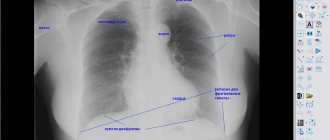
- chest x-ray,
- echocardiography,
- electrocardiography,
- cardiac catheterization
- angiography.
When the surgeon receives the test results, he makes a conclusion whether the operation can be performed. In preparation for surgery, medications and physical therapy
. The doctor will definitely ask you to get rid of bad habits, this will increase the body’s endurance and help the patient endure the operation more easily.
Diagnosis of heart valve defects
Often, patients learn about problems with heart valves after a full, adequate examination by a doctor, who, during auscultation, identified a heart murmur, after which the patient is prescribed a number of tests that have different diagnostic value:
- transesophageal echocardiography (more complex and more expensive study);
- ECG;
- transthoracic echocardiography (ultrasound of the heart) is the most informative, common and inexpensive way to diagnose pathology of the heart valve apparatus;
- aortoventriculography;
- chest x-ray;
- multislice computed tomography with contrast.
At the initial stage of the disease, the patient may be prescribed medications that help the heart compensate for the excessive load due to valve disease. The selection of optimal therapy in this case should be carried out by an experienced cardiologist. However, heart valve disease is a problem that over time may require surgery to repair or replace the damaged valve.
Indications for heart valve replacement surgery:
- progressive heart failure;
- hemodynamically significant valve lesions;
- ineffectiveness of drug therapy;
- risk of severe complications.
Surgical treatment: heart valve replacement
In recent years, significant progress has been made in the surgical treatment of heart valves. Improvements in technology (including artificial blood circulation machines), the development of uniform standards and protocols for both preoperative examination and the course of the operation have made it possible to reduce the risks of perioperative complications, making the operation on the heart valve apparatus safer than refusing surgery and trying to live with valve dysfunction.
There are basically two types of heart valve surgery:
- prosthetics with artificial or biological prosthesis,
- reconstructive interventions.
It is quite natural that a person’s own valve, after successful reconstruction, functions better than an artificial prosthesis.
Reconstruction of the valve apparatus
The purpose of valve repair is to eliminate the cause of its dysfunction. One of the options for plastic surgery of the mitral valve in case of mitral insufficiency is the removal of part of its posterior leaflet with subsequent annuloplasty or only annuloplasty (strengthening the fibrous ring of the valve using special support rings or reducing the diameter of the fibrous ring using special sutures - plastic according to De Vega, according to Batista, according to Alfieri).
In case of prolapse of the anterior leaflet of the mitral valve, it is possible to perform prosthetic replacement of the anterior leaflet chords, which normally ensure tight closure of the mitral valve. In case of aortic insufficiency, it is possible to perform an operation to normalize the closure of the valve leaflets - plastic according to El-Khoury. In case of valvular stenosis and the absence of calcification of the leaflets and fibrosis of the leaflets and subvalvular structures, it is possible to perform an open commissurotomy (separation of the areas of fusion of the valve leaflets).
The development of cardiac surgery over the past decades has significantly expanded the indications for correction of heart defects and improved the survival of patients in the long term after surgery. The number of operations for valvular heart disease performed under artificial circulation (CPB) has increased significantly. Due to a number of objective reasons, the number of repeated interventions is also increasing, which is associated with relapse of the defect after reconstructive plastic surgery, dysfunction of the mechanical prosthesis caused by endocarditis or inadequate anticoagulant therapy, degeneration of biological prostheses, etc. At the same time, repeated operations on heart valves are technically much more difficult and are considered by many authors as a risk factor [1, 3, 8].
The purpose of the study was to analyze the immediate results of repeated surgical interventions in patients who had previously undergone surgery for valve pathology under cardiopulmonary bypass.
Material and methods
For the period from 2003 to 2012, in the department of surgery of heart defects of the Federal State Budgetary Scientific Institution Russian Scientific Center for Surgery named after. acad. B.V. Petrovsky repeated interventions on the heart valve apparatus were performed in 90 (6.8%) patients. In 51 (56.7%) patients, the primary operation was performed in other medical institutions. The age of the patients ranged from 18 to 70 years (mean 54.6±9.5 years). Among the patients there were 33 (36.7%) men and 57 (63.3%) women (Table 1)
.
The study included patients admitted for re-intervention after successful primary correction. Patients who required reintervention as a result of a complication during their hospital stay after the primary operation were excluded from the study. The time between the first operation and re-intervention ranged from 2 months to 36 years, on average 11.6±8.5 years. Three patients in our study underwent heart valve surgery for the third time; the interval between the second and third interventions was 9, 12, and 15 years.
Among the primary interventions, reconstructive plastic surgery on the mitral valve was performed in 34 (37.8%) patients; isolated mitral valve replacement — 32 (35.6%); isolated aortic valve replacement — 20 (22.2%); mitral and aortic heart valve replacement was performed in 4 (4.4%). In 50 (89.2%) patients, mechanical prosthetic heart valves were used during the primary operation; biological prostheses were implanted in 6 (10.8%).
Indications for reoperation are presented in table. 2
.
Recurrence of heart disease in most cases (43 patients, 47.8%) was caused by the progression of the rheumatic process with fibrous deformation and calcification of the valve leaflets, subvalvular structures and fibrous ring. Over the past years after surgery, 10 patients developed a defect of rheumatic etiology on the unoperated valve. Among the causes of dysfunction of mechanical prosthetic valves, the following were identified: thrombosis (see figure on the color insert)
- 16 (17.8%) cases, pannus - 14 (15.6%), paraprosthetic fistulas - 13 (14.4%).
Figure 1. Mechanical heart valve obstruction by thrombus. In 10 patients, the formation of paraprosthetic fistulas was associated with prosthetic infective endocarditis; in 3, no signs of endocarditis were detected.
Resternotomy was performed using a standard technique with removal of wire ligatures and the use of an oscillating saw. This stage of the operation was greatly facilitated if the entire length of the pericardium was sutured during the primary operation. An adhesive process in the pericardium of varying severity was observed in all patients. Cardiolysis was carried out by blunt and acute methods. First of all, the structures necessary to connect the IR apparatus were identified. The IR apparatus was connected according to the aorta-vena cava scheme. In case of hemodynamic disturbances, cardiolysis was continued on parallel cardiopulmonary bypass and was carried out only to the extent necessary for the main stage of the operation, which made it possible to minimize blood loss at this stage of the operation.
The main stage was carried out under conditions of hypothermic perfusion, with cooling to 28-30 °C, and pharmacocold cardioplegia with a solution of Consol or Custodiol. The cardioplegic solution was delivered through the roller pump of the CPB device antegrade to the aortic root, selectively to the ostia of the coronary arteries, or retrograde through the coronary sinus. Cell Saver autologous blood return systems were used to reduce blood loss. To access the mitral valve, a left atriotomy was used (14 cases) along the old suture parallel to the interatrial groove, and for large sizes of the right atrium, preference was given to access through the right atrium and the interatrial septum (47).
And only in 5 cases was the Dubost approach performed, which has maximum visibility of the mitral valve in conditions of incomplete cardiolysis, but at the same time is the most traumatic. To access the aortic valve, an aortotomy was performed in a typical location.
During re-intervention, valve replacement was used in all cases after reconstructive plastic surgery. Isolated re-replacement of the mitral or aortic heart valve was performed in 36 (40%) patients, suturing of a paraprosthetic fistula with De Vega tricuspid valve repair was performed in 2 (2.2%). Repeated interventions on the mitral and aortic valves were performed in 14 (15.6%) patients, of which 6 (6.7%) underwent mitral and aortic valve replacement after primary repair. Replacement of the mitral and tricuspid heart valves was performed in 1 (1.1%) patient, replacement of the mitral and aortic valves - 2 (2.2%), replacement of the mitral valve and replacement of the aortic valve - 3 (3.3%), replacement of the aortic valve and mitral valve replacement — 2 (2.2%) patients. In addition, 3 (3.3%) patients additionally underwent coronary artery bypass surgery, 31 (34.4%) underwent tricuspid valve plastic surgery according to De Vega.
When replacing heart valves, bioprostheses were used in 11 (12.2%) patients, mechanical prostheses were implanted in 79 (85.9%).
CPB time averaged 113.6±34.7 minutes (from 50 to 212 minutes), aortic cross-clamp time was 80.4±29.3 minutes (32-172 minutes). Intraoperative blood loss averaged 1024.5±110.2 ml (400-2000 ml). The use of the Cell Saver system for the return of autologous blood, as well as the preoperative procurement of autoplasma, made it possible to avoid the use of donor blood components in more than half of the cases.
results
The average time in hospital was 15.5±9.4 days (8-45 days), the average length of stay in the intensive care unit was 2.5±1.4 days (from 1 to 16 days).
Mortality rate was 2.2% (2 patients). The causes of death were, in the first case, massive blood loss due to spontaneous rupture of the left ventricular myocardium on the 1st day after replacement of the mitral and aortic heart valves; in the second case, pulmonary embolism and arrhythmogenic cardiac arrest on the 1st day after replacement of the aortic and mitral heart valves .
Among non-lethal complications in the immediate postoperative period (Table 3)
Heart rhythm disturbances were the most common - in 35 (38.9%) patients.
In 6 (6.5%) patients, due to persistent junctional rhythm, a temporary pacemaker was installed for more than 10 days.
Atrial flutter developed in 9 (10%) cases, and cardioversion was successfully performed in 8 of them. In 1 patient, atrial flutter was refractory to electrical pulse therapy, and radiofrequency ablation was subsequently performed. Installation of a permanent pacemaker was performed in 5 patients; the indications were complete transverse block, nodal bradycardia, sinus node dysfunction and atrial fibrillation with pauses of more than 2.5 s. The second place in terms of development frequency was taken by heart failure, which was observed in 30 (33.3%) patients. In 10 (11.1%) patients, severe heart failure, which required cardiotonic support for more than 2 days, was due to the initial severe condition of the patients, the volume or the combined nature of the reintervention.
Respiratory failure was observed in 10 (11.1%) patients. Complications from the central nervous system in the immediate postoperative period were noted in 4 (4.4%) patients: encephalopathy was diagnosed in 3, acute cerebrovascular accident in the left middle cerebral artery system on the 2nd day after mitral valve replacement - in 1 (1 ,1%). Transient liver failure occurred in 2 (2.2%) patients, and renal failure occurred in 3 (3.3%) patients; in one observation, hemodialysis sessions were required. Infectious complications occurred in 2 patients: in one case, mediastinitis was diagnosed, in the second - suppuration of the soft tissues of the postoperative wound. Postoperative bleeding requiring resternotomy developed in 1 (1.1%) patient.
Discussion
The number of reoperations on heart valves increases every year. Repeat mitral valve interventions account for approximately 10% of all mitral valve surgeries in the United States [9]. In our center in 2012, repeated operations on valves accounted for 6.8% of all operations on the valve apparatus.
Revision cardiac surgery remains a challenge for cardiovascular surgeons. There are many risks associated with resternotomy beyond the surgery itself. Many surgeons prefer to perform thoracotomy to reduce the incidence of complications and reduce the length of stay of the patient in the intensive care unit and the overall length of hospitalization, especially in patients who have previously undergone open heart surgery [9].
Another controversial approach is reoperation through a minimally invasive approach. According to some authors [8], it allows one to avoid massive bleeding and injury to large vessels. The minimally invasive approach is mainly used for isolated re-operations on the mitral valve and after previous coronary artery bypass surgery. The goal of a minimally invasive approach is to reduce the risk of major bleeding during surgical access. A. Vleissis et al. [11] preferred to use a minimally invasive approach for repeated interventions on the mitral valve. According to them, in a group of 22 repeated operations on the mitral valve through a minimally invasive approach, there were no deaths or cases of postoperative wound infection.
F. Casselman et al. [1] published the results of a study of 80 patients who underwent endoscopic revision mitral valve surgery through a mini-thoracotomy approach without rib spread. The mortality rate in their series was 3.8%.
According to a study by R. Umakanthan et al. [10], including 90 patients, minimally invasive right thoracotomy without aortic cross-clamping may be a good alternative to traditional resternotomy when performing repeated interventions on the mitral valve. In their work, the authors drew conclusions about the safety and effectiveness of minimally invasive access, reducing surgical mortality in high-risk patients.
M. Ghoreishi et al. [5] prefer the resternotomy approach for repeated mitral valve interventions, but perform preoperative computed tomography of the chest to determine the risk of injury to large vessels during resternotomy. In a series of reoperations on the mitral valve, they divided patients into two groups based on the results of computed tomography: low risk (large vessels were located at a distance of more than 1 cm from the posterior surface of the sternum) and high risk (large vessels were located at a distance of less than 1 cm from the posterior surface of the sternum ). According to the authors, injury to large vessels during sternotomy has a significant negative impact on the results of surgical intervention. Intraoperative mortality was significantly higher when large vessels were injured. Patients in the high-risk group underwent cannulation of peripheral vessels before resternotomy to allow emergency transfer to CPB.
In the present study, we used only the transsternal approach. Most patients had concomitant heart valve pathology: in addition to aortic or mitral valve dysfunction, there was tricuspid valve insufficiency or a combination of aortic and mitral valve dysfunction. The minimally invasive approach for recurrent valve surgery has limitations for the simultaneous performance of concomitant cardiac interventions. When using a minimally invasive approach, it becomes difficult to perform interventions on two or three valves simultaneously, or “maze” surgery in patients with atrial fibrillation. To perform such operations, in our opinion, it is more justified to perform a standard resternotomy, which provides access to all structures of the heart to the greatest extent. To reduce the risk of injury to the heart and large vessels during resternotomy, we routinely suture the entire pericardium during the initial operation.
According to P. Ellman et al. [2], installation of an IR before resternotomy did not reduce the risk of intraoperative injury to large vessels. In the study of repeated heart valve surgeries, we did not perform peripheral vascular cannulation before resternotomy, and there were no cases of major bleeding during resternotomy.
According to the results of M. Murzi et al. [8], the volume of blood transfusion during repeated operations on heart valves is higher when using a minimally invasive approach compared to standard resternotomy (4.1 units versus 2.7 units). Surgical mortality with minimally invasive access was 5.7%, with resternotomy - 5.9%. The rate of conversion to standard resternotomy during minimally invasive valve reintervention was 1.7%. In the present study, when traditional resternotomy was used exclusively, the mortality rate was 2.2%.
In a 10-year single-center study, H. Vohra et al. [13] identified risk factors for mortality and postoperative complications, the most significant of which were: left ventricular ejection fraction less than 50%, the need for multivalve correction, and the urgency of the intervention.
In a study by P. Vogt et al. [12], which focused on reoperations for degeneration of biological aortic valve prostheses, risk factors for emergency reoperation were: active infective endocarditis before the primary operation, postoperative pneumonia after the primary operation, long-term dysfunction of the biological prosthesis, acute regurgitation on the bioprosthesis, and pulmonary hypertension. The independent risk factors for mortality during biological valve replacement surgery are: emergency surgery, high transvalvular gradient during the primary operation, and two- or three-vessel coronary artery disease. According to our data, repeat operations in the first 3 years after the primary operation were performed in 18 patients; they were associated with endocarditis before the primary operation, prosthetic valve thrombosis, paraprosthetic fistula and a high transvalvular gradient before the primary operation. Overall mortality within 30 days after aortic valve replacement was 5.2% (9 out of 172 patients). Moreover, the mortality rate in the group of planned reprosthetics was only 1.4% (2 out of 141), while emergency reprosthetics was accompanied by a mortality rate of 22.6% (7 out of 31) [12]. In our study, there were no deaths during emergency valve reinterventions. Both deceased patients underwent two-valve replacement.
According to research [5], simultaneous operations on two valves have a higher mortality rate. The main cause of early mortality in simultaneous mitral and aortic valve replacement was severe heart failure with low ejection fraction [7]. According to our data, the first cause of death was spontaneous rupture of the left ventricular myocardium of the 2nd type, while cardiolysis of the left parts of the heart was not performed, and there were no prerequisites for myocardial injury in the rupture zone. The cause of the second death was pulmonary embolism.
Paraprosthetic fistula was the reason for reoperation in 13 (10.9%) patients. In 10 of them, a paraprosthetic fistula occurred after mitral valve replacement and in 3 after aortic valve replacement. In 2 patients, after mitral valve replacement, it was possible to perform suturing of the paraprosthetic fistula, although the size of the paraprosthetic fistulas was more than 1 cm.
N. Fukunaga et al. [4] conducted a retrospective analysis of 118 reoperations of mitral valve replacement performed over 20 years. Worsened survival after repeat mitral valve surgery was observed in patients with tricuspid regurgitation grade 2+ or greater. Based on the results of the study, it was concluded that it is necessary to maintain the level of tricuspid regurgitation in the postoperative period no higher than 2+ to improve long-term survival. In our series of 90 recurrent heart valve surgeries, we performed De Vega tricuspid valve annuloplasty at the time of reoperation in 31 patients. Tricuspid valve annuloplasty was performed in patients with grade 2 or higher tricuspid regurgitation at the time of reoperation.
Conclusion
Repeated heart surgeries are one of the most difficult categories of surgical interventions, accompanied by high mortality and a large number of complications. In our group of patients, we were able to obtain good results in terms of mortality and the number of postoperative complications. We attribute these successes primarily to the coordinated work of all services providing surgical intervention and postoperative care. From a surgical point of view, the choice of access technique for visualizing cardiac structures is individual, however, we most often give preference to standard resternotomy, which has a number of undeniable advantages: it provides the most complete overview of all cardiac structures, allows for concomitant interventions on the heart and has a high level of safety with the correct technique its implementation.
In the 3-year period after the primary operation, the most common reasons for reoperation were: infective endocarditis, thrombosis of a mechanical prosthesis, and paraprosthetic fistula.
Types of valves used in prosthetics
Biological valves
They can be made from animal or human tissues (heterografts, homografts, autografts). Biological valves may contain some artificial components to provide valve support and placement. The main advantage of such a valve is that there is no need for lifelong anticoagulant therapy (constant strict use of drugs that significantly thin the blood and require constant blood tests), and the main disadvantage is its limited service life (10-15 years).
Mechanical valves
They consist entirely of mechanical elements (titanium and pyrolytic carbon) and are designed to replace the patient's own valve functions. The mechanical valve is very reliable and durable, designed for many years of full-fledged operation, which is the main advantage, but requires the patient to constantly take anticoagulants.
The Center for Cardiac Surgery and Cardiology provides assistance to patients with valvular pathology of varying complexity, including one-, two- and three-valve replacement with biological and mechanical prostheses of Russian and foreign production, elimination of valvular insufficiency using a variety of plastics (plasty on a support ring, tri- and quadriangular resection leaflets with annuloplasty according to De Vega, Batista) of the valve apparatus, restoration of the chordal apparatus by prosthetics of the mitral valve chords, etc. The main direction in surgery of valve pathology is aimed at increasing the number of reconstructive operations on the valve apparatus and reducing valve replacement.
The Clinic offers surgical treatment of heart valves in combination with other diseases, such as coronary heart disease, atrial fibrillation (atrial fibrillation).
Indications for replacement
A number of diseases of the cardiovascular system are associated with damage to the heart valves. Israeli experts recommend replacement in the following cases:
- the leaflets of the tricuspid or aortic valve are not able to provide the necessary tightness (valvular insufficiency);
- the tricuspid and mitral valves cannot open normally (stenosis); surgical intervention is also required for stenosis of the aortic mouth;
- heart valves are damaged as a result of rheumatism or atherosclerosis;
- valve pathologies are caused by infective endocarditis.
Such deviations, as a rule, can only be corrected surgically.
Side effects
After surgery, in addition to general weakness of the body, you may experience a deterioration in appetite or sleep disturbance. Visual disturbances, loss of concentration on an object, swelling of the lower extremities and some others may occur. All these sensations are exclusively temporary and will pass within one to two months. Having repaired your heart, you will not be able to harm other organs in your body, since the medicine at Ilyssa Medical Group is at a high level. Heart valve replacement in Israel will allow you to operate your body as if you were 16 again, without worrying that the operation will cause complications inside or outside the body.
Be sure to tell your attending physician about any of your feelings and problems that arise when you meet. Remember also that after a month you will need to undergo a routine examination, re-take tests and undergo research procedures: chest X-ray, ECG and EchoCG.
For more information, contact the specialists of the Ilyssa Medical Group medical center.
How is heart valve replacement surgery performed in Israel?
This is a long procedure that lasts three hours or longer. First, the patient is given anesthesia, and then the surgeon will make an incision in the middle of the sternum. After this, a heart-lung machine is connected, without which blood circulation is impossible (the operation is performed on a static heart).
minimally invasive techniques are actively used in Israel.
heart operations. During this procedure, a special catheter equipped with a video camera is inserted into a small incision. In this type of surgery, there is no need to install a heart-lung machine. Sometimes the catheter is inserted through the femoral artery, and in this case the procedure does not even require anesthesia, only local anesthesia is sufficient. A tiny conductor will carry the valve to the heart through the blood vessels.
When the operation is completed, the patient will be taken to a special ward for observation. After about five to six days, he is transferred to a regular ward; if the patient requires additional therapy, his stay will last up to 10 days. Specialists will monitor the patient’s condition for several more weeks. During this period, it is necessary to follow the doctor’s instructions and the regimen. For a speedy recovery after surgery, it is recommended to perform breathing exercises and physical therapy.
If your health worsens and pain occurs after heart valve replacement in Israel, you should immediately contact your doctor.
You need to pay special attention to your vital activity after surgery. You will be forced to forget about coffee and cigarettes and other bad habits. You need to develop a daily routine for yourself that will allow you to get enough sleep and eat well every day. Switch to a healthy diet: limit fatty and salty foods, add more fruits and vegetables to your diet. It is worth walking in the fresh air as often as possible and protecting yourself from stressful situations or factors that make you nervous or worried. It is important to avoid physical overload and regularly engage in physical therapy.