- home
- Articles about cholelithiasis
- This is useful to know
- About the structure of the human body
- Catalog of useful medical articles
›
›
›
›
There are two types of valves in the human heart.
These are leaflet and semilunar valves. In this article we will understand which valves are called leaflet and why, and which are semilunar. In addition, we will take a close look at the structure and operation of the semilunar valves. Sections of the article
Leaf and semilunar valves
Structure and operation of semilunar valves
Left semilunar valve
Right semilunar valve
The problem of the semilunar valves
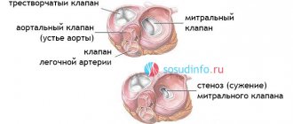
Leaf and semilunar valves
The leaflet valves are located around the right and left atrioventricular orifices. Those openings that connect the right atrium with the right ventricle, and the left atrium with the left ventricle.
Why casement?
Because each of these valves is a set of flat leaflets. The right atrioventricular has three valves, and the left has two.
Their valves are some kind of flat triangular petals. With their wide ends, these petals are tightly connected to a dense tendon ring surrounding the hole. And their narrow and movable ends hang freely into the cavity of the ventricle (this is in the opening position).
At the moment of closing, it is the flaps that close the opening, tightly adjacent to each other. You can read about each of the leaf valves in the articles:
- "Mitral valve"
- "Tricuspid valve"
The semilunar valves are located in the openings that connect the ventricles of the heart to the large arteries leaving the heart.
These are the aorta and pulmonary artery. The aorta comes from the left ventricle, and the pulmonary artery comes from the right.
Each hole is equipped with a valve consisting of three semicircular pockets.
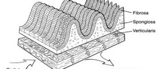
Semilunar valves, like leaflet valves, are formed by folds of the inner layer of the heart wall - the endocardium. But the structure of these “folds” is different.
Structure and operation of semilunar valves
As I already said, semilunar valves are not flat petals, but three small semicircular pockets.
When the blood flow rushes from the ventricle into the artery (aorta or pulmonary artery), the empty and therefore flat “pockets” “fall” into the cavity of the vessel and allow the flow to pass in the desired direction: from the ventricle to the artery.
All this happens at the moment when the ventricle contracts, that is, at the moment of ventricular systole.
When the ventricle relaxes or its diastole occurs, blood from the artery tries to get back into the ventricular cavity. But, as soon as the blood flow reverses, the beautiful semilunar pockets fill with blood. They become large and voluminous, and therefore cover the hole. Thus, the path of blood in the opposite direction is closed.
Both the right and left semilunar valves have three “pockets”.
Human circulatory system
The structure of the human heart
What does a human heart look like?
Where is the human heart?
What's Below the Heart?
Heart cavity
How many valves are there in the heart?
Valve apparatus of the heart
Structure of the heart wall
Wall of the heart: pericardial sac
Heart wall, myocardium
Structure of the heart
Heart chambers and valves
The heart has four chambers. Blood moves through contraction (which pushes blood out of the chambers) and relaxation of the heart (which forces blood into the chambers). The top two chambers are called the atria . Blood enters them through the veins. The atria contract with little force and force blood into the ventricles. The ventricles are the lower muscular chambers of the heart.
The ventricles contract with greater force and send blood into the body through the arteries. The right ventricle pumps blood to the lungs. Of all the chambers of the heart, the left ventricle works the most. Powerful contractions of the left ventricle send oxygenated blood to all organs of the body. The heart also has four valves . They ensure blood flow in one direction. The heart valves open at the right time and allow blood to move forward, and then close and prevent the blood from flowing back. The mitral and tricuspid heart valves control blood flow from the atria to the ventricles. The aortic and pulmonary valves control blood flow from the ventricles. The structure of the chambers and valves of the heart, see Fig. 1.
Rice. 1. Structure of the heart (front view)
Blood circulation throughout the body
Blood brings oxygen and nutrients to tissues and organs and carries away waste products. There are two circles of blood circulation.
The first circle of blood circulation goes from the heart to the lungs, where the blood mixes with oxygen and releases carbon dioxide. The second circle of blood circulation covers the entire body, and the blood gives off oxygen and nutrients and takes away carbon dioxide and waste products produced by the body. Blood returns to the heart through the veins, and enters the lungs and other organs of the body through the arteries.
Heart muscle, cardiac cycle and blood pressure
The heart muscle (myocardium) produces the force necessary to push blood through the ventricles. When the ventricles contract, blood is pushed out of the heart through the valves into the aorta (from the left ventricle) or the pulmonary artery (from the right ventricle). This phase of the cardiac cycle, when the ventricles contract and push blood out of the heart, is called " systole ".
After contracting, the ventricles must relax and receive the next portion of blood to pump. After the ventricles relax, blood enters them from the atria. This phase of heart activity is called " diastole ".
| Rice. 2. During systole, the heart contracts and pushes blood out of the ventricles | Rice. 3. During diastole, the heart relaxes and the ventricles fill with blood coming from the atria |
The alternation of contraction (systole) and relaxation (diastole) is called the cardiac cycle. You may have heard these terms when you had your blood pressure taken at an artery in your upper arm. Blood pressure is made up of two numbers that correspond to systole and diastole of your cardiac cycle. For example, your blood pressure might be 120/80. 120 is the systolic pressure, that is, the pressure created by contraction in the left ventricle. 80 is diastolic pressure, which is the blood pressure in the body when the left ventricle is relaxed and receiving blood.
Cardiac arteries
Like all other organs and muscles in the body, the heart has arteries that supply it with nutrients and oxygen. The heart arteries are called coronary arteries . There are three of them: the right coronary artery, the anterior descending branch of the left coronary artery and the circumflex branch of the left coronary artery. When these arteries become narrowed due to the formation of cholesterol plaques, it is called coronary artery disease. If any coronary artery is completely blocked, part of the heart muscle is deprived of blood supply and a heart attack occurs. During a heart attack, the heart muscle is damaged because some muscle cells die.
Normal, healthy heart valves
The valves of the human heart are amazing anatomical structures. These tissue-paper-thin flaps attached to the wall of the heart open and close with every heartbeat, day after day, year after year. With every blow, the valves demonstrate their extraordinary strength and flexibility.
There are four heart valves. The two valves are called atrioventricular (i.e. atrioventricular). They control blood flow from the atria to the ventricles. The atrioventricular valve on the right side of the heart is called the tricuspid valve , located between the right atrium and the right ventricle. It is called “tricuspid” because it has three doors. The atrioventricular valve on the left side of the heart is called the mitral valve ; The mitral valve controls blood flow between the left atrium and the left ventricle. It has two doors. The remaining two heart valves - the aortic and pulmonary - have three leaflets. These are exhaust valves that regulate blood leaving the ventricles and heart. The aortic valve acts as a “door” between the heart and the rest of the body. Every drop of blood leaving the left ventricle and ascending aorta must pass through the aortic valve. The aortic valve is located between the left ventricle and the ascending aorta, i.e. the upper part of the aorta. The pulmonary valve is located between the right ventricle and the pulmonary artery. The mitral and tricuspid valves are significantly larger than the aortic and pulmonary valves.
For the shape and location of the four heart valves, see Fig. 4 below. The characteristic sound of the heartbeat ("knock-knock") is caused by the closing of the heart valves; The first “knock” corresponds to the closing of the mitral and tricuspid valves, and the second “knock” corresponds to the closing of the aortic and pulmonary valves.
Rice. 4. Heart valves (top view)
Function of heart valves
A normal, healthy valve does not obstruct blood flow and allows blood to flow freely in only one direction. It closes completely and quickly and does not allow much blood to flow back through the heart valve (reverse flow of blood through the heart valve is called " regurgitation "). Although mild regurgitation, or backflow, may be present and well tolerated, severe regurgitation is always pathological.
When the heart valve opens completely and evenly, blood flows through it evenly. When a valve does not open completely or evenly, blood flow through it becomes chaotic and turbulent. When a valve is narrowed or does not open fully, it is said to be " stenotic ".
Both regurgitation (backflow) and stenosis (narrowing) increase the workload of the heart.
Rice. 5
Return to top
see also
| Heart valve diseases and their diagnosis | Congenital heart defects and their treatment |
Additional materials
| The structure of the human heart |
Left semilunar valve
Its second name is the aortic valve or aortic valve, since it opens the path of blood from the heart to the aorta.
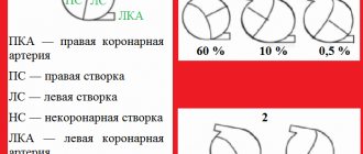
Each of the valve flaps gets its own name:
- right coronary
- left coronary
- non-coronary
At the end of each valve there are small seals - Arrentius nodes. These formations ensure a tighter closure of the valves.
The flaps of this valve are very tight, which is not surprising. After all, this valve is located on the border between the most powerful chamber of the heart and the largest artery in the human body. It opens the way for blood into a large, branched circle of blood circulation. Therefore, it must and can withstand a very heavy load.
Proper nutrition for diseases of the atrioventricular canal
A healthy diet and active lifestyle are necessary for every person, and especially for those who suffer from valve problems.
Patients with atrioventricular canal defects are offered a special diet. For obese people, one of the important points of the program is the recommendation to reduce weight and thus reduce the load on the myocardium and blood vessels.
To preserve the valves, experts recommend using table No. 10 in your diet.
Main nuances of diet No. 10:
- Confectionery sweets and baked goods are prohibited;
- animal fats are excluded;
- limited energy drinks: sugar, black tea, salt, coffee, nicotine;
- increasing the amount of protein products in the menu: lean meat and fish;
- adding dishes containing fresh herbs and vegetables to the daily diet;
- There should be dried fruits and raw fruits and berries on the table every day.
Soy products and fast food are excluded from the menu of patients of all ages. Alcohol in any quantity is completely prohibited.
There are some features of cooking for table No. 10.
Soups with concentrated broths are prohibited; first courses are prepared with vegetable and mushroom broths. To prepare the second, do not use garlic, sorrel, spinach, radishes and radishes, legumes, peas, and sauerkraut.
Not all fruits and berries are allowed; fruits with coarse fiber are excluded: pears, apples, blueberries, bananas, citrus fruits.
When switching to a diet, it is important to remember that it is medical, developed by specialists to restore the functionality of the heart, therefore, using the principle of therapeutic nutrition, you should not dilute it from time to time with fast food dishes, in which case it loses its meaning and there will be no effect.
You should not overeat, especially at night, and break prohibitions. How to choose the right products to restore the function of valves and the heart as a whole.
Right semilunar valve
Its second name is pulmonary valve. It regulates the flow of blood from the right ventricle to the pulmonary artery.
Its structure is no different from the structure of the aortic valve: it is also equipped with three beautiful “pockets”. But they got different names:
- front door
- back
- right
This valve is less dense than the aortic valve, which can be explained simply: the load on it is much less. The right ventricle is not as powerful as the left; the pulmonary circulation is called small because it is much shorter and less branched.