Heart valves are the basis of the internal frame of the heart, which are folds of connective tissue. Their functions boil down to delineating the volumes of blood in the atria and ventricles, allowing these chambers to alternately relax after pushing out blood at the time of contraction.
If the valve for some reason cannot perform its function, intracardiac hemodynamics, or internal blood flow, is disrupted
. Because of this, the heart muscle gradually wears out and heart failure develops. In addition, blood can no longer circulate normally throughout the body, since the pumping function of the heart is impaired, and blood stagnation occurs in the internal organs - kidneys, liver, brain. Sooner or later, if left untreated, congestion leads to dystrophy of all human organs, and, as a consequence, to death. Therefore, valve pathology is a fairly serious problem, in some cases requiring cardiac surgery.
There are two types of valve surgery – valve surgery and valve replacement. In the first case, the valve is reconstructed on a support ring and is used for heart valve insufficiency. The second type of operation involves complete replacement of the valve with a prosthesis. Heart valve replacement will be discussed in more detail below. Most often, the mitral and aortic heart valves are replaced.
Indications for surgery
{banner_banstat0}
The main indication for valve replacement in the heart is its gross organic damage with the formation of heart disease,
having a significant effect on hemodynamics. Valve defects can develop as a result of rheumatic fever (rheumatism) - a form of streptococcal infection characterized by damage to the joints and heart (usually occurs as a result of frequent tonsillitis, chronic tonsillitis).
The need for valve replacement is taken into account based on the stage of heart failure, as well as according to data obtained from ultrasound of the heart (echocardioscopy).
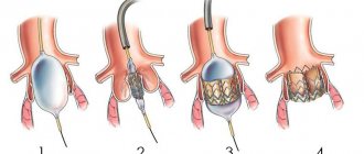
valvular structure of the heart and an example of mitral valve stenosis requiring replacement
So, clinical indications for surgery:
- Fainting, chest pain, shortness of breath in patients with aortic valve stenosis,
- Clinical manifestations of aortic stenosis in patients who have undergone coronary artery bypass grafting,
- Severe stages of chronic heart failure - severe shortness of breath at the slightest household activity and/or at rest, significant swelling of the limbs, face, whole body (anasarca) in patients with moderate or severe mitral valve stenosis,
- Initial signs of heart failure (shortness of breath during significant physical exertion, heart rhythm disturbances) in patients with mild mitral valve stenosis,
endocarditis is one of the causes of valve damage
Infectious, or bacterial endocarditis - vegetation of bacterial inflammation on the inner lining of the heart, including the valves.
Echocardioscopy data:
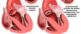
- Severe (critical) aortic stenosis, even in the absence of clinical manifestations - the area of the aortic valve opening is less than 1 cm2,
- Reduced ejection fraction (the volume of blood thrown into the aorta with each contraction of the left ventricle) less than 50%,
- The area of the mitral annulus is less than 1.5 cm2,
- Ejection fraction with mitral stenosis is less than 60%.
Structure
The tricuspid valve, according to the scientific literature, is also called tricuspid. When the heart moves, all processes occur synchronously. If a blockage occurs in one of the departments, a minor temporary one, the body immediately senses that the condition changes significantly. The state of health immediately worsens, it becomes difficult to breathe and it is impossible to move.
The tricuspid valve is located on the left side of the sternum. It is part of the pumping system for pumping blood. It is a kind of cap that opens under the pressure of ejected blood. Closing occurs due to the reverse forces of the liquid automatically due to pressure on the surface of the valves.
Incomplete closure of the tricuspid valve occurs due to relaxation of the heart muscle, when it is no longer able to function normally. Due to a violation of the redistribution of internal pressures, tissues begin to break down, which as a result threatens the formation of heart disease. Some diseases become provocateurs of such pathology.
Contraindications for surgery
{banner_banstat1}
Heart valve replacement surgery is contraindicated for the following diseases and conditions:
- Acute myocardial infarction,
- Acute cerebrovascular accidents (stroke),
- Acute infectious diseases, fever,
- Exacerbations and worsening of chronic diseases (diabetes mellitus, bronchial asthma),
- Extremely severe heart failure with an ejection fraction of less than 20% with mitral stenosis, in which case the attending physician should decide whether a heart transplant is necessary.
Stages of dysfunction
The tricuspid heart valve is located on the right side of the organ. Pathological changes develop gradually. In medicine, disorders of the functioning of the tricuspid valve are distinguished, taking into account the complexity of pathological changes and the possibility of their reversal.
There are the following stages of development of organ damage:
| Name | Description |
| Stage I | At this stage of pathological processes, minor disturbances occur that affect the movement of the pulmonary and systemic circulation. |
| Stage II | Pathological processes progress, but if treatment is started in a timely manner, degenerative changes can be reversed. |
| Stage III | Significant problems in the functioning of the tricuspid valve provoke the appearance of specific symptoms, for example, increased blood pressure. |
| IV stage | At the last stage of the disease, the valve structure is affected. The person’s condition is deteriorating, progressive symptoms require urgent hospitalization and strict supervision by the attending physician. |
At the last stage of valve damage, pathological processes are irreversible. Properly selected therapy will improve the patient’s quality of life and prevent complications.
Prosthetic heart valves – what are they?
{banner_banstat2}
Since the 1970s, the configuration of prosthetic valves has undergone some changes. Valves based on ball prostheses are considered one of the most outdated.
Later, valves based on hinged disc prostheses began to be used.
The most modern valves are those based on bicuspid hinged prostheses, which are currently used.
In addition, in patients with an increased risk of thrombosis, models obtained from the pig heart are used - biological prostheses, or xenografts.
The disadvantage of mechanical prostheses is the high rate of formation of blood clots on the valve leaflets
, which is associated with a high risk of pulmonary embolism, ischemic stroke, thrombosis of the femoral arteries with possible amputation of a limb, etc. In this regard,
in elderly people (over 65 years old), it is preferable to undergo valve replacement surgery with a biological prosthesis.
It is also possible to have an operation with prosthetic replacement of the aortic valve with the patient’s own pulmonary valve with simultaneous replacement of the latter with a biological prosthesis.
The disadvantage of biological prostheses is the high risk of re-development of bacterial inflammation on the installed porcine valve.
The service life of the valves in the absence of complications is from 10 to 15 years; if the valve wears out, it is possible to perform a second operation to replace it.
Preparing for surgery
{banner_banstat3}
After a diagnosis of heart disease or infective endocarditis has been made, the decision about the need to replace the affected valve should be made as soon as possible. After this, the patient undergoes the required minimum of clinical studies and is referred by the attending physician to the cardiac surgery center. Typically, surgery can be performed within a few months of diagnosis. If a patient submits an application to the regional health department for a quota (budgetary allocations from the federal budget to provide high-tech assistance to the population), then a response to the quota can be received within 20 days.
For admission to the cardiac surgery department, the following documents and examinations are required:
- Passport, insurance policy, SNILS,
- Referral from the treating cardiologist or therapist,
- Extract from the previous place of hospitalization (department of cardiology, therapy) with the examination methods performed,
- If the patient has not been hospitalized, it is necessary to perform on an outpatient basis general clinical blood and urine tests, a biochemical blood test, determination of blood group and blood coagulation ability, ultrasound of the heart, ECG, 24-hour monitoring of ECG and blood pressure, chest x-ray, exercise tests (treadmill test, bicycle ergometry),
- You may need to consult an ENT doctor, gynecologist, urologist and dentist to exclude foci of chronic infection.
Causes and symptoms
Among the established causes of weakening of the tricuspid valve are the following:
- Carcinoid syndrome.
- Consequences of developed rheumatism.
- With endocarditis of infectious origin.
- Mechanical damage to the papillary muscles or rupture of the chordae.
- Consequence of myocarditis.
- After cardiomyopathy.
- A consequence of severe conditions of thyrotoxicosis.
Congenital pathologies often occur together with other abnormalities in the structure of the heart. Tricuspid valve stenosis can lead to chest retraction, which the doctor detects by palpation. Also, when listening to the heartbeat, significant noise appears during systole (the ejection of blood from the ventricle).
However, murmurs can only be detected in acute failure. Less severe symptoms are often ignored. For an accurate diagnosis, instrumental examination using devices is required.
How is the operation performed?
{banner_banstat4}
Preoperative preparation is limited to the prescription of sedatives and hypnotics. The operation is performed under general anesthesia on the same or the next day after hospitalization using a heart-lung machine, which performs the functions of pumping blood throughout the body during manipulations.
After putting the patient into deep sleep, a median sternotomy is performed - a longitudinal incision of the skin and sternum. Next, an incision is made in the left atrium for mitral valve replacement and in the aortic wall for aortic valve replacement. After this, the prosthesis ring is fixed with continuous sutures and the dissected part of the heart is sutured.
After installing the prosthesis, electrodes for temporary cardiac stimulation must be applied, and the surgical wound is sutured. Wire sutures are used to fuse the edges of the sternum.
In the early postoperative period, the patient is in the intensive care unit with artificial ventilation, the cessation of which is possible only when the patient is completely stabilized and spontaneous breathing is restored.
The operation time is from three to six hours, and the hospital stay is determined by the general condition of the patient and ranges from two to four weeks.
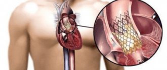
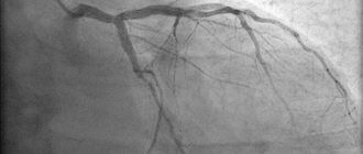
In addition to open heart operations, it is currently possible to perform minimally invasive operations, in particular, with a mini-access from an intercostal incision on the right or left without dissecting the sternum, as well as with endovascular intervention.
minimally invasive aortic valve replacement
The latter is used only for aortic valve replacement and is carried out by introducing a biological prosthesis through the femoral vein into the right and then into the left atrium with further location in the aorta.
Endovascular heart valve replacement is primarily preferred for individuals for whom open heart surgery is contraindicated.
Video: report on valve replacement surgery
{banner_banstat5}
Main functions
Throughout a person’s life, the heart provides a closed path of blood flow, delivery of oxygenated blood to organs and tissues, and venous outflow of carbon dioxide and breakdown products. The cardiovascular system consists of blood circulation circles. The large one originates in the left ventricle and ends in the right atrium, the small one starts in the right ventricle and goes to the left atrium.
The tricuspid valve is actually an element of a small circle that performs the following functions:
- During heart contractions, it prevents reverse regurgitation (blood flow from the lower ventricle into the atrium).
- Directly involved in blood circulation, ensures the delivery of venous blood to the vessels of the lungs.
- Through which the process of gas exchange in the alveoli of the lung tissue and heat transfer takes place.
Cost of the operation
{banner_banstat6}
In most cases, surgery to replace heart valves is performed free of charge, thanks to quotas from the Russian healthcare system under the compulsory medical insurance system. However, if for some reason it is not possible to obtain a quota, there is always the option of carrying out the operation at your own expense.
The cost of the operation itself, the prosthesis and rehabilitation in the early postoperative period ranges from 90 to 300 thousand rubles
, and the price is higher, the more complex the operation is, for example, the simultaneous replacement of the aortic valve and pulmonary valve is higher than one of them.
Heart valve replacement surgeries are carried out in all major cities of Russia, and now such interventions are not rare or inaccessible to the population.
Diagnostic methods
Systolic murmur during inspiration is important for diagnosis. This indicates tricuspid valve insufficiency. However, it should be remembered that this phenomenon is not permanent and may disappear completely for some time. To confirm the preliminary diagnosis, an electrocardiogram is taken.
In the resulting pathology graph one observes:
- deviation of the electrical axis to the right;
- an increase in the size of the P wave (in the area of the second and third chest leads).
Radiography may also be used. The image shows dilatation of the ventricle or atrium. Deviations are also noticeable in echocardiography images, where abnormal movements of the heart septa are established. When analyzing the patient’s condition, the following points are taken into account:
- Type of noise and area of its manifestation.
- The size of the heart, it is often enlarged.
- The presence of stagnation in the blood circulation.
- The magnitude of venous pressure.
- Liver size.
- Condition of the chest.
- Right atrial pressure.
Complications
{banner_banstat7}
The most serious complications after the introduction of a prosthesis are thromboembolic ones. Prevention of their development is lifelong antithrombotic therapy with the help of anticoagulants and antiplatelet agents - drugs that “thin” the blood. Such drugs include:
- Subcutaneous injections of heparin in the early postoperative period,
- Continuous use of warfarin under monthly monitoring of INR (international associated ratio) - an important indicator of the blood clot-forming system; normally it should be within 2.5 - 3.5,
- Constant use of aspirin (thromboAss, acecardol, aspirin Cardio, etc.).
No less dangerous consequences are the development or recurrence of infective endocarditis, the prevention of which is the rational prescription of antibiotics in the postoperative period, as well as their further use during any operations and minimally invasive interventions (tooth extraction, gynecological and urological manipulations, etc.).
Main functions
Throughout a person’s life, the heart provides a closed path of blood flow, delivery of oxygenated blood to organs and tissues, and venous outflow of carbon dioxide and breakdown products. The cardiovascular system consists of blood circulation circles. The large one originates in the left ventricle and ends in the right atrium, the small one starts in the right ventricle and goes to the left atrium.
The tricuspid valve is actually an element of a small circle that performs the following functions:
- During heart contractions, it prevents reverse regurgitation (blood flow from the lower ventricle into the atrium).
- Directly involved in blood circulation, ensures the delivery of venous blood to the vessels of the lungs.
- Through which the process of gas exchange in the alveoli of the lung tissue and heat transfer takes place.
Lifestyle
A person’s further life after surgery comes down to the following points:
- Regular visits to the doctor - monthly in the first year after surgery, every six months in the second year and annually thereafter, with constant monitoring of the functions of the cardiovascular system using ECG and echocardioscopy,
- Regularly taking prescribed medications (anticoagulants, antibiotics),
- Treatment of residual heart failure with the constant use of digoxin and diuretics (indapamide, veroshpiron, diuver, etc.),
- Adequate physical activity
- Compliance with the work and rest schedule,
- Following a diet - excluding fatty, fried, salty foods, eating large amounts of vegetables, fruits, dairy and cereal products,
- Complete elimination of bad habits.
Forecast
{banner_banstat8}
The prognosis after surgery is undoubtedly higher than without it,
since with heart defects severe heart failure develops, which not only impairs the tolerance of normal physical activity, but also leads to death. In patients after surgery, mortality is much lower, and is mainly associated with the development of thromboembolic complications (0.2% of deaths per year). Therefore, surgery to replace heart valves is an intervention that significantly prolongs the patient’s life and improves its quality.