Year after year, the number of patients with cardiovascular diseases, which are the main causes of disability and mortality among older people, is growing.
In addition to atherosclerotic lesions of the vessels of vital organs (heart and brain), which can cause dangerous complications in the form of stroke and heart attack, another scourge for patients aged 70 years and older is damage to the heart valves and, in particular, the aortic valve.
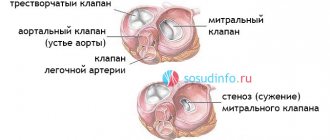
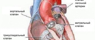
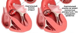
The aortic valve is a figurative “gate of entry” through which blood from the heart enters the aorta and is then distributed throughout the body. Damage or narrowing of this valve leads to poor blood circulation in the body.
The aortic valve is especially susceptible to pathological changes (most often narrowing) in older people.
The main reasons for this problem are:
- progressive sclerotic changes in the valve with age (>50%)
- inflammatory (including rheumatic) changes (10-15%)
According to WHO, sclerotic changes in the aortic valve are observed in a third of patients over 65 years of age and in more than 37% of people over the age of 75 years. Of these, approximately 15% have significant valve narrowing (stenosis).
1.What happens during normal surgery?
If surgery is required to treat heart valve disease, it can be performed traditionally, minimally invasively, or minimally invasive balloon valvuloplasty.
Traditional surgery to treat heart valve disease is performed under general anesthesia. The surgeon makes an incision in the center of the chest, thereby allowing direct access to the heart (in this case, actually open heart surgery is performed). The surgeon then operates on the abnormal valve, eliminating its defects, or replaces the heart valve.
A must read! Help with treatment and hospitalization!
PMC degrees
There are three degrees of valve damage.
With the first, the sash sagging is less than 5 mm. This phenomenon is typical for people with congenital pathology. In the first degree, the reflux of blood into the ventricle reaches the level of the anterior leaflet. As a rule, the patient does not feel signs of problems with the valve system.
In the second degree, the sealing increases to 9 mm. The reflux of blood into the ventricle reaches the middle of the atrium. People with this feature may periodically experience signs of valve dysfunction throughout their lives: shortness of breath, fatigue. If present, physical activity should be limited. Second degree secondary conscription is not a reason for exemption from military service, but it does influence the choice of troops. Women with prolapse when planning pregnancy should notify the doctor about their condition so that the specialist can prescribe the necessary examinations.
The third degree of compaction is a deflection of 10 millimeters or more. Regurgitation reaches the opposite wall of the atrium. Patients with this pathology experience signs of cardiac dysfunction:
- shortness of breath;
- abnormal pulse rate: tachycardia, bradycardia;
- intolerance to sports and other stress;
- dizziness;
- chest pain.
A person needs mandatory treatment, which can be not only therapeutic, but also surgical. Young people with a third-degree deformity are exempt from compulsory military service based on a medical report.
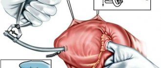
2.How is minimally invasive surgery performed to treat heart valve diseases?
The point of minimally invasive surgery is that surgery is performed through a much smaller incision
. This type of surgery reduces blood loss, trauma, and shortens hospital stay. In any case, surgeons will tell you whether minimally invasive surgery is right for you. Often, to determine how the heart valves are functioning before and after surgery, the surgeon and cardiologist will perform a transesophageal echocardiogram, in which an ultrasound probe is inserted into the esophagus.
During heart surgery, the valve may be "repaired."
This most often occurs when treating a mitral heart valve. But to correct the functioning of the aortic, pulmonary and tricuspid valves, some of the operations are also performed.
If the valve can be "repaired" without replacement, the surgeon may perform one of the following procedures:
- Commissurotomy.
During this heart surgery, the adhesions of the valve leaflets (petals) are separated, thereby widening the valve opening. - Decalcification.
This is a procedure to remove calcium deposits from the valve leaflets, thereby increasing their flexibility and ability to close tightly. - Change in the shape of the valve flaps.
This procedure is also called quadrangular resection. The idea is that if one of the valve flaps falls back when closing, a segment is cut out of the valve and the flaps are stitched together, allowing the valve to close more tightly. - Valve support ring.
Heart surgery is performed when the ring of tissue that supports the valve is too wide. In this case, it can be tightened using a ring made of fabric or synthetic material. - Repairing ruptures and holes in heart valves.
What are the advantages of heart surgery when repairing a valve? The fact that you will not need to take blood thinning medications for life
(anticoagulants). In addition, this intervention helps preserve the muscle strength of the heart.
Visit our Cardiology page
How to recognize prolapse?
Since in the first and second degrees the disease can occur without symptoms, you need to contact a good cardiologist in Rostov (Western) to diagnose it. A doctor can determine congenital prolapse by a number of external signs, including: above average height and elongated limbs, as well as hypermobile joints.
When visiting a cardiologist, the patient can list the following complaints, after which the doctor will prescribe an ultrasound scan:
- Frequent dizziness and faintness;
- The appearance of shortness of breath;
- Arrhythmia and interruptions in cardiac function;
- Fatigue and panic attacks;
- Chest pain.
In this case, the cardiologist will write a referral and tell you how much an ultrasound will cost to make a diagnosis. You will also need to do an x-ray and an ECG. Daily monitoring of cardiac activity and bicycle ergometry are prescribed.
The reason for clarifying the cost of ultrasound will also be noises that the doctor may detect at the appointment. To establish their cause, you will also need to do ultrasound diagnostics.
3.What to do if heart valve repair cannot be done?
In cases of aortic or pulmonary valve disease, heart surgery to replace the valve is usually performed.
During surgery, the abnormal heart valve is removed and a new valve is transplanted in its place. It could be:
- Mechanical heart valve.
The valve is made entirely of mechanical parts and materials that are well tolerated by the body. Most often this is a valve made of two carbon flaps in a ring, covered with polyester fabric. The advantage of mechanical valves is their durability and they last for many years without problems. But there are also disadvantages. Because the materials used to make the valve are artificial, you must continue to take anticoagulants (blood thinners) to prevent clots from forming in the valve after valve replacement. Clots can increase the risk of stroke. Another feature is the quiet ticking sound that is sometimes heard when the valve blades open and close. - A biological valve (bioprosthetic valve)
is made from human or animal tissue. Pig or cow tissue can be used for a bioprosthesis. Tissue valves may also have some artificial parts that help position it or create a frame. The advantage of a biological valve is that you will not need to take anticoagulants after valve replacement. But such valves are less durable and usually require replacement of the valve after about 10 years. However, recent studies have shown that some biological valves remain effective for at least 17 years. - Allograft valve.
This is an aortic or pulmonary valve obtained from a human donor. It is considered an ideal option for transplantation, especially when the patient has aortic disease or infection. When a valve is transplanted from a donor, the complete anatomy of the heart is preserved. But this valve replacement option is not always available.
About our clinic Chistye Prudy metro station Medintercom page!
Diagnosis of valve dysfunction
The first step to diagnosing the problem is to listen to the tone of the heartbeat and detect any murmurs. Auscultation of the heart makes it possible to make a preliminary diagnosis, with which the patient is sent for further examination.
ECG, ECHO-CG of the heart and chest x-ray are the next stage of examination, allowing you to determine whether there is an increase in the heart chambers.
X-ray shows distortion of the heart shape and its enlargement.
ECHO shows the deformation of the valves, the inability to completely close or open, and also helps to find the cause of problems with the valve, the degree of its insufficiency and the possibility of compensation from the body.
At the next stage of diagnosis, a catheter is inserted for coronography and ventriculography.
4.Is it possible to treat heart valve diseases without surgery?
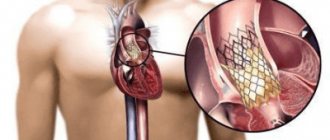
There are alternative options to traditional surgery for treating heart valve diseases. For some patients with mitral valve stenosis (narrowing) or pulmonary valve stenosis, an air (balloon) valvotomy procedure may be recommended.
It helps open a narrowed artery.
During this procedure, a special catheter is placed into a blood vessel in the groin area and guided towards the heart. The tip of the catheter is inserted into the narrowed valve. Once there, the tiny balloon at the end of the catheter expands (inflates) and deflates several times to widen the valve opening. After this, the balloon is removed. During balloon valvotomy, an echocardiogram may be performed to view the valve more completely.
Treatment
Patients with mitral valve prolapse are treated with medication. The combination of drugs is determined by the attending physician. Taking medications is indicated if the disease manifests itself with severe symptoms. If they are absent, no treatment is required
To eliminate signs of pathology use:
- Sedatives. They have a calming effect and reduce anxiety. Preference is given to plant-based products, which are produced using extracts of hops, valerian, and lemon balm.
- Antiarrhythmic drugs. Their task is to stabilize the heart rhythm, which eliminates the feeling of interruptions in the functioning of the heart.
- Adrenergic blockers. They act on adrenergic receptors and slow down the pulse.
- Antihypertensive medications for blood pressure control (amlodipine, atenolol, anaprilin, enalapril, etc.)
Differentiated approach in the treatment of aortic valve insufficiency
Aortic valve replacement through an incision in the aortic wall
Diagnosed asymptomatic mild aortic insufficiency does not require specialized treatment, however, limiting physical activity and annual visits to a cardiologist are strongly recommended. The indication for conservative therapy is moderate aortic insufficiency in the absence of symptoms; in this case, it is worth visiting a cardiologist at least once every six months.
In case of severe aortic valve insufficiency and the absence of symptoms of insufficiency, constant drug therapy, examination by a cardiologist every six months, and echocardiography once or twice a year are prescribed. Indications for surgical treatment are severe aortic insufficiency in the presence of clinical symptoms and the development of left ventricular heart failure.
Finally, it is important to remember that the success of recovery most often depends on your emotional state.
Diagnosis of acquired valvular insufficiency
Doppler echocardiography
- The first step in the diagnostic search, if an acquired heart defect is suspected, the doctor resorts to physical diagnosis, which is primarily an examination of the patient and auscultation of the heart. Auscultation reveals altered heart sounds and murmurs.
- The second stage of diagnosis is research methods, including electrocardiography (ECG), chest X-ray and echocardiography (EchoCG). The ECG visualizes signs of enlargement of the left chambers of the heart. An x-ray allows you to see changes in the size and shape of the heart, as well as pathological processes in the lungs. With the help of EchoCG, you can see a decrease in the size of the hole and changes in the valve leaflets; EchoCG also allows you to determine the cause of insufficiency, its degree, the presence of complications and the compensatory capabilities of the body.
EchoCG is the best method of primary diagnosis and dynamic monitoring of the patient’s condition.
- The third stage of diagnosis is invasive research methods, namely cardiac catheterization followed by ventriculography and coronography.
Valve insufficiency clinic
Fatigue and shortness of breath
In the initial stages of the disease, patients may not complain at all. This period is called the compensation stage. In the future, patient complaints depend on the severity of the disease and whether the valvular heart disease is isolated or combined. When complaints appear, the stage of decompensation of the process begins, which over time can lead to heart failure.
With moderate mitral regurgitation, the patient is concerned about fatigue and shortness of breath. In more serious cases, scanty hemoptysis may occur due to pulmonary edema. Due to the progressive enlargement of the left atrium, compression of the nerve innervating the larynx occurs, which is clinically manifested by hoarseness of the voice.
With aortic valve insufficiency, the initial symptoms are shortness of breath, rapid heartbeat and chest pain. Arterial hypotension and pulmonary edema can occur with severe aortic insufficiency. In the absence of timely surgical intervention, there is a high risk of death.
Acquired heart defects
Acquired heart defects are a group of diseases accompanied by disruption of the structure and function of the heart valve apparatus and leading to changes in intracardiac circulation.
Acquired heart defects develop as a result of acute or chronic (long-term) diseases and injuries that impair the function of the valves and cause changes in intracardiac hemodynamics (blood movement through the vessels).
More than half of all acquired heart defects are due to lesions of the mitral (located between the left atrium and the left ventricle) valve, about a third are lesions of the aortic (separates the left ventricle and aorta) valve, the rest are represented by combined (changes affecting several valves) defects.
Symptoms of acquired heart disease
- Dyspnea.
- Marked weakness.
- Changes in skin color - constant pallor or, conversely, pinkishness.
- Feeling of heartbeat.
- Possible pain in the heart area during physical activity.
- Headaches, dizziness, fainting (loss of consciousness).
Forms
Acquired heart defects are classified into several categories.
According to etiology (cause of occurrence) there are:
- rheumatic (occurs as a result of rheumatism - a systemic inflammatory disease of connective tissue with predominant damage to the heart);
- endocardial (due to endocarditis - inflammation of the inner lining of the heart);
- syphilitic (due to syphilis - a systemic disease that is predominantly sexually transmitted and affects many organs and systems) and so on.
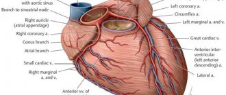
Based on the type of valve affected, the following are distinguished:
- aortic;
- mitral;
- tricuspid valve defect;
- valve defect of the pulmonary artery trunk.
By the number of affected valves:
- isolated, or local (damage to 1 valve),
- combined defect - insufficiency and stenosis (narrowing of the lumen) simultaneously occur on one valve;
- combined defect - changes affect several valves.
Functionally:
- stenosis - narrowing of the lumen of the opening as a result of post-inflammatory (occurs after the inflammatory process) cicatricial adhesions of the valve leaflets;
- insufficiency - incomplete closure of the heart valve leaflets;
- prolapse - protrusion, protrusion or eversion of the valves into the cavity of the heart.
According to the severity of the defect and the degree of hemodynamic disturbance (blood movement through the vessels) of the heart:
- does not have a significant effect on intracardiac circulation;
- moderately expressed;
- pronounced.
According to the state of general hemodynamics:
- compensated heart defects - without circulatory failure (a condition in which the heart is not able to adequately supply blood to all organs and tissues);
- subcompensated - with transient (temporary) decompensation (inability to compensate for impaired blood flow) caused by excessive physical exertion, elevated body temperature, pregnancy, and so on;
- decompensated - with developed circulatory failure.
Reasons for purchasing
The most common causes of acquired heart defects are:
- rheumatism is a systemic inflammatory disease of connective tissue primarily affecting the heart;
- infective endocarditis (inflammation of the inner wall of the heart);
- atherosclerosis is a chronic disease characterized by hardening and loss of elasticity of the walls of the arteries, narrowing of their lumen due to the so-called atherosclerotic plaques (formations consisting of a mixture of fats (primarily cholesterol (a fat-like substance that is a “building material” for the body’s cells) and calcium ));
- heart injuries (bruises and wounds of the heart muscle);
- syphilis is a systemic disease, transmitted primarily through sexual contact and affecting many organs and systems;
- sepsis (blood poisoning) and others.
Diagnostics
- Analysis of the medical history and complaints - when (how long ago) and what kind of complaints appeared, whether the patient consulted a doctor, underwent examination and treatment, with what results, and so on.
- Analysis of life history - past infectious diseases and chest injuries are clarified.
- Analysis of family history - it is found out whether any of the close relatives have heart diseases, what kind of diseases they are, and whether there have been cases of heart defects in the family.
- Medical examination. Wheezing in the lungs, murmurs in the heart are determined, the level of blood pressure is measured, the boundaries of the heart are determined by percussion (by tapping) (to determine hypertrophy (increase in size)), heart sounds and sounds are listened to to determine the type of defect, the lungs are auscultated and the size of the liver is determined ( to diagnose heart failure, a condition in which the heart cannot provide adequate blood flow to all organs).
- General blood test - allows you to detect signs of inflammation in the body (increased levels of leukocytes (white blood cells), increased levels of ESR (erythrocyte sedimentation rate (red blood cells), a nonspecific sign of inflammation)) and identify complications and possible causes of heart disease.
- A general urine test can detect complications of heart defects.
- Biochemical blood test - determination of the level of total cholesterol (a fat-like substance that is a “building material” for the body’s cells), “bad” (promotes the formation of atherosclerotic “plaques” (cell clots)) and “good” (prevents the formation of “plaques”) cholesterol, level of triglycerides (fats, the source of cell energy), blood sugar.
- Electrocardiography (ECG) is a method of recording the electrical activity of the heart on paper. Allows you to diagnose changes in heart rhythm, determine the type of arrhythmia and signs of ischemia (insufficient blood supply to the heart muscle).
- Phonocardiography is the recording of sound signals of the beating heart: murmurs and tones. The method allows you to assess the duration, intensity, nature, origin of heart murmurs and tones, and record the third and fourth heart sounds, which are indistinguishable by ear, which ultimately allows you to determine the type and nature of the heart defect.
- Echocardiography (EchoCG) is an ultrasound examination of the heart. Allows you to diagnose the defect itself, the area of the atrioventricular orifice (connecting the atrium and ventricle of the heart), the severity of regurgitation (backflow of blood), the condition and size of the valves, and determine the pressure in the vessels.
- Chest X-ray with intravenous contrast agent (angiocardiography) - assesses the condition of the lungs, the size of the heart and its chambers. The method helps to identify specific changes in the vascular bed in such patients.
- Multislice computed tomography - cardiography (MSCT of the heart) is a method of layer-by-layer scanning of heart structures, based on recording an X-ray beam passing through tissue using several rows of ultra-sensitive detectors. Cardiac MSCT enables 3-dimensional reconstruction of the heart and is used to identify valve defects.
- Cardiac magnetic resonance imaging (MRI) is a method of obtaining diagnostic images based on the physical phenomenon of nuclear magnetic resonance, thus it is safe for the body.
- It is also possible to consult a therapist or cardiac surgeon.
Treatment of acquired heart disease
Conservative (drug) treatment of acquired heart disease is prescribed only with the goal of stabilizing the heart rhythm, preventing heart failure (a condition in which the heart is unable to provide normal blood flow to all organs), complications and relapses (recurrences) of the underlying disease that caused the heart disease.
The main treatment method for acquired heart defects is surgery.
Valve defect correction:
- valvotomy (dissection of fused heart valves);
- valvuloplasty (restoration of valve function by cutting the valve walls and subsequent suturing of new leaflets).
- Prosthetic valve replacement (replacement with an artificial one).
Complications and consequences
- The development of heart failure (a condition in which the heart is unable to adequately supply blood to all organs and tissues).
- Heart rhythm disturbance (any heart rhythm other than normal).
- Thromboembolic complications (complications in which blood clots (blood clots in a vessel) can enter any vessel in the body through the bloodstream, block its lumen and cause organ dysfunction).
- Disability of patients.
- Lethal outcome (death).
Prevention of acquired heart disease
Prevention of all acquired heart defects consists in preventing the underlying disease that causes the defect (for example, timely treatment of tonsillitis (an infectious disease primarily affecting the palatine tonsils) prevents the development of rheumatism (a systemic inflammatory disease of connective tissue primarily affecting the heart)).