Duplex scanning of head and neck vessels is a highly informative and safe method based on ultrasound technology. Ultrasound waves “reveal” the arteries and vessels of the head and neck and make it possible to assess the state of blood flow according to several basic signs, as well as examine the lumen of the vessel for the presence of a wide range of pathologies. These include aneurysms, blood clots, changes in wall thickness and protrusion, plaques, and tortuosity of blood vessels. In comparative diagnostics, duplex scanning reveals changes in the condition of the walls and blood flow speed, which helps to adjust treatment or decide on its initiation if the patient has previously been under observation.
Doppler ultrasound examination is non-invasive, painless, and does not require prior preparation. The procedure is repeated the required number of times without restrictions to confirm a complex diagnosis.
General description of the study
The essence of the duplex scanning method is to combine the capabilities of ultrasonic waves, which visualize the structure of the vascular walls, and the Doppler effect, which reflects the nature of blood movement using multi-color coding (for clarity).
The software combines the two images and displays the data in a clear form for medical analysis.
Thus, scanning is carried out in 2 modes:
- two-dimensional (B-mode) - vessels and adjacent tissues are viewed, complete data on blood supply is not obtained in this mode;
- duplex (Dopplerography) - a two-dimensional color drawing is constructed, from which an expanded description of the condition of the vessels can be made.
The Doppler effect reflects the change in wavelength for an observer when a signal is reflected from a moving object, which is different types of blood cells. Ultrasound is reflected from them, helping to assess the direction of blood flow and calculate its speed and dynamics. In this case, pathologies will be indicated by turbulence, retrograde movement, and slowdown.
There are 3 main areas of research:
- transcranial ultrasound Doppler ultrasound - examines the blood vessels of the brain;
- Doppler ultrasound of the brachiocephalic vessels - the area of study lies in the neck area;
- Doppler ultrasound MAG is an examination of the main arteries of the head, which include two vertebral and two carotid, forming a circle at the base of the brain (their changes affect the entire body).
What is a diagnostic method
This method of vascular diagnostics provides complete information about the state of the human vascular system. Using ultrasound, you can evaluate and analyze the speed and nature of blood flow, as well as determine the presence of blood clots or other vascular pathologies.
The study usually proceeds in two stages: two-dimensional mode and duplex study.
Content:
- What is a diagnostic method
- Why is the procedure necessary?
- What is the difference between duplex and triplex scanning?
- Who needs the procedure
- Preparation for the procedure and contraindications
- How the procedure works
- Advantages and disadvantages of the technique
In two-dimensional mode, you can see the vessels and adjacent tissues, limited only by this information. It does not provide any information about blood supply.
With a duplex study, the doctor sees a complete picture of what is happening in the vessels, with a detailed description and a color two-dimensional picture.
Who is prescribed such a study?
The study is prescribed to identify the causes of headaches, with elevated cholesterol levels in tests, for early diagnosis of atherosclerosis, with pathologies of the spine, suspected tortuosity of the vessels supplying the brain, and frequent dizziness. To make a full diagnosis, an ultrasound examination of the vessels is performed both at the level of the neck and inside the skull.
Symptoms for which duplex scanning is indicated:
- pain in the head that appears with alarming and noticeable frequency;
- memory loss for a short period of time;
- dizziness;
- bleeding from the nose for no apparent reason;
- high cholesterol;
- mechanical damage in the neck area;
- noise in ears;
- the presence of certain diseases - stroke, heart attack, TIA, diseases caused by circulatory disorders, weakened blood flow, diabetes.
When assessing the results of a duplex study, a specialist considers a lot of characteristics and data, comparing them with normal indicators. What is important for the doctor is the thickness of the vessel wall (normally 0.12 cm), the location of the arteries relative to each other, surrounding tissues and organs, the speed of blood in different branches, the nature and presence of branches in certain areas, etc.
Duplex scanning not only reveals pathological processes when symptoms have already appeared, but also shows vascular pathologies at an early stage of their development. This allows you to prevent the disease in time and quickly begin treatment.
If the wall is thickened, it can be assumed that fat accumulates on it and connective tissue grows. If, with a thickened wall, the vascular layers are poorly distinguishable, this indicates vasculitis. When observing characteristic plaques in the vessels, we can talk about atherosclerosis (data on the location and size of plaques are of great importance for treatment).
What is Doppler ultrasound (Doppler ultrasound)
Doppler ultrasound (Doppler ultrasound) of the veins of the lower extremities is one of the most accessible research methods, based on the Doppler effect (named after the Austrian physicist and mathematician Doppler), which consists in recording a measurement of the speed of reflected sound waves from moving blood cells. Allows you to study only one function - the patency of the vessel based on the graph of the blood flow.
Ultrasound Doppler for examining the veins of the lower extremities
It is performed blindly, the sensor is placed on the approximate projection points of the vessel. If a violation of the patency of a vessel is detected, it is impossible to clarify the cause of this violation, because there is no visualization of the vessels. Currently, it is practically not used during a consultation with a phlebologist, as it is not very informative.
What is usually revealed during the study
Duplex affects mainly extracranial vessels, and to a lesser extent intracranial ones. The doctor pays special attention to the following vessels of the neck - the common carotid arteries (and bifurcation areas), the extracranial part of the carotid arteries, part of the brachiocephalic trunk, external carotid arteries, veins next to the skull, venous vertebral plexus, temporal arteries, etc.
For transcranial duplex scanning, do an ultrasound taking into account access through the cranial bones - they suppress a large number of ultrasonic waves. Access is through natural openings - the greater occipital, temporal bones, and orbits. An ultrasound scan of a baby's brain can be performed through the fontanelles - they provide a significant overview and help to collect more information. During this type of duplex scanning, the carotid and vertebral arteries inside the skull, the basilar and cerebral arteries are examined.
Duplex scanning is a method that detects a wide range of pathologies of the arteries and veins in the head and neck. If the diagnosis is already known, the doctor is able to monitor the patient’s condition (after surgery, stroke, head and neck injuries).
In patients with pathologies, ultrasound of the head and neck reveals:
- atherosclerosis;
- arteriovenous malformation of the brain;
- compression of blood vessels by neoplasms;
- diseases of the central nervous system;
- blood clots;
- endarteritis;
- stenosis and expansion;
- aneurysms;
- plaques in people with high cholesterol in tests
- bends, loops.
Atherosclerosis can be suspected if you are overweight, have high levels of cholesterol and triglycerides in tests, or have a predisposing heredity. Endarteritis has autoimmune causes, in which the artery wall is damaged, and the damage is visible on the screen. Aneurysms are detected for the first time (with characteristic patient complaints) or confirmed if they were already noticed on a previous ultrasound, CT scan or angiography. On the duplex, they look like thinned and stretched walls of arteries (less often veins), protruding outward.
Some patients require regular vascular monitoring. Once a year, to assess the effectiveness of treatment and create an individual prognosis, duplex is recommended for patients with diabetes mellitus, surges in blood pressure, degenerative changes in the discs in the cervical spine, and strokes in the past.
In addition to patients with diseases, regular diagnosis is indicated for smokers, people over 40 years of age (for men) and over 45 (for women). If you have relatives with vascular pathologies and diseases that provoke them, you should also be examined regularly.
Indications for use
There are three groups of indications for diagnosis.
Suspicious symptoms, the origin of which needs to be clarified. Among them:
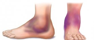
- Pain of unknown origin in the legs.
- Heaviness in the lower extremities.
- Dilated veins. Tortuosity of blood vessels. The appearance of spider veins. The described signs are early visual symptoms of varicose veins.
- Exercise intolerance.
- Intermittent claudication. A typical symptom of atherosclerosis. When an attack of acute pain begins in response to physical activity.
- Edema. In the morning or evening, after a hard day at work.
- Inadequate temperature perception. Chilliness or excessive sweating of the feet, a feeling of heat regardless of the ambient temperature.
- Cramps.
- Paresthesia. Feeling of goosebumps, tingling, numbness of the skin.
- Pallor of the skin of the legs.
The second group of indications are controversial diagnostic findings obtained as a result of other examinations. Including laboratory tests: blood biochemistry, blood lipid studies.
Phlebologists and vascular surgeons prescribe a systematic examination for patients from high-risk groups:
- Persons over 35 years of age.
- People who smoke and drink.
- Patients with diabetes mellitus, hypertension.
- People with a family history.
- Persons with physical inactivity and lack of physical activity.
- Patients who are overweight.
The frequency of preventive examinations depends on the severity of the clinical case. On average, we are talking about a procedure every 6-12 months.
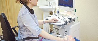
How does the procedure work?
There is no preparation for duplex scanning. Before diagnosis, it is enough to remove clothing or jewelry in the area where the device will be used. The patient lies comfortably on the couch and relaxes. The doctor applies a special conductive gel to the skin, which ensures better contact of the sensor with the skin, and applies the sensor to the body. Continuously changing sections appear on the screen, which the specialist studies in real time.
As a rule, the sensor is applied to the area of the ultrasound window (for example, 2-3 cm above the zygomatic bone). At this point, hard tissue absorbs ultrasound waves the least, which helps to examine blood flow in several arteries. After duplex scanning is completed, the gel is easily wiped off the scalp and neck.
Ultrasound scanning of the vessels of the neck and head takes about 30 minutes, and the patient feels calm and does not experience discomfort.
During the examination, the patient does not experience pain. The duplex scanning method feels practically no different from a regular ultrasound. The duplex scanning method feels comparable to a conventional ultrasound. The results are entered into the patient’s electronic database if he is treated in our clinic, or given in hand and as a printed copy. No special recommendations are given after the duplex procedure - the patient can immediately return to his usual rhythm of life.
Price list for ultrasound examination of vessels
| № | Name of service | price, rub. |
| 1 | Color duplex scanning of the veins of the lower extremities/upper extremities. | 2 100 |
| 2 | Color duplex scanning of the arteries of the lower extremities / upper extremities. | 2 100 |
| 3 | Ultrasound color duplex scanning of the vessels of the lower extremities (veins and arteries). (UZDG, UZDS, UZAS, DS of vessels...) | 3 200 |
| 4 | Triplex scanning of the vessels of the upper extremities (veins and arteries). | 3 200 |
| 5 | Duplex scanning of the brachiocephalic vessels of the neck for children over 1 year old and adults. (BCA of veins and arteries, USDG of extracranial, extracranial parts of the brain). | 2 100 |
| 6 | Transcranial color duplex scanning of brain vessels. TCD for children over 1 year old and adults. Intracranial examination of the vessels of the head. | 2 500 |
| 7 | Duplex scanning of the vessels of the brain and neck with functional tests for children from 1 year and adults. | 3 200 |
| 8 | Renal vessels and abdominal aorta (duplex scanning of veins and arteries). | 1 800 |
| 9 | Triplex scanning of the abdominal aorta and inferior vena cava | 1 500 |
| 10 | Ultrasound of the pelvic vessels (Doppler study of blood flow in the uterus and appendages). Transvaginally. | 1 700 |
| 11 | Neurosonography with Doppler analysis. NSG or ultrasound of the brain for infants. | 1 500 |
| 12 | Ultrasound video recording on DVD. | 500 |
| 13 | You can order duplex scanning of blood vessels and other studies at home, see the page “Ultrasound at home” |
Common advantages and disadvantages of the presented method
Duplex scanning has a number of obvious advantages:
- is highly informative;
- does not require the use of ionizing radiation;
- a cheaper and more accessible research method than alternative ones;
- carried out in real time, does not require waiting for a conclusion;
- helps to reflect the condition of soft tissues that are not sufficiently visualized on x-rays;
- non-invasive, performed without contrast (to which allergies are sometimes found), does not bring any unpleasant sensations;
- Suitable for children and pregnant women.
Any method, along with positive ones, also has negative sides, and in some cases it makes sense to choose other types of research. The decision is made by the doctor on an individual basis, after evaluating the results of other diagnostic procedures and tests, examination, and questioning of the patient.
The disadvantages of the method include:
- difficulty in examining small vessels;
- the possibility of diagnosis only during the research process, and not after it based on the results;
- narrow area to be examined because the bones of the adult skull impede the passage of ultrasound.
An important role in the quality of the diagnostics performed is played by the level of equipment and the professionalism of the doctor. This is one of the diagnostic methods to which increased demands are placed on the professionalism of doctors, so you should choose a clinic for duplex scanning especially carefully, because the result can differ significantly.
How is the study carried out and how long does it last?
The study is carried out as a regular ultrasound:
- The patient lies down on the couch.
- The doctor applies the gel to the skin.
- Applies the sensor.
- Explores the structural and functional features of arteries and veins.
If necessary, special tests are carried out. The specialist asks the patient to bend or straighten his leg and take a certain position. To artificially provoke possible disorders and evaluate changes in blood flow.
The procedure takes 10-20 minutes. The patient receives a diagnostic protocol and a doctor’s conclusion.
Price policy
The cost of a duplex depends on:
- profile of the clinic;
- location;
- rating;
- personnel qualifications;
- technical equipment of the diagnostic room.
The accuracy of diagnosis and timely detection of even minor injuries depend on the quality and experience of the doctor. Do not save on duplex scanning of the head and neck, even if the cost may vary, we guarantee the result and quality of the examination performed.
If necessary, the cost of the study can include a consultation with a neurologist, phlebologist and other specialized specialists. Our medical center has the best doctors, and we also train and improve the qualifications of staff from other clinics.
In our clinic you will receive one of the best services and high quality research. You can also get treatment here. The cost of duplex scanning of the head and neck can be found in the “Price List” section. The service is provided by appointment and 100% prepayment.
Cost of ultrasound (USDG, ultrasonography) of the veins of the lower extremities in Moscow
The cost of ultrasound examination of veins in Moscow, as for many high-tech modern services, does not always correlate with quality. You can undergo an examination at a very high cost, but with a fairly low information content. This is possible in some commercial medical centers where there is no modern phlebology. This approach can be called “ultrasound examination for the sake of ultrasound examination.” Things are far from being the best in the public sector. Patients often wait months in line for an ultrasound examination at a public clinic. And very often the results of such diagnostics leave much to be desired. The thing is that in both of these cases, the ultrasound examination is carried out by an ultrasound doctor who does not himself treat venous pathology. The ultrasound examination, which is carried out by the phlebologist himself, is performed in strict accordance with accepted European standards. The results of such an examination may differ radically from those obtained by an ultrasound specialist. The most paradoxical thing in this situation is that ultrasound examination of veins by a phlebologist in Moscow will almost always cost less in relation to commercial medical organizations. At the Moscow Innovative Phlebological Center, we do not logistically separate specialist consultation from ultrasound scanning. An ultrasound of the veins is performed during a consultation with a phlebologist. This is the only way to quickly and accurately obtain comprehensive information about the patient’s venous system.
| Service | Treatment category | Price |
| Appointment with an expert phlebologist with ultrasound scanning of the veins of the lower extremities | 2900₽ 3500₽ | |
| Appointment with an expert phlebologist without ultrasound scanning of the veins of the lower extremities (based on ultrasound results from another clinic) | 2000₽ 2400₽ | |
| Appointment with an expert phlebologist with ultrasound examination based on the results of treatment for one year | for free |
For the convenience of our patients, we work with banks that offer credit lines. Also in our center there is a flexible system of discounts and installments. Payment by Visa and Mastercard is possible. At the end of the course of treatment, the patient is given a package of documents to submit to the tax office for tax deduction.
The price at the Moscow Innovative Phlebological Center for such a specialist consultation with expert-level ultrasound scanning is 2,900 rubles. It should be noted that the price of this examination, which meets the best European standards, has been maintained in our Phlebology Medical Center since 2015.