General information
For some reason, almost all people are sure that arterial blood is the type that flows in arterial vessels. In fact, this opinion is wrong. Arterial blood is enriched with oxygen, which is why it is also called oxygenated. It moves from the left ventricle to the aorta, then goes through the arteries of the systemic circulation. After the cells are saturated with oxygen, the blood turns into venous and enters the veins of the BC. In a small circle, arterial blood moves through the veins.
You may be interested in: Aromatase inhibitors: purpose and list of drugs
Different types of arteries are located in different places: some are deep in the body, while others allow you to feel the pulsation.
Venous blood moves through the veins into the CD and through the arteries into the MC. There is no oxygen in it. This liquid contains a large amount of carbon dioxide, decay products.
Anatomy
Veins and arteries consist of three layers:
- Tunica adventitia : The outer layer of a blood vessel, composed of collagen and elastin. This layer allows the vessel to expand or contract, depending on the type of vein or artery. This function is important for controlling blood pressure.
- Tunica media : This is the middle of the blood vessel. Elastin and muscle fibers form the shell of the carriers. The amount of elastin or muscle varies depending on the type of blood vessel. For example, elastic arteries contain few muscle fibers in their membranes.
- Tunica Intima: This name refers to the inner layer of a blood vessel. It mainly contains elastic membranes and tissues and may include valves that help blood move in the right direction.
Function
Blood has specific and general functions. The latter include:
- nutrient transfer;
- transport of hormones;
- thermoregulation.
Venous blood contains a lot of carbon dioxide and little oxygen. This difference is due to the fact that oxygen enters only the arterial blood, while carbon dioxide passes through all vessels and is contained in all types of blood, but in different quantities.
The cardiovascular system
The cardiovascular system connects the heart and blood vessels together. The system forms a closed circuit of vessels that transport blood throughout the body.
The cardiovascular system is essential for maintaining life. This is the first major network of organs to develop in embryos.
All body tissues need oxygen and nutrients to survive. They also require the removal of waste, which is a byproduct of metabolism.
Blood is needed both to provide oxygen and nutrients and to remove waste from tissues.
The heart pumps blood throughout the body. It must work constantly and with sufficient force so that all tissues of the body receive enough blood to function. Cardiovascular disorders can have serious consequences.
Cardiovascular diseases are a group of disorders that affect the heart and blood vessels, such as coronary heart disease.
These diseases are the leading cause of death worldwide, accounting for approximately 17.9 million deaths in 2021.
Color
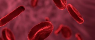
Venous and arterial blood have different colors. In the arteries it is very bright, scarlet, light. The blood in the veins is dark, cherry-colored, almost black. This is related to the amount of hemoglobin.
When oxygen enters the blood, it enters into an unstable combination with the iron contained in red blood cells. Once oxidized, iron colors the blood bright red. Venous blood contains many free iron ions, which is why it becomes dark in color.
What are arteries
These are vessels that transport oxygen from the heart to the internal organs. By contracting the myocardium, blood circulation is ensured at a speed of 20 cm/s. Purified blood, full of oxygen and nutrients, is necessary for metabolism.
Passing through organ tissue saturates it with carbon dioxide, excreted through venous hematopoiesis.
They are divided into three types:
- diameter;
- structural features;
- topographic principle.
The diameters are:
- large;
- small.
Large in diameter, unlike other components of the vascular system, are: aorta, carotid and subclavian.
The aorta arises from the left ventricle of the heart along the spinal column, dividing into the left and right iliac branches. It begins a large circle of blood circulation, supplying oxygen to the organs and tissues of the body.
General sleep supports the functioning of the brain, providing it with oxygen and microelements necessary for metabolism.
The subclavian vessel supplies blood to the occipital parts of the brain, medulla oblongata, cerebellum and cervical spine. The left arch departs from the aorta, bending around the pleura and, passing through the upper aperture of the chest, exits to the neck and lies in the space of the first rib.
Arterioles are small in diameter. Their task is to regulate blood flow in the SMC.
- Why do the veins in the arms hurt in the elbow bend?
Arteriolar tone determines peripheral resistance, which, along with the stroke volume of the heart, affects blood pressure.
There are three types:
- elastic;
- muscular;
- mixed.
The first type includes mainly the aorta. Its structure is characterized by the predominance of elastic fibers over muscle fibers.
The muscle type contains smooth muscle fibers and is characterized by a weak outer elastic membrane. An example is arterioles.
The muscular-elastic type is distinguished by the presence of muscle and elastic fibers in the structure of the vessel.
Blood movement
When wondering what the difference is between arterial blood and venous blood, few people know that these two types also differ in their movement through the vessels. In arteries, blood moves away from the heart, and through veins, on the contrary, towards the heart. In this part of the circulatory system, blood circulation is slow as the heart pushes fluid away from itself. Valves located in the vessels also affect the reduction in movement speed. This type of blood movement occurs in the systemic circulation. In the pulmonary circle, arterial blood moves through the veins. Venous - through the arteries.
In textbooks, on schematic representations of blood circulation, arterial blood is always colored red, and venous blood is always colored blue. Moreover, if you look at the diagrams, the number of arterial vessels corresponds to the number of venous ones. This image is approximate, but it fully reflects the essence of the vascular system.
The difference between arterial blood and venous blood also lies in the speed of movement. The arterial is ejected from the left ventricle into the aorta, which branches into smaller vessels. Then the blood enters the capillaries, nourishing all organs and systems at the cellular level with useful substances. Venous blood collects from capillaries into larger vessels, moving from the periphery to the heart. When a fluid moves, different pressures are observed in different areas. Arterial blood pressure is higher than that of venous blood. It is ejected from the heart under a pressure of 120 mm. rt. Art. In the capillaries the pressure drops to 10 millimeters. It also moves slowly through the veins, since it has to overcome the force of gravity and cope with the system of vascular valves.
Due to the difference in pressure, blood for testing is taken from capillaries or a vein. Blood is not taken from the arteries, since even minor damage to the vessel can provoke extensive bleeding.
Incomplete separation of arterial and venous blood
Incomplete separation of arterial and venous blood occurs with heart defects and large vessels , as well as lung diseases:
- atrial septal defect;
- a hole in the septum between the ventricles;
- collapse of the walls (closing) of the alveoli of the lungs (atelectasis), filling with fluid (edema, pneumonia);
- fistula between a vein and an artery (congenital, less commonly acquired);
- transposition of the great vessels (aorta and pulmonary artery change places);
- underdevelopment of the heart chambers (two- and three-chamber);
- open arterial (ductus Botallus, connects the aorta and pulmonary artery).
Dark blood from a vein: what does it mean?
The dark color of blood from a vein means that the organ from which it flows is actively functioning. The higher the rate of metabolic processes, the more oxygen is absorbed from the blood, which means its color will be darker.
We recommend reading the article about venous congestion in the legs. From it you will learn about the causes and symptoms of the pathology, conservative and surgical treatment.
And here is more information about vascular injury.
- What color is venous blood and why is it darker than arterial blood?
Venous blood flows from the organs, collects in the large vena cava and then into the right half of the heart. Through the pulmonary arteries it approaches the alveoli and turns into the arterial artery. The properties of blood from a vein are darker, thicker, and clot quickly. It is used for laboratory diagnostics, as it contains metabolic products, toxins, microbes, and hormones. Venous bleeding is accompanied by a slow and uniform release of blood; it is easier to stop than arterial bleeding.
Bleeding
When providing first aid, it is important to know which blood is arterial and which is venous. These species are easily identified by their flow patterns and color.
With arterial bleeding, a fountain of bright scarlet blood is observed. The liquid flows out pulsatingly and quickly. This type of bleeding is difficult to stop, which is the danger of such injuries.
When providing first aid, it is necessary to raise the limb and compress the damaged vessel by applying a hemostatic tourniquet or pressing it with finger pressure. In case of arterial bleeding, the patient must be taken to the hospital as quickly as possible.
Arterial bleeding may be internal. In such cases, a large amount of blood enters the abdominal cavity or various organs. With this type of pathology, a person suddenly becomes ill, the skin turns pale. After some time, dizziness and loss of consciousness begin. This is due to lack of oxygen. Only doctors can provide assistance with this type of pathology.
With venous bleeding, dark cherry-colored blood flows out of the wound. It flows slowly, without pulsation. You can stop this bleeding yourself by applying a pressure bandage.
First aid
When providing first aid for bleeding, it is important to determine its type and, depending on this, act. If an artery of the arm or leg is affected, it is necessary to apply a tourniquet above the affected area
While the tourniquet is being prepared, press the artery above the wound to the bone. This is done with a fist or by pressing hard with your fingers. Elevate the injured limb
If an artery in the arm or leg is affected, a tourniquet must be applied above the affected area. While the tourniquet is being prepared, press the artery above the wound to the bone. This is done with a fist or by pressing hard with your fingers. Elevate the injured limb.
Place a soft cloth under the tourniquet. You can use a scarf, rope, or bandage as a tourniquet. The tourniquet is tightened until the bleeding stops. You need to place a piece of paper under the tourniquet to indicate the time of application of the tourniquet.
If a tourniquet cannot be applied, for example, if the iliac artery is injured, make a tight tampon with a sterile or at least clean cloth. The tampon is wrapped with bandages.
In case of venous bleeding, a tourniquet or tight bandage is applied below the wound. The wound itself is covered with a clean cloth. The affected limb needs to be raised higher.
For these types of bleeding, it is good to give the victim painkillers and cover him with warm clothes.
In case of capillary bleeding, the wound is treated with hydrogen peroxide, bandaged or covered with a bactericidal adhesive plaster. If it seems to you that the blood is darker than a normal wound, then the venule may be damaged. Venous blood is darker than capillary blood. Proceed as if you had damaged a vein.
The health and sometimes life of a person depends on proper assistance during bleeding.
Blood is designed to carry substances necessary for cell function
, tissues and organs. Removal of decomposition products also occurs with the help of this liquid. These two different functions within the same system are carried out through arteries and veins. The blood flowing through these vessels contains different substances, which leaves its mark on the appearance and properties of the contents of the arteries and veins. Arterial blood and venous blood represent different states of the unified transport system of our body, which ensures the balance of biosynthesis and destruction of organic matter in order to obtain energy.
Venous and arterial blood move through different vessels
, but this does not mean that they exist in isolation from each other. These names are conditional. Blood is a liquid that flows from one vessel to another, penetrates into the intercellular space, returning again to the capillaries.


