Leukemia, which is often called blood cancer or leukemia, is a serious cancer. It affects the hematopoietic organs and causes changes due to which the number of leukocytes in the patient’s blood sharply increases. As the disease progresses, the blood gradually ceases to perform its functions in the body, which causes death if left untreated. The disease affects not only adults, but also children, and is found with equal frequency in men and women.
Types of disease
The modern classification of leukemia includes several principles of division into types. First of all, it is worth mentioning the division of cases of the disease according to the nature of the course into:
- acute, characterized by rapid development and the formation of a large number of immature blood cells - blasts;
- chronic, which are characterized by a long course and pathological production of mature leukocytes with altered properties.
Another principle for classifying leukemia is based on the level of differentiation of the affected cells, which can be:
- undifferentiated;
- quotational;
- blast.
In addition, oncologists distinguish many types of leukemia according to the types of cells whose pathological changes underlie the disease. According to this principle, lymphoblastic, monoblastic, myeloblastic, lymphocytic, myeloma and other types of disease are distinguished.
Palliative care
There are a large number of treatment methods available for the treatment of acute leukemia. This could be chemotherapy, targeted therapy, immunotherapy. All these methods can lead to remission even after multiple relapses. Therefore, as such, palliative therapy is rarely prescribed and, as a rule, in older patients who cannot tolerate severe treatment.
In this case, chemotherapy, drug therapy and radiation therapy are used.
Chemotherapy
Chemotherapy in palliative treatment is not carried out with the goal of achieving remission, but to keep the tumor clone from rapidly multiplying. At this stage, standard cytostatic drugs in lower dosages, immunotherapy, targeted therapy and other methods of antitumor treatment can be used.
Radiation therapy
Radiation therapy can be used to relieve pain in severe bone damage, as well as in the presence of extramedullary lesions.
Symptoms
The course of different types of disease is not accompanied by specific symptoms of leukemia. On the contrary, its manifestations are the same for all types of pathology and are poorly diagnosed in the initial stages. These include:
- increased susceptibility to colds and seasonal ailments;
- night sweats;
- joint and bone pain;
- swelling of the lymph nodes;
- instability of body temperature, constant sharp changes;
- pallor, grayish skin tone;
- general malaise, weakness;
- long-term healing of even minor injuries;
- causeless loss of body weight.
In the later stages of the disease, more distinct signs of leukemia appear:
- characteristic yellowness of the skin and eye whites;
- deterioration of vision, up to complete dysfunction;
- severe headaches;
- difficulty breathing, pneumonia;
- frequent nausea, vomiting;
- problems with cardiac activity;
- numbness of the facial muscles, partial or complete.
In later stages, due to increased fragility of blood vessels, subcutaneous and internal hemorrhages often develop. Ulcerative-necrotic tissue lesions and severe tonsillitis may appear.
What processes affect the patient’s well-being?
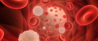
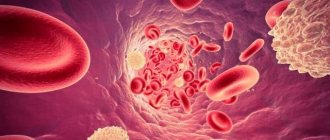
In a person with leukemia, immature lymph and leukocytes, devoid of protective functions, are released into the blood. The level of red blood cells drops and proper oxygen exchange does not occur in the tissues. Peripheral blood supply is disrupted, platelet levels decrease, blood stops clotting, internal organs enlarge and swell.
Oncology develops against the background of hereditary and genetic abnormalities. Leukemia progresses under the influence of alcohol, nicotine, harmful foods, and carcinogens on the body. A special factor influencing the development of blood cancer is radiation exposure of a woman during pregnancy or the effect of large doses of radiation on the body of an adult.
Causes and risk factors
A specific relationship between certain exposures and the occurrence of blood cancer has not yet been discovered, but risk factors that increase the likelihood of developing the disease are well known to oncologists.
- Heredity. If there are cases of the disease in the family, the risk increases 3-5 times. In addition, genetic diseases are a significant risk factor.
- Radiation. Significantly more often than usual, symptoms of leukemia occur in people exposed to radiation.
- Chemical substances. Dozens of chemical compounds that people come into contact with during their professional activities have pathogenic effects that provoke mutation of blood cells. In particular, these include gasoline vapors.
- Smoking. In people who smoke, the risk of acute myeloblastic leukemia increases several times.
The presence of one or even several of the listed factors does not mean that a person will necessarily get sick, but the likelihood of blood cancer is significantly increased.
Causes
As with any cancer, the main cause of malignancy is a violation of apoptosis (programmed cell death), which leads to the proliferation of pathological tumor cells. All this happens against the background of a weakened immune system.
Predisposing factors
- Genetic predisposition - if close relatives suffered from similar diseases.
- Primary or secondary immunodeficiency.
- Exposure to ionizing radiation. People injured during the accident at the Chernobyl nuclear power plant and liquidators of the consequences were exposed to such irradiation. People who have passed
- courses of radiation therapy against the background of other oncological diseases.
- Exposure to chemical carcinogens. This group also includes people who receive chemotherapy for other cancers.
Stages
Acute and chronic blood cancer differ somewhat in the nature of the disease. Oncologists distinguish the following stages of acute leukemia:
- full clinical development, in which the patient experiences bleeding, inflammatory processes in various organs, the number of red blood cells decreases, and hemoglobin decreases;
- remission, when clinical manifestations decrease and the number of immature cells in the bone marrow does not exceed 5%;
- relapse, when the patient’s well-being worsens with an increase in the number of immature leukocytes in the bone marrow and blood;
- terminal, in which the tumor progresses, and almost all blood cells become pathological, which is why ulcerative-necrotic processes develop in many organs.
The stages of remission and relapse can be repeated cyclically several times, with relapses becoming more and more severe, and remissions becoming short and less pronounced.
Chronic leukemia is divided into the following stages:
- initial, lasting up to a year, during which the number of neutrophilic leukocytes in the blood increases and the alkaline phosphatase level drops several times;
- developed, lasting up to five years, with a clinical picture characteristic of the disease, when the bone marrow gradually ceases to produce normal blood cells, the spleen increases in size, and the altered cells gradually penetrate into the peripheral bloodstream;
- terminal, the duration of which does not exceed six months, characterized by the spread of cancer cells to all vital organs through the bloodstream.
Chronic lymphocytic leukemia
Because chronic lymphocytic leukemia progresses slowly, many people do not need treatment for several years until the lymphocyte count begins to increase, the lymph nodes become enlarged, and the number of platelets and red blood cells decreases. Anemia is treated with red blood cell transfusions and injections of erythropoietin (a medicine that stimulates the formation of red blood cells). If the platelet count decreases, the corresponding blood component is transfused, and if an infectious disease develops, antibiotics are prescribed. Radiation therapy is used to slow the growth of the lymph nodes, liver and spleen when this is accompanied by discomfort. Medicines
, which are used to treat leukemia itself, do not cure the disease and do not prolong life, and in addition can cause severe side effects.
Over-treatment is more dangerous than under-treatment. When the lymphocyte count becomes very high, your doctor may prescribe anticancer drugs, sometimes in combination with corticosteroids (hormonal drugs). Prednisolone and other corticosteroids may provide significant and rapid improvement in people with advanced leukemia. However, this reaction is usually short-lived, and corticosteroids cause many side effects with long-term use, including an increased risk of severe infections. Drug therapy for
B-cell leukemia involves alkylating agents, which destroy cancer cells by interacting with their DNA. Interferon alpha is highly effective for the treatment of hairy cell leukemia.
Forecast
Most forms of chronic lymphocytic leukemia progress slowly. A patient's chances of recovery depend on how far the disease has progressed. Determination of the stage of the disease is based on indicators such as the number of lymphocytes in the blood and bone marrow, the size of the spleen and liver, the presence or absence of anemia, and platelet content. People who have early stages of B-cell leukemia often live 10 to 20 years after diagnosis and usually do not need treatment. Patients who have severe anemia and a platelet count of less than 100 x 109 per liter of blood (normal range is 180-320 x 109) have a worse prognosis than those who do not have severe anemia and have a normal platelet count. Typically, death occurs because the bone marrow stops functioning: it cannot produce enough normal cells to deliver oxygen to the body's cells, fight infections, and prevent bleeding. People who have T-cell leukemia have a slightly worse prognosis. For reasons likely related to changes in the immune system, patients with chronic lymphocytic leukemia are more likely to develop other cancers.
Diagnostics
The main procedures in diagnosing leukemia are laboratory blood tests: general, biochemical, genetic.
Equally important are bone marrow and cerebrospinal fluid puncture studies:
- cytogenetic, identifying atypical chromosomes characteristic of altered cells;
- cytochemical, which makes it possible to detect the acute form of the disease at an early stage;
- immunophenotypic, as a result of which it becomes clear what types of cells have undergone malignant changes;
- myelogram, which allows you to determine the stage of the disease and the dynamics of the process.
At the same time, the patient is prescribed instrumental studies of internal organs - ultrasound, CT, MRI to determine leukemic infiltration.
Classification of CLL
Chronic leukemia is classified by stages and risk groups.
The stage of the disease is determined based on a clinical examination and blood test results:
- Stage A - hemoglobin level more than 100 g/l, platelets more than 100 × 109/l, and less than 3 areas of lymph nodes are affected.
- Stage B - hemoglobin level more than 100 g/l, platelets more than 100 × 10 9 / l, and more than 3 areas of lymph nodes are affected.
- Stage C hemoglobin level less than 100 g/L or platelet level less than 100 × 10 9 /L.
Classification by risk groups
For this classification, an international prognostic index was developed that takes into account the following parameters:
- TP53 mutation (17p).
- IGHV mutation.
- β2-microglobulin level >3.5 mg/l.
- Stage B/C.
- Age over 65 years.
Each of these parameters is assigned a certain number of points; when summed up, the patient is assigned to one of 4 risk groups:
- 0-1 point - group with a low risk of progression.
- 2-3 points - intermediate risk of progression.
- 4-6 points - high risk of progression.
- 7-10 points - a very high risk of progression.
Treatment
After leukemia is detected, treatment is selected in accordance with the form and stage of the disease, the level of damage to vital organs, age, and general condition of the body. The treatment method is chosen by a specialist - an oncologist, an oncohematologist, and his goal is to restore the normal hematopoietic process, achieve long-term remission, and ideally, a complete cure.
The choice of method largely depends on the form of the pathology. In acute cases of the disease, chemotherapy has a good effect; in cases of chronic disease, the most effective method is often a bone marrow transplant or stem cell treatment. Adverse symptoms are relieved by detoxification and hemostatic therapy, the introduction of healthy platelets and leukocytes, and courses of antibiotics.
Rehabilitation
The list of clinical recommendations for acute leukemia during the rehabilitation period includes:
- measures to improve immunity;
- balanced diet;
- detoxification therapy;
- restoration of intestinal microflora;
- anti-stress psychotherapy;
- improving sleep quality.
During the recovery period, it is important to follow all the orders of the attending oncologist and complete the recommended treatment courses on time and in full.
Forecasts
The success of leukemia treatment depends on many factors, but in most cases the prognosis is favorable. Acute forms are more complex, since the rapid course of the disease leaves less time for the doctor to act. In the chronic form, favorable outcomes, according to statistics, account for up to 85% of cases.
In any case, there remains a chance for a long-term remission and complete relief from the disease. In adults, the probability of a positive outcome is at least 40%, in children - up to 90%.
Pathophysiology
Under the influence of the above etiological factors, somatic mutations of the precursor cells of hematopoietic and lymphoid tissues occur in the human body.
https://onlinelibrary.wiley.com/doi/abs/10.1046/j.1526-0968.2002.00402.x
The first stage of leukemia formation begins with a mutation in the parent cell, which acquires the ability to rapidly proliferate. As a result of this process, cells are formed that are their clone.
At the stage of formation of the first copy, the tumor retains the ability to further differentiate (benign tumor growth). Over time, numerous mutations and changes in structure occur in the primary leukemia clone. As a result, they not only actively proliferate, but also lose the ability to differentiate (malignant tumor growth).
In both children and adults, acute leukemia can present with extremely high blast counts; a phenomenon known as hyperleukocytosis. Respiratory failure, intracranial hemorrhage and severe metabolic abnormalities frequently occur in acute hyperleukocytic leukemia (AHL) and are the main determinants of the observed high early mortality (20% to 40%).
The process leading to these complications has long been known as leukostasis, but the biological mechanisms underlying its development and progression remain unclear. Traditionally, leukostasis is associated with the accumulation of leukemic blasts in the microcirculation, and its treatment is aimed at quickly reducing leukocytosis. https://onlinelibrary.wiley.com/doi/abs/10.1046/j.1526-0968.2002.00402.x


