Circulation is the movement of blood in the human body. It consists of three main parts: blood, blood vessels (arteries, veins, capillaries) and the heart.
We decided to prepare educational material so that each of you is aware of all the nuances of the cardiovascular system. This is important so that you can understand in time what problem you could or may encounter in the future, and also so that the terminological expressions of our specialist during a face-to-face consultation do not seem like a foreign language to you.
The heart is the basis of the circulatory system
The heart is a muscular organ about the size of a human fist that is located on the left side of the chest, just in front of the lungs. This organ is actually a powerful double pump with four chambers that pumps blood and keeps it moving throughout the body.
The right side of the heart consists of an upper (atrium) and lower (ventricle) chamber. The atrium receives processed venous blood, saturated with carbon dioxide, and then sends it to the ventricle. From there it enters the pulmonary arteries, where it is again saturated with oxygen. “Fresh” blood circulates to the left upper chamber (atrium), from where it enters the aorta and begins renewed transportation throughout the body.
The heart muscle beats more than 3 billion times during a lifetime.
VENOUS THROMBOEMBOLIC COMPLICATIONS: WHO IS TO BLAME AND WHAT TO DO?
Venous thromboembolic complications, which include: deep vein thrombosis, saphenous vein thrombosis, thrombophlebitis and pulmonary embolism, have been a major clinical problem for a long time.
According to statistics, about 100 thousand patients in the Russian Federation die annually from sudden pulmonary embolism. Thromboembolism is represented by blood clots that form in the lumen of blood vessels and spread with the blood flow throughout the body. Most often, blood clots form in the lumen of the veins of the lower extremities, and then enter the right half of the heart and then into the arteries of the lungs.
In the Russian Federation, about 80,000 new cases of venous thromboembolic complications are recorded annually. Within a month after deep vein thrombosis, 6% of patients die from pulmonary embolism, but even a successful outcome of the acute period does not mean the disappearance of this problem. After thrombosis, in the long-term period, post-thrombotic disease is formed, caused by organic damage to the deep veins and manifested by a violation of the venous outflow of blood. After thromboembolism of the pulmonary arteries, occlusion or stenosis of the arteries develops - chronic post-embolic pulmonary hypertension, which is fraught with the development of chronic pulmonary heart disease. After massive pulmonary embolism, 10-15% of patients die over the next 5 years.
The mechanism of intravital formation of blood clots inside blood vessels is described by a triad discovered in 1856 by the German morphologist Rudolf Virchow, which includes damage to the vascular wall, slowing down blood flow and increasing blood viscosity (clotting ability).
The most significant factors for the occurrence of venous thrombosis are hemodynamic disorders (slow blood flow) and hypercoagulation (increased blood clotting).
The likelihood of venous thrombosis increases if a person has congenital or acquired thrombophilia, i.e. a condition characterized by a tendency to thrombus formation. Many cases of “unexpected” venous thrombosis and pulmonary thromboembolism (particularly those occurring in young people who do not have serious clinical risk factors) may be associated with the presence of thrombophilia. Thrombosis in patients with thrombophilia can be initiated by surgical interventions, trauma, pregnancy and childbirth, i.e. those conditions that are accompanied by tissue damage, changes in vascular tone, and hormonal levels.
The main risk factors for thrombosis include:
- - thrombophilia (hereditary or acquired);
- — episodes of VTEC in the anamnesis;
- — current oncological process;
- — injuries and surgical interventions;
- - increasing age (over 40 years), the older the person, the greater the likelihood of thrombosis;
- — heredity (presence of blood relatives with a history of thrombosis before the age of 50);
- - overweight and obesity;
- - varicose veins of the lower extremities;
- - taking estrogen-containing drugs (oral contraception, hormone replacement therapy in the postmenopausal period);
- - pregnancy and the immediate postpartum period (6 weeks) are characterized by significant hormonal changes, the predominance of estrogens in the blood, hemoconcentration, which leads to a significant increase in blood coagulation activity;
- - prolonged bed rest, immobilization (plaster, skeletal traction) for more than 3 days, long air or road travel (more than 8 hours) lead to venous stagnation due to the lack of activity of the muscular-venous pump of the lower leg;
- - acute inflammatory diseases (pneumonia, colitis), sepsis;
- - compression of the veins by a volumetric formation (most often at the pelvic level), which leads to a significant disruption of venous blood flow and blood stasis;
- — chronic diseases: diabetes, arterial hypertension.
The main symptoms of thromboembolic complications:
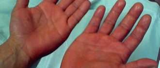
- — with thrombosis of the superficial veins of the lower extremities, movement-limiting pain is observed along the thrombosed veins, a band of hyperemia and a local increase in skin temperature in the projection of the vein;
- — with thrombosis of the deep veins of the lower extremities, severe pain often occurs in the lower leg and foot, a change in the color of the skin below the site of thrombosis is possible, this is due to the fact that when the vein is blocked, the blood does not flow from the limb, leading to bursting pain.
Pulmonary embolism has manifestations of varying severity. Sometimes, due to the insignificance of symptoms, pulmonary embolism of small branches may remain unrecognized, leading to complications from the lungs and heart, for example, to the development of chronic thromboembolic pulmonary hypertension. Typically, pulmonary embolism of small branches is accompanied by attacks of dry cough or hemoptysis with pain in the chest of various localizations. Often the patient experiences attacks of sudden shortness of breath and a feeling of lack of air. Massive pulmonary embolism is characterized by severe pain in the chest, shortness of breath, hemoptysis and cyanosis (blue discoloration) of the skin of the face, neck, earlobes and chest strictly up to the horizontal line between the nipples. Clinical death can occur instantly, which without treatment turns into biological death.
Only a specialist can make a correct diagnosis, establish the cause of thromboembolic complications, determine management tactics after thrombosis has occurred or prescribe thromboprophylaxis for existing risk factors, and assess the risk of thrombotic events before starting hormonal therapy or planning surgical interventions.
The GAU RO OKDC employs hematologists and hemostasiologists who have undergone special training in the pathology of the blood coagulation system and have modern high-quality laboratory diagnostics at their disposal. Doctors at the hemostasis pathology center can determine patient management tactics; select appropriate therapy; reduce the risk of possible complications; determine the need for laboratory monitoring of the effectiveness of therapy.
Specialists from the Center for Pathology of Hemostasis at the State Autonomous Institution RO OKDC will help you promptly recognize disturbances in the hemostasis system and, if necessary, select an effective treatment suitable in each specific case.
Tatyana Nikolaevna Abramova, physician-hemostasiologist of the highest category.
GAU RO "OKDC", Rostov-on-Don, st. Pushkinskaya, 127. 8(863) 227-0000. rokdc.ru
share information
0
Social buttons for Joomla
Blood vessels
Blood vessels have different shapes, structures and volumes, depending on their role in the body.
1. Arteries are the strongest vessels in the human body. Their walls are dense and elastic, consisting of three layers - endothelium, smooth muscle fibers and fibrous tissue. The task of the arteries is to saturate all organs and tissues with blood enriched with oxygen and nutrients. An exception is the arteries of the pulmonary circulation, through which venous blood flows from the heart to the lungs. The largest arterial vessel is the aorta.
2. Veins perform the function of transporting waste blood, saturated with carbon dioxide, back to the heart. This vein fluid is obtained from capillaries. Like an artery, a vein consists of several layers - endothelial, soft connective, dense connective and muscle. Venous walls are several times thinner and more vulnerable than arterial walls. For this reason, as you move away from the heart, the movement of venous blood may be disrupted - the pressure in the capillaries is almost equal to atmospheric pressure, and a normal flow is not created. Therefore, in hemodynamics, the vessels are assisted by the venous valves and the venous pulse.
3. Capillaries are the thinnest vessels, similar in volume to human hair. They are branches of large peripheral arteries. It is through them that tissues and organs are supplied with oxygen and nutrients. They also communicate with the veins to carry cellular waste. Consequently, these tiny vessels are at the same time the breadwinners and caretakers of our body.
Normal blood circulation within the vascular system is ensured by blood pressure.
Differences between venous and capillary blood analysis
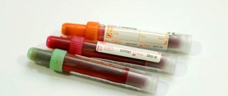
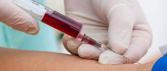
For most tests, which are prescribed by doctors of various specializations to diagnose diseases and monitor the progress of their treatment, the biomaterial is venous blood.
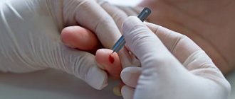
Capillary biomaterial is sometimes used to perform OAC. In any case, donating blood from a vein is preferable, since in this case the research results will be more informative. When performing OAC with different types of biomaterial, the results may differ. Therefore, to obtain reliable information about the patient’s health status over time, it is recommended to submit the same type of biomaterial. Capillary blood analysis
The main advantage of testing blood taken from a finger is the ease of taking biomaterial. This is why this type of test is often prescribed to young children. Research on this biomaterial has a significant number of limitations and disadvantages. Most laboratory tests do not use capillary blood. The research results are not highly accurate. There is a high probability of their distortion due to the formation of microclots, tissue fluid entering the blood, and platelet aggregation. Features of the procedure for taking biomaterial also lead to a decrease in the information content of research results. If questionable results are obtained, the study is usually repeated, and venous blood is used as a biomaterial.
Venous blood analysis
Blood from a vein is used for a variety of studies (for example, hormonal). Drawing blood by an experienced nurse takes a few minutes and does not cause significant discomfort. The results of such studies are characterized by high accuracy and great information content. To obtain the most accurate results and reduce the risk of their distortion by external factors, it is important to strictly follow the recommendations for preparing for the study. Preparation rules depend on the type of analysis. In addition, the attending physician can, if necessary, give the patient individual recommendations.
Preparing to donate blood
Before taking the test, it is important to exclude all factors that could affect the result. Such factors include physical activity (for example, climbing stairs, brisk walking), significant emotional arousal, and stress. Therefore, before donating blood, it is recommended to sit near the office or in the waiting room for 15-20 minutes. It is advisable to avoid heavy physical work and intense training a few days before the test.
If for any reason a patient cannot visit a clinic or private medical center to donate biomaterial, it is necessary to use the services of a laboratory that provides home testing services.
In most cases, blood is donated before starting medication or 2 weeks after finishing it. If it is necessary to evaluate the concentration of the drug in the blood, their use is continued in accordance with the doctor’s recommendations. To avoid misinterpretation of the results, it is important to inform your doctor about taking any medications, as well as dietary supplements.
If the patient has undergone X-rays, physical therapy, or a rectal examination, blood donation is usually delayed for some time. The final decision on the time of testing is made by the doctor for each patient individually.
It must be taken into account that in girls and women of reproductive age, the results of hormonal studies are significantly influenced by physiological factors. Blood must be donated for testing strictly at the phase of the cycle recommended by the doctor.
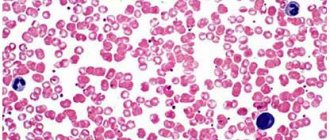
Cellular structure of blood
Blood consists of two components: plasma (50-60%) and suspended formed elements (40-50%).
The second category includes:
· Erythrocytes (red blood cells) are the most numerous of the formed elements. According to official studies, one drop of blood contains about 5 million red blood cells. Red blood cells are responsible for transporting gases - oxygen and carbon dioxide. They contain the protein hemoglobin, which binds oxygen molecules in the lungs. Red blood cells deliver oxygen to all tissues and organs, after which they absorb carbon dioxide and carry it to the lungs. It is removed from the body during respiration.
· Leukocytes (white blood cells) - elements that protect our body from foreign bodies and compounds, are part of the immune system. White blood cells recognize and attack pathogens through the production of antibodies and macrophages. When an infection enters the body, the production of leukocytes increases significantly. Normally, their quantity is inferior to the concentration in the blood of other formed elements.
· Platelets (blood platelets) are cells that provide coagulation (clotting) of blood flowing from a damaged vessel and protect the body from heavy blood loss. They stick to the hole in the damaged vessel, forming a “sealing” plug to stop bleeding. It is the platelets that can stick together and form pathological blood clots inside the vessels, called thrombi.
All formed elements are synthesized by the bone marrow and distributed through plasma, the liquid part of the blood.
When do you need to take a General blood test without leukocyte formula (venous blood)?
- To assess general health (routine medical examinations, screening examinations, etc.);
- For diagnostics at the first stage of a wide range of diseases that may manifest themselves as changes in the characteristics of peripheral blood;
- Diagnosis of anemia, polycythemia along with the study of hemoglobin and hematocrit;
- Monitoring ongoing drug treatment for patients suffering from bleeding or chronic anemia;
- Determination of the activity of erythrocyte formation processes in the bone marrow.