Signs of heart disease
Diagnosis of cardiac disease is difficult for a person far from medicine. Sometimes heart disease occurs unnoticed by the patient. On the contrary, individual malfunctions in the body - osteochondrosis, neuralgia, diseases of the lungs, gastrointestinal tract - can cause acute painful spasms. You need to know how the heart hurts, the signs, and where it occurs.
It should be remembered that heart disease does not appear suddenly. The acute condition is preceded by several months, sometimes years, by warning symptoms. Here is a list of the first signs, the appearance of which suggests cardiac pathology:
- pressing, squeezing pain behind the sternum;
- irradiation to the left arm, neck, shoulder blade, jaw;
- pain occurs and intensifies during physical activity, and goes away during rest;
- uneven heartbeat, unstable pulse;
- associated conditions: fatigue, shortness of breath, nausea, sweating;
- the appearance of swelling of the feet and hands;
- insomnia, sleep disorders;
- apnea, snoring during sleep.
These symptoms should alert you and prompt you to be examined without waiting for a health-threatening condition.
High blood viscosity syndrome (HBS), which occurs as a result of various factors, can cause many life-threatening pathologies. To prevent the development of complications, you need to know how to thin the blood at home, quickly and without negative consequences. Read more in the article: “quickly thin the blood at home.”
Heart rhythm and conduction disorders
The normal heart rhythm is called sinus rhythm.
The heart has its own electrical (conducting) system, consisting of an electrical impulse generator - the main pacemaker - and conductive pathways connecting the entire electrical circuit. The main pacemaker, located in the right atrium, generates regular electrical impulses at a certain frequency, like a metronome. In response to each impulse, the chambers of the heart contract in strict sequence.
First, a wave of electrical excitation covers the atria, as a result of which they simultaneously contract, throwing blood into the ventricles. Having passed through the atria, the wave does not immediately pass to the ventricles, since they are separated from the atria by tissue that is unable to conduct electrical impulses. In only one small area, a single “bundle of wires” passes through this tissue, through which, after a short delay, the electrical impulse can travel to the ventricles and cause the same wave-like contraction as in the atria. This bundle is called the atrioventricular junction (AV node), and the delay between the contraction of the atria and ventricles is necessary so that the atria have time to “push” blood into the ventricles before the latter begin to contract.
Normally, the atrioventricular junction is the only place in the heart where the transfer of electrical excitation to the ventricles occurs. After this, the electrical impulse spreads through both ventricles, causing them to contract. At the same time, the blood is pushed out of them into the arteries, providing blood supply to all organs of the body and the heart itself.
Thus, a normal heart rhythm differs from an abnormal one in two main features: regularity and a certain frequency. Any disturbance in heart rhythm is always a consequence of dysfunction of the conduction system.
Painful symptoms in ischemic heart disease
Coronary heart disease is a pathology caused by impaired blood supply to the myocardium. The most common cause of coronary artery disease is atherosclerosis of the coronary arteries. There are two acute clinical forms of the disease: angina pectoris and myocardial infarction.
Myocardial infarction and angina attack are life-threatening conditions that require urgent intervention!
How to recognize angina pectoris
Angina is the most common cause of heart pain. It mainly manifests itself in the elderly, but the disease is getting younger, increasingly affecting people of middle age and younger.
Manifestations of angina pectoris are characterized by:
- intensifying and subsiding attacks;
- compressive, less often burning pain;
- retrosternal localization, as well as the area of the scapula, shoulder girdle, neck, jaw;
- provoked by physical activity, emotional stress;
- there is no connection with the moments of inhalation and exhalation;
- often occurs at night, in a supine position;
- accompanied by shortness of breath, sweating;
- blood pressure increases;
- after taking a nitroglycerin tablet, the spasm stops within 1-3 minutes.
Where does it hurt during a heart attack?
Advertising:
Myocardial infarction is a dangerous disease that leads to partial necrosis of the heart muscle. The insidiousness of the pathology is that the symptoms of a heart attack are varied. Pain during a heart attack can be retrosternal (anginal form), expressed in suffocation (asthmatic) or radiating to the epigastric region (gastralgic form).
You can’t fall asleep in the evening or, having woken up at night, you can’t close your eyes until the morning - this is insomnia. What to do at home to restore sleep. Causes of insomnia Most often, sleep disturbance - insomnia - occurs in older people. Read more in the article: “how to deal with insomnia at home.”
Manifestations of a heart attack in men and women are different.
Symptoms of heart attack in men:
- sharp, burning pain in the chest;
- intensifies due to movement;
- suffocation, feeling of squeezing;
- panic, fear of death.
Painful spasms during a heart attack are not eliminated after taking nitroglycerin.
Symptoms of heart attack in women:
- retrosternal pain (with the classic form);
- dizziness, pain in the back of the head;
- shortness of breath, bronchospasm;
- abdominal pain, nausea, heartburn.
Manifestations of a heart attack in women are sometimes expressed in an erased form. The patient may experience discomfort for a long time, and only upon examination will she learn about the presence of post-infarction scars.
Inflammatory processes in the heart
Advertising:
Pain syndrome is sometimes caused by inflammatory processes. Inflammatory pathologies include pericarditis and myocarditis.
The difference between inflammatory pain and attacks of ischemic heart disease:
- prolonged, aching, monotonous pain;
- often occur after an infection, such as influenza;
- low-grade (slightly elevated) temperature;
- sometimes combined with inflammation of the joints;
- cannot be treated with nitroglycerin.
With pericarditis, pain is concentrated in the center of the chest, radiating to the arm, neck, and back. The intensity increases when inhaling, coughing, and also lying down. A sitting position of the body with a forward bend alleviates the condition.
Inflammatory processes in the heart are often diagnosed in children and adolescents.
Myocarditis causes aching, squeezing, and sometimes stabbing pain, independent of body position. Discomfort intensifies the day after physical activity.
How does it all work? A few words about the structure of the heart and its parts
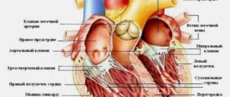
The structure of the heart in mammals with 2 circles of blood circulation is approximately the same. The heart consists of two atria (the first chambers along the path of incoming blood), two ventricles, valves between these chambers, and vessels entering and leaving the heart with valves at their beginnings.
Between the right atrium and the right ventricle is the right atrioventricular
, or a
trioventricular
, which consists of 3 valves.
Therefore, it is called tricuspid
, or
tricuspid
.
Between the left atrium and the left ventricle is the left atrioventricular valve
, which consists of two valves and is called
mitral
.
Valves
, located at the mouths of the vessels extending from the heart, or the main vessels, namely
the aorta
and
pulmonary artery
, are called
“aortic”
and
“pulmonary”
.
Atrioventricular valves
– casement, i.e. their structure resembles doors on the wings: opened and closed, down - up.
Aortic valves
and
the pulmonary artery are different in structure
. Each of them consists of 3 semilunar valves that close in the center. When they open, they press against the wall of their vessel (aorta or pulmonary artery), and close, completely closing the lumen of the vessel. Moreover, their appearance resembles a brand name.
The tissue of the valves themselves, both atrioventricular and semilunar, is thin, even transparent in children, but amazingly elastic and durable, designed by nature for continuous rhythmic work, amounting to billions of monotonous actions.
Between the cavities of the heart, or its chambers, there are partitions that separate the flow of venous and arterial blood. This is the interatrial septum, i.e. between the right and left atria, and the interventricular septum - between the right and left ventricles. In a normal, formed heart, they are completely closed, there are no holes or defects in them
and thus blood never flows from one half of the heart to the other.
Let us dwell in more detail on the anatomical structure of the heart and its chambers. After all, even those that have the same name (atria or ventricles) are structured completely differently and perform different functions.
The shape of the heart resembles a pear lying somewhat on its side, with the top located to the left and below, and the base to the right and above. The apex of the heart is the part of the heart whose movements can be felt if you place your palm on the chest in the fifth intercostal space to the left of the sternum. You can easily feel its push both in yourself and in your child. These are the movements of the apex of the heart with each contraction. The contractions are almost synchronous with the pulse, which can also be easily felt on the arm (where the forearm meets the hand) or on the cervical vessels. The pulse is the filling of the vessels with a wave of blood coming from the heart with each contraction. Pulse frequency and its rhythm are an indirect and easily accessible reflection of the activity of the heart itself.
The apex is the most mobile part of the heart, although the whole thing, all its departments, are in constant motion.
The work of the heart, its movement, consists of two alternating phases - contraction (systole) and relaxation (diastole).
The rhythmic, constant alternation of these phases, necessary for normal functioning, is ensured by the occurrence and conduction of an electrical impulse through a system of special cells - through the nodes and fibers of the conduction system of the heart
.
Impulses arise first in the uppermost, so-called sinus node
, then pass to the second,
atrioventricular node
, and from it - along thinner fibers - to the muscles of the right and left ventricles, causing contraction of all their muscles.
Right atrium
accepts venous blood from the vena cava, i.e. from the whole body and in addition the venous blood of the heart itself. This is the largest chamber in volume and, perhaps, the most stretchable chamber of the heart. If necessary, it can accommodate several times more blood than under normal conditions, i.e. has a gigantic “reserve” of volume. The wall of the right atrium consists of a layer of thin muscle fibers. In addition to the function of “receiving” venous blood, the right atrium performs the function of a cardiac pacemaker. Both main nodes of the conduction system of the heart lie in its walls.
The right atrium connects or, more precisely, opens into the right ventricle through the atrioventricular foramen
regulated
by the tricuspid valve
. This hole is wide enough to allow the entire volume of blood to pass from the atrium into the right ventricle during the period of relaxation of its muscles, i.e. in the diastole phase, and fill its cavity.
The right ventricle is a much thicker-walled muscular structure than the atrium. This is the most anterior part of the heart, lying immediately under the sternum. It is relatively stretchable when needed. The shape of its cavity resembles a new moon appearing in the sky. If you look closely, you can see how the luminous stripe of the month in a semicircle covers a large dark ball of the unlit part of the Moon. Likewise, the right ventricle flows around the powerful cylindrical left ventricle with its cavity.
Inside, this ventricle consists of two cones that extend into each other: the inlet cone and the outlet cone. They converge at their apices at the apex of the heart and are separated at the top by a muscular ridge, the so-called supraventricular crest.
The right ventricle opens into the pulmonary artery, which, together with the aorta, is the so-called main, or “great” vessel. At the transition from the ventricle to the pulmonary artery there is a tricuspid, semilunar valve of the pulmonary trunk, which allows blood to pass in one direction - to the lungs.
The left atrium is the most posterior of the cardiac chambers. It receives oxygenated, arterial blood from the pulmonary veins. There are only four veins and they flow into the posterior wall of the left atrium. The chamber of this atrium is much smaller than the right one, and its ability to stretch is significantly less.
The left atrium opens through the atrioventricular orifice into the left ventricle. This hole contains a bicuspid - mitral - valve, the opening and closing of which regulates the process of filling and emptying of the ventricle in the systole and diastole phases.
The left ventricle is the main one in the heart, and in the entire circulatory system. This is a powerful muscular chamber, the walls of which are 3-4 times thicker than those of its right neighbor. It is a compact cone with the inlet (with the mitral valve) and outlet (with the tricuspid aortic semilunar valve) lying next to each other and closely interconnected.
In order for this entire complex system to work harmoniously and efficiently, it must receive constant necessary nutrition in the form of oxygen and nutrients, and waste products must be removed. For this purpose, there are arterial and venous systems of the heart itself.
The arterial system of the heart itself consists of two - the left
and
the right
-
coronary (coronary) arteries
, which depart at the very beginning, at the mouth of the ascending aorta. These are its first branches. They immediately divide into smaller ones and distribute blood to all parts of the continuously moving heart. The “spent” blood, which has given up oxygen, flows through numerous small veins, which gather into one large one - the coronary sinus - and flow into the cavity of the right atrium. Thus, the heart feeds itself, and its function directly depends on the correct position and condition of the coronary arteries.
So, let's summarize. Anatomically, the heart is a powerful muscular organ with four chambers and four valves. The structure of the chambers and valves is different from each other, because subordinated to performing various tasks. The right parts of the heart are separated from the left by partitions and do not communicate with each other.
Quoted from the book by G. E. Falkovsky, S. M. Krupyanko. The heart of a child. A book for parents about congenital heart defects
How to get treatment at the Scientific Center named after. A.N. Bakuleva?
Online consultations
Arterial pathologies of the heart
Painful sensations with lesions of the aorta are characterized by the following symptoms:
- localized at the top of the chest;
- continuous, not stopping for several days;
- aggravated by load;
- in case of aortic rupture - sharp unbearable pain, loss of consciousness.
Signs of pulmonary artery damage:
- acute pain when trying to inhale, concentrated in one place;
- labored breathing;
- tachycardia;
- a sharp decrease in blood pressure;
- blue skin.
Arterial pathologies require urgent treatment in a hospital. Contact your doctor immediately if you have these symptoms.
Non-cardiological pathologies
With some diseases, a person experiences chest pain of various origins. For example, compression of the intervertebral nerves during osteochondrosis causes a reflex pain reaction.
Advertising:
Possible reasons:
- intercostal neuralgia;
- manifestations of cervical and thoracic osteochondrosis of the spine;
- diseases of the gastrointestinal tract (acute pancreatitis, spasm of the bile ducts of the liver, stomach ulcer);
- diseases of the nervous system;
- pathologies of the respiratory system (pleurisy, pneumonia).
Painful spasms in non-cardiac diseases, unlike an attack of angina, are not relieved by nitroglycerin.
Diseases that cause chest cramps
| Characteristics | Intercostal neuralgia | Osteochondrosis | Gastrointestinal diseases | Neuroses | Pleurisy, pneumonia |
| Nature of pain | piercing, sharp | aching, dull, sometimes tingling | long lasting | various | acute |
| Where does it hurt | clearly localized (between the ribs on the left or right) | radiates to the back, shoulder blades, arm, neck, upper abdomen | epigastric region, stomach | No clear localization | in the area of inflammation |
| What does it depend on | worsens with inhalation and exhalation, sudden movements | worsens with inhalation, straining, turning, bending | associated with food intake | independent of movement | worsens with inhalation, coughing |
| Associated symptoms | — | hand numbness, headache, dizziness | nausea, vomiting, bloating, stool upset | anxiety, sleep disturbances, sweating or chilliness, headache | pulmonary symptoms, fever |
| How to film | painkillers: Diclofenac, Nimesulide, Ibuprofen | supine position, oral and local anesthetics | antispasmodics | antidepressants | — |
So, you suspect a non-cardiac pathology. Of course, the cause of your discomfort must be diagnosed and eliminated: some diseases threaten your life.
Preventive actions
As with any other disease, it is better to try to prevent it than to treat it later. And here the issue of preventing “bull’s heart” is important.
When an enlarged heart occurs, the consequences can manifest themselves in different ways, so with the least risks you should pay attention to your health. Ideally, you should adhere to the principles of a healthy lifestyle and eliminate bad habits such as alcohol and smoking. A person who does not want to face the consequences of an enlarged heart should constantly monitor blood pressure, consume less salt, engage in suitable physical activity, and lose excess weight. The recommendations are simple, but they will help you keep yourself in good shape.
What to do when it hurts
Do you suspect you have heart disease or have you already been diagnosed? If you experience severe chest pain, take the following steps.
Advertising:
- Don't panic, get into a sitting position.
- Unbutton tight clothing, shirt collar.
- Ask to open a window for fresh air.
- Carefully change your body position. If the pain subsides, the cause is most likely due to a non-cardiac disease.
- If the intensity increases and compresses, the possible cause is angina pectoris.
- Dissolve a nitroglycerin tablet under your tongue. The angina attack subsides after a few minutes.
- If the spasm does not stop within 15 minutes, take a second tablet and call an ambulance - there is a possibility of a heart attack.
Even if you managed to relieve the pain on your own, do not relax. You need to be examined by medical specialists.