Definition
Heart flutter is a generalized colloquial definition of one of two pathological conditions.
- Flutter of the ventricles of the heart.
- Atrial flutter.
Ventricular flutter is a life-threatening condition in which the heart is unable to pump blood through its vessels. It is accompanied by loss of consciousness, rapidly increasing symptoms of tissue hypoxia, and without immediate resuscitation, it leads to death within a few minutes.
Therefore, in general practice, heart flutter is most often understood as atrial flutter.
Atrial flutter (AFL) is a cardiac arrhythmia characterized by a rate of 240–400 beats per minute, usually with some degree of atrioventricular (AV) node conduction block. In the most common form of atrial flutter (typical AFL), electrocardiography (ECG) shows a negative sawtooth pattern in leads II, III, and aVF.
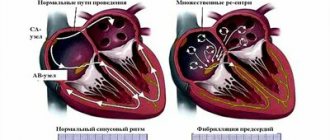
Typical (or classic) AFL involves a single reentrant circuit with activation of a circle in the right atrium around the tricuspid annulus. The front of an electric wave most often propagates counterclockwise. Atypical TP occurs according to a different pattern. It may involve the right or left atrium.
TP is associated with a variety of heart diseases. In most studies, approximately 60% of patients with atrial flutter have coronary artery disease (CAD) or hypertension; 30% do not have heart disease. Unusual forms of atrial flutter were noted during long-term follow-up in 26% of patients with surgical correction of congenital heart defects.
Symptoms in patients with AFL usually reflect decreased cardiac output as a result of increased ventricular rate. The most common symptom is rapid heartbeat. Other symptoms include fatigue, shortness of breath and chest pain. An ECG is important for making a diagnosis. Transthoracic echocardiography (TTE) remains the initial imaging modality of choice for the evaluation of AFL.
In this condition, it is very important to control the ventricular response rate or return the patient to sinus rhythm. First, immediate electrical cardioversion is considered for hemodynamically unstable patients. Catheter ablation remains the most appropriate therapy for patients with typical AFL if they meet the contraindication criteria.
Atrial flutter is similar in many ways to fibrillation (underlying disease, pathological factors, complications and treatment). Some patients even experience both flutter and atrial fibrillation. However, the underlying mechanism of AFL makes this arrhythmia amenable to treatment with percutaneous catheters.
Cardiac health and arrhythmia treatment
To prevent heart disease, you need to lead a healthy lifestyle.
Remember that as soon as you notice the presence of symptoms of any type of arrhythmia, contact your physician or cardiologist. If you feel a sharp deterioration in your health, call an ambulance.
Classification and mechanism of occurrence of disorders
In humans, the most common form of atrial flutter (typical) involves a single reentrant circuit with cycle activation in the right atrium around the tricuspid annulus (most often in a counterclockwise direction) with a region of slow conduction located between the tricuspid valve, the annulus, and the coronary sinus ostium (sub-Eustachian isthmus).
Typical counterclockwise atrial flutter has caudocranial activation (counterclockwise around the tricuspid annulus when viewed on a left anterior oblique fluoroscopic image) of the interatrial septum.
A typical AFL may also have a reverse activation sequence (clockwise around the tricuspid annulus). This option is much less common. When electrical activity moves clockwise, the electrocardiogram (ECG) will show positive flutter waves in leads II, III, and aVF, and they may appear somewhat sinusoidal. This arrhythmia is still considered to be a typical isthmus-dependent flutter. It is usually called reverse typical TP.
Atypical AFL is less studied and electroanatomically characterized. It can happen:
- from the right atrium as a result of surgical scars;
- from the left atrium, in particular from the pulmonary veins (focal entry site);
- from the mitral ring.
Left atrial flutter is a common complication after linear ablation procedures (for atrial fibrillation).
Why is arrhythmia dangerous?
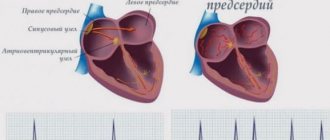
The disease occurs as a result of disturbances in the conduction system of the heart, which ensures sinus rhythm - regular contractions of the myocardium. Contractions of the heart muscle originate in the sinus node and spread through the myocardium of the right atrium to the atrioventricular node. It turns out to be less excitable, so the atria contract first, only then the ventricles. Without timely diagnosis and treatment, arrhythmia causes angina attacks, heart failure, pulmonary edema and even cardiac arrest.
Causes
TP is associated with a variety of heart diseases. In most studies, approximately 30% of patients with atrial flutter have coronary heart disease, 30% have hypertensive heart disease, and 30% have no underlying heart disease. Rheumatic heart disease, congenital heart disease, pericarditis, and cardiomyopathy can also cause flutter. In rare cases, mitral valve prolapse or acute myocardial infarction are associated with this condition.
In addition, the following conditions are associated with TP:
- hypoxia;
- chronic obstructive pulmonary disease;
- pulmonary embolism;
- hyperthyroidism;
- pheochromocytoma;
- electrolyte imbalance;
- alcohol consumption;
- obesity;
- digitalis glycosides;
- myotonic dystrophy in childhood.
AFL may be a consequence of open heart surgery. After cardiac surgery, it can occur as a result of the appearance of natural barriers - incisions in the atria and cardiac scars. Some patients develop atypical left atrial flutter after pulmonary vein isolation procedures for atrial fibrillation.
Although there are no clearly defined genetic conditions that cause atrial flutter, in many cases there is likely a genetic predisposition to its occurrence. Genome-wide association studies in recent years have identified genes associated with atrial flutter.
The PITX2 (paired homeodomain2) gene at the chromosome 4q25 locus is known to play an important role in left-right cardiac asymmetry and has been found to have a strong association with atrial fibrillation and an even stronger association with typical AFL. But there are no clinically available genetic tests that could identify people at increased risk of LT.
What is cardiac arrhythmia?
Arrhythmia is the name of a group of all pathologies that are characterized by disturbances in heart rhythm. Arrhythmias themselves can differ radically from each other in terms of their occurrence, complications, effects on the body, and manifestations. Let's figure out what arrhythmia is, what causes it, what symptoms it manifests, what types of arrhythmia there are, and how to treat the “motor” of your body.
Symptoms and accompanying manifestations
Symptoms in patients with AFL usually reflect decreased cardiac output as a result of increased ventricular rate. At the same time, contrary to popular belief, the feeling of “heart fluttering” does not occur. A condition that some patients tend to describe as “heart fluttering” occurs with paroxysmal ventricular tachycardia.
Typical symptoms of TP include:
- dizziness;
- fatigue or poor exercise tolerance;
- slight shortness of breath;
- pre-fainting state.
Less common symptoms include angina, severe shortness of breath, or fainting resulting from poor left ventricular function. With this arrhythmia, thromboembolic phenomena are possible.
In addition, patients may have symptoms of conditions that cause AFL. They may be non-cardiac (such as hyperthyroidism or lung disease) or cardiac.
Often, arrhythmia in these conditions occurs under the influence of provoking factors - alcohol, pneumonia, acute myocardial infarction and surgical procedures. Stimulants such as ginseng, cocaine, ephedra or methamphetamine can also trigger an attack.
For AFL lasting more than 48 hours, anticoagulation with warfarin or transesophageal echocardiography is necessary to exclude left atrial thrombus before cardioversion to sinus rhythm.
The AFL rhythm itself is unstable and usually reverts to either atrial fibrillation or sinus rhythm.
Remaining stable chronic AFL is uncommon, but possible. A history of preexcitation syndrome (Wolf-Parkinson-White) puts the patient at risk for 1:1 flutter waves, which can cause ventricular fibrillation and death.
Patients who are simultaneously diagnosed with new-onset rapid atrial fibrillation or atrial flutter and decreased left ventricular ejection fraction tend to have a high incidence of left atrial appendage thrombus.
Sinus arrhythmia. Diagnosis and treatment
Characterized by uneven pauses between heartbeats. That is, there is a feeling that at first the heart is about to jump out, and then it seems to freeze for a while. Sinus arrhythmia is present in people after suffering stress, eating food, physical activity, etc.
Symptoms of sinus arrhythmia
This type is characterized by the possible presence of all the symptoms of arrhythmia. When the heart rate accelerates, there is a feeling of lack of air, shortness of breath, and chest pain. When the heart rate slows down, weakness, dizziness and headaches appear. Severe sinus arrhythmia may manifest as loss of consciousness. In this case, you must urgently call an ambulance.
Diagnostics
Electrocardiography (ECG) can provide important information to distinguish “typical” from “atypical” AFL. Transthoracic echocardiography (TTE) is considered the initial diagnostic modality of choice for evaluating atrial flutter.
History and physical examination findings guide laboratory testing. Although hyperthyroidism remains a rare cause of AFL, asymptomatic hyperthyroidism, especially in older patients, may present as fibrillation or AFL and should be excluded during thyroid function testing.
A complete blood count is performed if anemia is suspected or if the patient has a history of recent or current blood loss associated with presenting symptoms.
Chest radiography may be helpful in assessing lung disease and pulmonary vasculature. Chest radiographic findings are usually normal in patients with AFL, but radiographic evidence of pulmonary edema may be noted in subacute cases.
Treatment
The general goals of treatment for symptomatic AFL are similar to those for the treatment of atrial fibrillation and include five goals.
- Ventricular rate control
- Restoration of sinus rhythm
- Preventing recurrent episodes or reducing their frequency or duration
- Prevention of thromboembolic complications
- Minimizing side effects from therapy
However, these goals must be selected based on the needs of each patient. In the acute setting of anticipated hemodynamic collapse, immediate electrical cardioversion is considered for hemodynamically unstable patients according to the Advanced Adult Life Support (ACLS) algorithms for the treatment of atrial fibrillation and AFL.
The main difference between atrial fibrillation and atrial flutter is that most cases of AFL can be treated with radiofrequency ablation (RFA). In all available studies, catheter ablation is superior to heart rate and rhythm control strategies with antiarrhythmic drugs.
In Belgium and other European Union countries, catheter ablation has become the first-line treatment for patients with typical atrial flutter in the absence of contraindications. Ablation is usually performed as an elective procedure, but it can also be done for sudden AFL.
Given its high efficacy and low complication rate, RFA is superior to drug therapy. Successful ablation reduces or eliminates the need for long-term use of antiarrhythmic drugs and anticoagulants (unless the patient also has atrial fibrillation).
Drug therapy
As in patients with atrial fibrillation, the decision about the need for post-conversion anticoagulation is made after consideration of each patient's individual thromboembolic and bleeding risks. Data from transesophageal echocardiography (TEE) studies suggest that anticoagulant therapy is recommended after conversion because adnexal flow velocity becomes very low immediately after conversion and is slow to recover.
For episodes of AFL of undetermined duration or more than 48 hours, anticoagulant therapy should be mandatory. Rate monitoring and therapeutic anticoagulation are required at least 4 weeks before cardioversion. If cardioversion is needed earlier, anticoagulant therapy with intravenous (IV) heparin is given as close to the time of cardioversion as possible. Anticoagulation therapy is also required for at least four weeks after cardioversion. If a thrombus is observed or suspected based on TEE results, cardioversion is postponed.
Contraindications to catheter ablation in patients with AFL are a reason to change treatment. Rate and rhythm control strategies are effective in these patients. Due to the risk of arrhythmia, drugs such as ibutilide, sotalol and dofetilide should be prescribed in hospital settings. Complications may occur after transition to sinus rhythm. The risk of proarrhythmia is probably greatest during the first 24 to 48 hours after starting antiarrhythmic drugs.
Preferred medications that slow atrioventricular (AV) node conduction include beta blockers (labetalol, atenolol, metoprolol, propranolol) and calcium channel blockers (eg, verapamil, diltiazem). These medications are used to control ventricular rhythm. They are also used in patients taking Class IA or IC antiarrhythmic drugs (to prevent the rapid ventricular response that may occur when the atrial rate slows).
Consideration of the use of anticoagulants in this patient population (at least until sinus rhythm is maintained) is imperative. Anticoagulant therapy (eg, warfarin) is indicated, especially if AFL lasts more than 48 hours or its onset is unknown.
Patients should be maintained at a therapeutic international normalized ratio (INR) for 3 weeks before conversion and at least 4 weeks after conversion to sinus rhythm.
In patients with chronic AFL, continuous long-term anticoagulant therapy is recommended. Particular caution is required when adding additional medications (including antibiotics) as they may dramatically change the INR in patients taking warfarin.
In patients requiring cardiac surgery, altering the atrial incision and creating a cryothermal lesion similar to the lesion created by radiofrequency catheter ablation may prevent recurrent postoperative arrhythmia.
Causes of cardiac arrhythmia in women and men
It is worth noting that the causes of arrhythmia are not divided by gender and are practically the same in women and men. The only thing is that women may experience pathology due to premenstrual syndrome (PMS) and menopause. Undoubtedly, there are many reasons for the occurrence of arrhythmia. Let's list the main ones.