All organs, tissues, structures and systems of the human body can be susceptible to inflammatory processes. This even applies to the system of blood vessels, where, it would seem, there is nothing to become inflamed - except the vascular walls. In medicine, there are several general terms to refer to vascular inflammation (vasculitis, angiitis), as well as a number of specific terms indicating the nature or localization of the affected vessels (arteritis, phlebitis, etc.).
The eyeball is “braided” with a network of thin blood vessels that nourish and saturate with oxygen all intraocular elements and media. The ocular vascular system, in its organization, is essentially no different from any other: blood enriched in the lungs enters the arteries, is delivered and distributed in the tissues by the smallest capillaries, then is discharged, along with metabolic products, through the veins. Normally, the vascular network of the eye is practically invisible; if we notice swollen, inflamed or, in common parlance, “burst” eye vessels, this is always a pathology and always a symptom.
The blood supply system of the eye is called the “uveal tract” and consists of the iris (anterior section), ciliary body (middle section, also responsible for accommodation) and choroid (posterior section, which can be called strategic: this is the choroid itself, providing nutrition and oxygenation and regeneration of the photosensitive retinal tissue that covers the inside of the fundus of the eye).
In accordance with the rules of word formation accepted in medicine, inflammation of the vascular system of the eye is designated by the term “uveitis”. It is believed that the original name “uvea” (ancient Greek “grape”) was inspired by the grape-shaped appearance of the ocular choroid, which association becomes especially clear during inflammation.
Causes of uveitis
The statistical share of uveitis in the clinic of ophthalmological diseases is not so large, but noticeable: it is 5-7%. Depending on the location of the lesion, anterior uveitis (inflammation of the iris and/or ciliary body); posterior uveitis , or choroiditis (inflammation of the “back” choroid of the retina, the retina itself and the optic nerve); as well as panuveitis - total inflammation of the entire circulatory system of the eyeball.
Speaking about the causes of uveitis, as well as any other vasculitis, it should be noted that not everything in the etiology and pathogenesis of vascular inflammation has been studied and explained to date.
The dominant cause of any (not only vascular) inflammation is the vital activity of pathogenic microorganisms - viruses, bacteria and fungal cultures, however, in this regard, their sudden activation does not always seem natural and explainable, to which neither the body’s immune defense nor even timely medication has time to fully respond counterattack.
Accordingly, the causes of purely infectious uveitis are toxoplasmosis, syphilis, chlamydia, brucellosis, tuberculosis and other bacterial invasions, pathologically rapid proliferation of fungi (for example, the candida genus), herpes viruses, measles, cytomegalovirus, etc.; in some cases - infection by parasites.
However, a connection has also been established, at least statistically (which indicates a reliable cause-and-effect relationship, which remains to be clarified) between uveitis and background systemic inflammatory processes, such as rheumatism, ankylosing spondylitis (Bechterew-Strumpel-Marie disease), complex infectious-allergic syndromic Reiter triad (urethritis-conjunctivitis-arthritis). Sometimes uveitis develops after and, presumably, as a result of ophthalmic trauma. One of the risk factors is genetic predisposition.
Finally, in some cases the causes of uveitis remain simply unknown.
Forecast
If you consult a doctor in a timely manner, the prognosis will be favorable. The acute form of anterior uveitis has severe symptoms. It can be cured within 3-6 weeks .
It all depends on the complexity of the lesion. The chronic form of the disease is dangerous because it is recurrent in nature - with any exacerbation it begins to develop. Such forms of pathology often cause the development of serious complications.
Therefore, it is important to contact an ophthalmologist on time.
Symptoms of uveitis
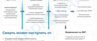
The clinical picture of ophthalmovascular inflammation is determined by a number of conditions: the nature of the pathogenic factor, the localization of the primary lesion, the speed and intensity of development. Accordingly, the symptoms of uveitis are very diverse.
Thus, more or less typical manifestations of anterior uveitis are subjective sensations of “veil before the eyes” and heaviness in the eyeball; decreased visual acuity and clarity; noticeable redness of the sclera; aching, “pulling” or sharp pain; miosis (constriction of the pupil) with weakened or absent response to light, or, on the contrary, a painful reaction of the eye to bright light; hyperlacrimation (increased lacrimation); surge in IOP (intraocular pressure). The extreme, limiting symptom of uveitis in some cases is complete loss of vision.
Posterior uveitis (choroiditis) is dangerous due to a long period of asymptomatic development, when there are neither visible signs nor subjective painful sensations. A decrease in visual acuity and quality also develops, as a rule, too slowly and gradually for the patient to notice it in the early stages. Seeking help is usually due to obvious “fogging” or the appearance of scotomas (blind spots) in the field of vision.
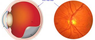
Photo
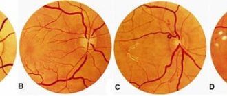
In this picture you can clearly see the condition of a healthy eye and one affected by uveitis.
This is a real photo of a patient who experienced such inflammation:
Diagnosis and treatment of uveitis
Of course, the chances of radical therapeutic success in uveitis depend decisively on how quickly and adequately its treatment is prescribed. If the inflammatory process manages to go far enough, the risk of developing complications, including severe ones, increases - up to cataracts, glaucoma (if the drainage of intraocular fluids is disrupted), fusion of the pupil and the appearance of posterior synechiae (fusions, adhesions), loss of transparency of the vitreous body, damage optic nerve and/or retina (neovascularization, i.e. formation of new vascular networks, detachment). In cases where only one eye is primarily affected, if there is a delay in starting therapy, the second (healthy) eye may become involved in the inflammatory process.
However, early diagnosis and treatment of uveitis is not always possible, and the reasons for this are not only due to the subjective factor (late presentation, absence of symptoms in the initial stages of posterior uveitis). In some cases, uveitis is objectively difficult to diagnose: biomicroscopy of the anterior ocular structures, ophthalmoscopic examination of the fundus, ultrasound scanning or more complex instrumental diagnostic methods is required (and is not always performed). But even with a full-scale and relatively timely examination, it is not always possible to establish the direct cause of inflammation: according to available medical and statistical information, in 30% of cases of uveitis, its pathogenic factor remains unknown. In this case, accordingly, etiopathogenetic treatment (targeted not only at the symptoms, but also at the direct cause of the disease) can only be prescribed thanks to the doctor’s intuition or simple luck of the patient. The general treatment protocol for etiologically unclear uveitis includes anti-inflammatory measures (both local and systemic), antibiotic and immunostimulating (antiviral) drugs, vasodilators, enzyme and physiotherapy.
Medicines of all pharmacological forms are used: drops, ointments, gels, conjunctival and parabulbar injections, tablet preparations, etc. Particular importance is given to mydriatics - drugs that block accommodation and dilate the pupil - to prevent adhesions. In cases where uveitis is accompanied by increased intraocular pressure, hirudotherapy (placement of medicinal leeches) is often highly effective.
It is very difficult to heal a disease without knowing its causes and internal patterns; in many cases, healed uveitis recurs. Therefore, a strategically correct step, the need for which is extremely important to bring to the patient’s consciousness, is to continue an in-depth and comprehensive examination even after symptomatic improvement has been achieved.
What is the minimum examination that may be needed?
General examination by an ophthalmologist with pupil dilation, and, if necessary, optical tomography and retinal angiography.
At the appointment, the ophthalmologist will definitely ask about complaints, collect information about the duration and characteristics of the disease, general status, and may ask you to fill out a special form. questionnaire for a patient with uveitis, conducts a general routine (and, if necessary, in-depth) ophthalmological examination, including autorefractometry (determining the type of vision), visometry (testing visual acuity), tonometry (measurement of intraocular pressure), biomicroscopy (examination of all media of the eye), ophthalmoscopy (detailed examination of the fundus of the eye), if indicated - gonioscopy (examination of the invisible part of the eye - the angle of the anterior chamber).
Diagnosis, treatment and determination of management tactics for uveitis is carried out only by an ophthalmologist. For additional diagnostics, the doctor may prescribe various blood tests, an X-ray (or CT scan) of the chest, and refer you for a consultation with a related specialist (for example, a rheumatologist).
Congenital aniridia
| A disease in which the iris is absent (see structure of the eye). At the same time, behind the cornea there is a picture of a maximally dilated pupil, that is, blackness. Sometimes a rim is visible - a remnant (rudiment) of the iris root and ciliary processes (see.
|
You can find out more about surgical treatment here
Prevention
There is no special prevention for this pathology. The risk of development can be reduced only with timely treatment of chronic diseases . This is especially true for: tuberculosis, syphilis, diabetes, rheumatoid arthritis. These diseases have a very negative effect on the organs of the visual apparatus.
The acute form of uveitis manifests itself with severe symptoms. Therefore, you should not ignore them, but contact an ophthalmologist as soon as possible. With timely treatment, successful results can be achieved. Lack of treatment for the acute form will lead to the development of the chronic stage of this pathology . In this case, drug therapy will be complex and lengthy.
This disease has different etiologies. It can occur suddenly in patients of any age. Therefore, it is important to follow simple prevention: hygiene standards, proper use of medications, avoidance of allergens . If activities involve chemicals, precautions must be taken to avoid eye burns. Prevention will reduce the risk of developing pathology.
Coloboma of the iris
| Typically, iris coloboma is located inferiorly at 06.00. clock, resembling a pear or a keyhole (see.
|
You can learn about plastic restoration of the pupil in our video
Install Flash Player to watch the movie.