1.What is mitral valve stenosis?
Mitral valve stenosis
, or
mitral stenosis
is a narrowing of the left atrioventricular orifice, which impedes the natural outflow of blood.
The basis of this disease is the thickening of the valve leaflets and limitation of their motor function. Mitral valve stenosis
– a curable heart defect in patients of any age, subject to proper and timely treatment.
A must read! Help with treatment and hospitalization!
2. Causes of stenosis
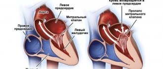
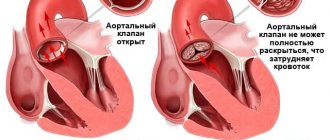
The mitral valve is located between the left ventricle and the left atrium. It opens in diastole. Arterial blood from the right atrium then enters the left ventricle through the atrioventricular foramen. Blood enters the left ventricle through the mitral valve, which consists of two leaflets. Under blood pressure during contractions of the left ventricle during systole, it closes, preventing the reverse outflow of blood. With mitral valve stenosis, the atrioventricular orifice narrows, which leads to disruption of the flow of blood into the left ventricle from the left atrium.
The main cause of mitral stenosis is rheumatism
– a systemic inflammatory disease that affects the heart tissue and joints. Streptococcal infectious diseases contribute to the development of rheumatism: scarlet fever, tonsillitis, erysipelas, etc. Children aged 7 to 15 years are at risk. In medical practice, there are cases when, despite numerous cases of sore throat, people considered themselves absolutely healthy, but at the same time had scars on the inner lining of the heart - the first sign of fusion of the valves and the development of mitral valve stenosis.
Visit our Cardiology page
Mitral stenosis: causes and consequences of the disease
The vitality of the human body directly depends on the proper functioning of the heart. The mitral valve is located between the left atrium and ventricle. Its function is to pass arterial blood into the left ventricle and prevent its reverse outflow. If the valve leaflets thicken and fuse together, the size of the atrioventricular opening becomes smaller. This narrowing is called mitral orifice stenosis, or mitral stenosis. Causing a large number of problems, the disease can cause irreversible consequences.
Causes and risk factors for developing the disease
The causes of the disease are varied, and although it has been scientifically proven that 4/5 of the cases of patients had a rheumatic etiology for the development of mitral stenosis or congenital, which is detected during the intrauterine development of the child. However, the remaining 25% includes diseases such as:
- heart injury and atherosclerosis;
- left atrial myxoma;
- intracardiac thrombi;
- heart surgery;
- aortic insufficiency;
- streptococcal infection;
- infective endocarditis and syphilis.
Heart defects play an important role in development; mitral stenosis affects other heart valves.
Due to the fact that a person’s appearance and condition do not undergo any changes for a long time, the disease can reveal itself 10-30 years after its onset. A malfunction of the organ catalyzes the launch of a compensation mechanism, which facilitates the passage of blood and reduces the effect of the disease on hemodynamics inside the heart: the pressure in the atrium cavity increases by 15-20 mm Hg. Art., systole lengthens in the left atrium, and at the same time myocardial hypertrophy develops in it.
8
24/7
However, with further progression of the disorders, the contractile function of the ventricle decreases, it expands, the load on the right atrium increases, which ultimately becomes the cause of a malfunction of the entire organ.
Degrees and stages of the disease
In a normal state, the size of the mitral orifice does not exceed 4-6 cm2, so its narrowing immediately affects intracardiac hemodynamics.
Mitral stenosis depends on the area of narrowing and has several degrees.
| Degree | Characteristic |
| First | Holes > 3 cm2. It is a minor violation and is not noticeable without a special examination. |
| Second | Sholes = 2.3-2.9 cm2. A moderate type of change does not have a strong effect on the functioning of the organ. |
| Third | Sholes = 1.7-2.2 cm2. The disease is severe and characteristic symptoms appear. |
| Fourth | Sholes = 1, -1.6 cm2. Critical mitral stenosis of the heart requires urgent hospitalization of the patient. |
The gradual progression of the disease affects the process of blood circulation within the large and small cardiac circles. Doctors distinguish only 5 stages of pathology.
- First. Mitral stenosis is completely compensated by the atrium. Signs of the disease are poorly detected.
- Second. Disturbances appear in the pulmonary circulation. During physical activity, initial symptoms are revealed.
- Third. The improper flow of blood in the large circle makes itself felt, and stagnation in the small circle is clearly expressed.
- Fourth. Atrial fibrillation appears, which is a consequence of pronounced signs of blood stagnation in both circles.
- Fifth. Dystrophy corresponding to stage 3 of heart failure is detected.
Changes in cardinal hemodynamics and the degree of narrowing are of great importance when prescribing treatment and in the further prognosis of the development of the disease. This is especially important for people with congenital aortic mitral stenosis, since it is most often combined with other heart pathologies.
Symptoms
For each stage of the development of the disease, specific symptoms are typical, which can be used to describe the clinical picture of the formation of the pathology.
In the initial stage of pathology, it can be identified through a complete clinical examination. The patient has no complaints and is in excellent health.
With the onset of the second stage, a slight increase in blood pressure and shortness of breath after physical activity may occur.
Stagnation is clearly expressed in both circles of blood circulation at the third stage of development:
- persistent shortness of breath with cough and foamy sputum;
- significant increase in pressure;
- swelling of the limbs;
- the liver and spleen are enlarged.
At the fourth stage, the size of the heart becomes noticeable:
- the liver and spleen are felt upon palpation;
- venous pressure is much higher than permissible norms;
- swelling of the extremities is clearly expressed;
- small ascites;
- initial form of atrial fibrillation.
The last fifth stage is characterized by all the symptoms in a pronounced form, in addition, shortness of breath does not go away even at rest, and the liver extends beyond the costal arch.
Drug treatment is possible up to the fourth stage, inclusive, after which it is pointless.
8
24/7
Diagnosis methods
Mitral stenosis detected at an early stage makes it possible to control and restrain the development of the disease. However, the body's compensatory capabilities prevent symptoms from appearing. Therefore, almost all cases of timely diagnosis recorded by statistics are a consequence of the discovery of the disease during preventive examinations.
Mostly, people come to doctors with complaints due to decompensation, when many systems of the body are modified due to mitral stenosis, treatment will no longer help, and urgent surgical care is required.
The specialist at the first moment pays attention to a certain type of patient’s complaints. Then he begins to collect an anamnesis, in which the patient’s previous illnesses and injuries play an important role.
An objective examination may reveal:
- diastolic murmur when listening to the heart;
- increased size of the liver and spleen;
- edema and ascites.
To clarify the diagnosis, complete laboratory tests of blood, urine, ultrasound and ECG are performed
- in general tests, deviations in ESR indicators will appear, leukocytosis and anemia may appear;
- biochemical studies show deviations from the norm only if the cause of the disease is atherosclerotic;
- antistreptolysin is detected in ASLO results;
- An ECG will indicate hypertrophy of the heart;
- During an ultrasound, the degree of mitral stenosis is revealed, the presence of thrombosis is determined by the stage of the disease.
In addition, to exclude calcification and vegetative changes in the valve, probing of the heart cavities is done, and radiography allows one to study the pulmonary pattern and other signs of the disease.
Treatment in early and late stages
The methods of treatment prescribed by a cardiologist depend not only on the stage of the disease, but also on the cause of its occurrence.
| Degrees | Methods |
| Diet therapy is used, physical activity is reduced, and a therapeutic course of medications is prescribed to suppress the etiological factor. Then drugs are gradually added to relieve excess stress on the heart muscles. In this case, the patient may need to take them for life. The emergence of atrial fibrillation makes it necessary to add new drugs to prevent thrombosis. |
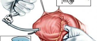
| If the patient’s well-being deteriorates, the mitral orifice narrows by more than 1.2 cm2 | Initially, an operation is performed to dissect the fused valve leaflets. But in the case when the clinic indicates the futility of commissurotomy in mitral stenosis, the diseased valve is completely replaced with an artificial one. |
A positive prognosis is given with young patients and early diagnosis.
Surgical intervention, according to experts, is preferable to therapeutic methods, since, despite a certain risk, it is possible to guarantee the absence of re-development of stenosis.
The age category of the patient leaves a certain imprint on the development of the disease. In older people, starting from 60 years of age, the development of mitral valve disease is considered a characteristic clinical feature. Despite its constant slow progression, survival rates without surgery are very high.
Disease prevention
To prevent the disease from coming suddenly, it is necessary to monitor your health and undergo regular preventive examinations. A visit to a specialist’s office is especially important if:
- shortness of breath and hemoptysis appear with any physical activity;
- lying in a horizontal position, attacks of suffocation begin;
- when working, fatigue occurs quickly and muscle weakness is felt;
- one feels the uneven functioning of the heart, and also dull aching pains appear in it.
In addition, it is important to carry out measures aimed at preventing the development of pathology:
- increase the level of natural immunity and adaptive capabilities of the body through hardening;
- the food consumed must be balanced and contain the required amount of microelements, vitamins and nutrients;
- is often outdoors, doing physical activity and walking;
- Avoid contact with patients with tonsillitis and pharyngitis; in case of illness, promptly treat these pathologies.
People who have already been diagnosed with mitral stenosis need to take the medications prescribed by the doctor, in strict accordance with the prescription, and be examined at the appointed time.
It should be remembered that only timely diagnosis of pathology allows one to avoid life-threatening consequences. You cannot put off visiting a doctor - every day can become a decisive factor in drawing up a further prognosis.
8
24/7
3. Symptoms of the disease
Symptoms of mitral valve stenosis are usually mild. In most cases, this disease occurs in a latent form for many years. The very first sign of the development of mitral stenosis is shortness of breath and fatigue, even in the morning. Other main symptoms of this disease include:
- increased fatigue;
- feeling of heartbeat;
- shortness of breath and feeling tired;
- swelling of the legs;
- frequent respiratory diseases;
- severe cough, as well as chest pain and discomfort.
About our clinic Chistye Prudy metro station Medintercom page!
4. Diagnosis and treatment of mitral valve stenosis
If you notice symptoms of stenosis, you should definitely consult a doctor, as symptoms of stenosis may worsen over time. For example, during significant physical activity and stress. Diagnosis of heart problems begins with a general examination, in particular with auscultation - listening to murmurs in the heart and lungs using a phonendoscope. Based on the data obtained, the doctor decides whether additional examination is necessary. The doctor may prescribe:
- echocardiography - sound examination of cardiac tissue;
- electrocardiography and Holter monitoring;
- chest x-ray;
- cardiac catheterization.
Diagnosis of mitral stenosis is the first step in treating this disease.
, because it is on the basis of the data obtained during the examination that the doctor will draw up an individual treatment regimen for mitral stenosis for you. Currently, there are two main forms of treatment for mitral valve stenosis: surgical and conservative.
- Surgical treatment of mitral valve stenosis involves heart surgery and is not suitable for all patients.
- Conservative treatment of mitral valve stenosis is based on drug therapy and is not able to cure the stenosis directly. But medications can eliminate the complications of this defect: heart failure, arrhythmia, etc.
Treatment of mitral stenosis, like all other heart diseases, is carried out by a cardiologist and includes regular examinations to record indicators of heart condition and monitor the course of the disease. People diagnosed with mitral valve stenosis should understand that this disease will be with them for the rest of their lives. To improve your well-being and prevent complications of heart disease, it is important to take care of your health and undergo regular medical examinations, as well as avoid strong physical and emotional stress and hypothermia.
Diagnosis of mitral-tricuspid disease
Recognition of multivalve defects is extremely difficult and requires a careful analysis of the patients’ complaints and medical history, a thorough and consistent physical examination of the cardiovascular system, and the use of a set of instrumental methods (ECG, phonocardiography, sphygmography, echocardiography, radiography). In difficult cases, to diagnose the defect, they resort to methods of probing the cavities of the heart and ventriculography.
The key point in diagnosing mitral-tricuspid disease is the presence of a long history of rheumatic fever. Therefore, the diagnosis of mitral-tricuspid disease should be carried out jointly by a cardiologist, cardiac surgeon and rheumatologist.
An approximate examination scheme looks like this:
- Oral questioning of the patient regarding complaints. In the early stages there are none, which complicates the work of doctors.
- Anamnesis collection. The nature of everyday life, family history, duration of manifestations, somatic diseases current and suffered in the past. These are all important factors. They must be recorded in writing.
- Listening to the sound of beating. The so-called auscultation. The tones are dull, regular, pathological premature contractions occur, as with extrasystole. With minor mitral stenosis, a diastolic murmur is heard immediately after the opening tone of the mitral valve (in protodiastole) and usually indicates a significant pressure gradient.
- Measuring blood pressure levels. Also heart rate.
- Daily monitoring. A more subtle technique. Determines heart rate dynamics against the background of a normal, habitual lifestyle for the patient. Considered the gold standard in early diagnosis. Can be carried out repeatedly, as needed.
- Electrocardiography. The second most important event. Deciphering the results requires high qualifications, which is why errors, sometimes fatal ones, are likely and frequent.
- Echocardiography. Effective at stage 2 and beyond. Since at advanced stages organic changes in the heart are observed. Thickening or hypertrophy of the ventricles is the most common sign.
- Ultrasound assessment of blood flow velocity.
- Load tests. They are carried out, but with great caution, since cardiac arrest is possible due to intense stress. Doctors must be ready and provide first aid to the patient in a timely manner. This is a big risk; whether to resort to the technique or not is a moot point. You need to weigh all the points. As a rule, they are prescribed when other methods provide little information.
- MRI or CT. Allows visualization of cardiac structures in detail. Iodine or gadolinium based contrast is used as needed.
Other methods are shown as part of advanced diagnostics.