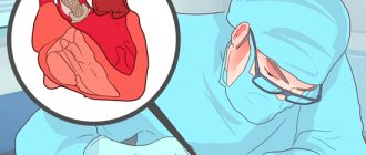
Heart valve prolapse is a disease accompanied by a malfunction of the valve located between the ventricle and the left atrium and is a common and relatively harmless anomaly, during which an unnatural protrusion of the valve leaflets appears when the heart contracts.
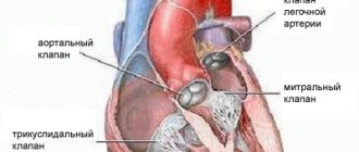
A heart valve is a movable valve consisting of individual elements that blocks the openings for blood circulation from one part of the heart to another. The valves' job is to control blood flow. There are four valves in the heart - mitral, tricuspid, aortic and pulmonary, which allow blood to flow in one direction and prevent it from returning back. When the heart muscle contracts, pressure is created, and blood is ejected from the heart, and the valves that regulate the movement of blood in this direction at the time of muscle contraction open. After the muscle contracts, the pressure in the heart drops, the valve closes, and blood cannot return to the heart. Among other types of prolapse, the most common is mitral valve prolapse, which is caused by congenital weakness of the connective tissue that makes up the heart valves.
What is MVP (mitral valve prolapse)?
Mitral valve prolapse (MVP) is a cardiac anomaly and one of the most common heart pathologies.
In the presence of this pathology, the mitral valve leaflets sag/bulge into the atrium cavity; this occurs at the moment of contraction of the left ventricle of the heart. As a result of this cardiac abnormality, regurgitation can occur, where some blood flows back into the left atrium. This pathology is benign (unlike heart defects, for example), but, unfortunately, it can also have serious unpleasant consequences. Severe regurgitation can cause disability. In some cases, mitral valve prolapse requires serious treatment (medical or even surgical), in others, doctors limit themselves to only monitoring the patient; the person must undergo regular examinations and monitor his lifestyle. Very often a person does not know that he has PMH and this anomaly is discovered during a medical examination on some other issue, since the symptoms of this disease are often non-specific: headache, fatigue, dizziness, cardiac pain. Therefore, it is very important if you have health problems, even if it is mild fatigue or malaise, which is long-lasting, not to put off visiting a doctor.
Mitral valve prolapse: dangerous or safe?
During a routine examination, did the doctor listen to a heart murmur? What could it be connected with?
Galina Petrovna Episheva, a therapist at the Expert Clinic Kursk, told us about one of the ailments with such manifestations, often detected by chance. Mitral valve prolapse is the topic of our conversation.
— Galina Petrovna, what is mitral valve prolapse?
This is a pathology characterized by dysfunction of the bicuspid heart valve, located between the left ventricle and the left atrium.
What happens to the heart during prolapse? Normally, during contraction of the left ventricle, the valve leaflets close so that blood moves only into the aorta and does not flow back into the atrium. With prolapse, there is some bending of the valve (or valves) in the direction of the atrium, and a certain amount of blood is thrown there.
— Is this a separate disease that is coded in the International Classification of Diseases or is it a syndrome?
This is a disease.
Mitral valve prolapse is one of the heart defects
— Is mitral valve prolapse divided into stages and degrees?
Yes. According to the classification, there are 3 degrees. In the first case, the protrusion of the leaflets towards the left atrium is 3-6 mm. With the second - up to 9 mm. With the third - more than 9 mm.
— How does mitral valve prolapse differ from heart disease?
Heart defects are a whole group of pathologies. Prolapse, in fact, is one of the defects.
— Is mitral valve prolapse an accidental finding or are there signs by which it can be identified?
For the most part, it is discovered by chance, since most often it is not accompanied by obvious symptoms - in particular, the first and second degrees. The main symptom is a murmur when listening to the heart. Usually, after this, the doctor refers the patient to an ultrasound of the heart (echocardiography), where this diagnosis is confirmed (or excluded).
— Does mitral valve prolapse require treatment?
If we are talking about the first degree and there are no symptoms, then everything can be limited to observation. In other cases, based on diagnostic results, therapy may be prescribed.
— For what reasons does mitral valve prolapse occur?
The issue has not yet been fully studied. A known role is played by the pathology of connective tissue that develops in the fetus. In this case, they talk about the primary nature of prolapse.
The cause of secondary prolapse can be some heart diseases - for example, rheumatism, coronary heart disease, endocarditis.
— When is prolapse dangerous and when is it safe?
This depends, in particular, on the volume of blood thrown back into the atrium. The larger this value, the more dangerous the defect. Possible consequences:
- increased pressure in the pulmonary vascular system;
- disturbance of heart rhythm, blood supply to the brain (up to stroke);
- circulatory failure;
— perforation of valve flaps;
- cardiac arrest.
People with this condition are more likely to have a mitral valve infection. Therefore, timely treatment of any foci of infection in the body (for example, tonsils in chronic tonsillitis, carious teeth), prevention of colds, sore throats is especially important for them.
— If a young man has mitral valve prolapse, will he be taken into the army?
This pathology may be the reason for a delay or complete exemption from military service. For example, with the first degree and the absence of symptoms, a young man falls into category “B” and can serve. At the same time, the commanding staff and the leading cardiologist of the military unit are warned about his illness. Such a conscript may be prohibited from physical and psycho-emotional stress.
At higher levels, guys are exempt from service.
— Is it possible to play sports with mitral valve prolapse?
Physical exercise is permitted. In this case, the protrusion of the valve leaflets should not exceed 6 mm. Of course, the level of load for any degree of prolapse is selected strictly individually.
A person with mitral valve prolapse should be under the supervision of a cardiologist
— Galina Petrovna, if mitral valve prolapse is diagnosed, does this mean that the heart of such a patient needs close attention? How often should you visit a cardiologist for prolapse?
Yes, such a person should be under the supervision of a cardiologist and follow his recommendations. The frequency of doctor visits is 1-2 times a year. It is necessary to undergo cardiac ultrasound with the same frequency.
It is important to visit a dentist and/or otolaryngologist in a timely manner (for preventive purposes and to eliminate chronic foci of infection). Give up bad habits and caffeine-containing products. Perform adequate physical activity.
The editors recommend:
Heart murmurs in a child: looking for reasons
How to keep your heart healthy?
Medical examination before starting classes in the gym
For reference:
Galina Petrovna Episheva
Graduate of the Faculty of General Medicine of Kursk State Medical University in 1990.
In 1991, she completed her internship in the specialty “Therapy”. Doctor of the highest category.
Currently, she is a therapist at Clinic Expert Kursk. Receives at the address: st. Karl Liebknecht, no. 7.
Mitral valve prolapse: causes
There are 2 types of mitral valve prolapse - primary and secondary, depending on the origin of the pathology.
The causes of primary mitral valve prolapse are genetic defects. This issue has not yet been fully studied, but there is evidence that MVP is a disease that is inherited. In addition, negative factors that have a negative effect on the fetus while still in the womb can affect the possibility of a person developing MVP. Such factors include viral diseases of the mother during pregnancy, gestosis, and poor environmental conditions.
Primary MVP is divided into:
- Mitral valve prolapse as an independent disease.
- Mitral valve prolapse as a manifestation of connective tissue development disorders (connective tissue dysplasia, Marfan syndrome, Ehlers-Danlos syndrome). In this case, the pathology, in addition to MVP, can be manifested by hypermobility of the joints, curvature of the spine, deformation of the chest; a person often experiences dislocations and subluxations of the joints, and other symptoms.
The causes of secondary MVP are other diseases, and it is against their background that secondary cardiac prolapse occurs:
- congenital heart defects;
- acquired heart defects;
- ICHD (coronary heart disease);
- myocarditis;
- rheumatic heart disease;
- cardiomyopathy.
Pathogenesis of MVP.
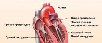
The mitral valve separates the cavities of the ventricle of the heart and its left atrium. When the mitral valve is normal, its leaflets are “deviated” downward at the moment the myocardium relaxes and the cavities of the heart are filled with blood, and vice versa – the leaflets are “raised” upward at the moment of atrial contraction, the left atrioventricular orifice is closed. With MVP, the valve does not work correctly: during the phase of atrial contraction, the valve leaflets “bend” into the cavity of the left atrium, but this should not be the case. Indeed, for this reason, the atrioventricular opening is completely or partially blocked and a reverse movement of blood occurs from the left ventricle to the left atrium (mitral regurgitation).
If a person has mitral valve prolapse, the ability of the heart muscle to contract may decrease, which in turn can lead to circulatory failure; The presence of arterial hypotension, borderline pulmonary hypertension, metabolic disorders and dysfunction of the autonomic nervous system may also be noted.
The heart of an expectant mother: what are the dangers of mitral valve prolapse?
The human heart is a tireless worker. On average, this muscular organ weighs only about 300 grams, but daily pumps 2 thousand liters of blood through 90 thousand kilometers of blood vessels.
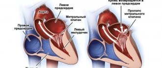
The heart consists of four chambers - the right and left atria and ventricles. Its right and left halves perform different functions. Normally, they are isolated from each other, but the atria and ventricles of the same name are separated from each other by special valves. The right atrium and right ventricle have a tricuspid valve, and the left-sided sections have a bicuspid, or mitral, valve. The purpose of these valves is to ensure proper blood flow. Namely: when the atria contract, blood should freely enter the ventricles, and when the ventricles contract, it should be sent further into the blood vessels. That is, the valves prevent reverse blood flow (regurgitation). Venous blood (saturated with carbon dioxide) enters the right atrium, and then into the right ventricle. Then it goes to the lungs, where it is enriched with oxygen and becomes arterial, acquiring a bright scarlet color. This is the so-called pulmonary circulation. Arterial blood is pumped into the left atrium and, bypassing the mitral valve, ends up in the left ventricle. From here, thanks to powerful muscle contractions, blood flows into the aorta, the largest artery in the body. Further, through the system of blood vessels, oxygen-rich blood is sent to all tissues and organs. This is a large circle of blood circulation. But if there is damage to the blood vessels, heart or valves, then malfunctions may occur in this ideal mechanism.
Mitral valve prolapse: “flapping” leaflet
The mitral valve consists of two leaflets - anterior and posterior, which separate the cavities of the left ventricle and left atrium. This ensures proper blood flow - when the heart muscle contracts, blood should only move in one direction. With mitral valve prolapse (other names for this pathology are “flapping” or “sailing” valve, Barlow’s syndrome, late systolic murmur syndrome), its leaflets do not close completely, as if “sagging”. In this case, the left ventricle and left atrium experience a constant increased load caused by excess blood volume. In severe cases, such long-term effects on the heart can lead to serious problems.
Looking for the causes of mitral valve prolapse
What is the cause of this anomaly? Very often - heredity, namely, a violation of the development of connective tissue (connective tissue dysplasia). The fact is that connective tissue makes up half of the body’s weight and performs many functions: protective, supporting, mechanical, etc. In other words, it forms the skeleton, joints, tendons, ligaments, outer integument, forms the walls of blood vessels, and is included in all organs and systems of the body. The proper functioning of connective tissue largely depends on how the child developed in the womb. Violation of the formation of connective tissue in the fetus is the cause of many congenital diseases, including mitral valve prolapse. And very often, prolapse is accompanied by other “companion diagnoses” caused by insufficiency of connective tissue, varicose veins, prolapse of internal organs, myopia, and disorders of the nervous system.
What causes connective tissue dysplasia? The answer from doctors is unanimous: one of the main reasons is a deficiency of microelements, in particular potassium and magnesium. How can you enrich your diet with essential microelements? Include fresh meat dishes, milk, buckwheat, dried apricots, baked potatoes, spinach, bananas, and nuts in your menu. Completely eliminate simple carbohydrates - sweet pastries, sugar, as well as tea and coffee. If connective tissue dysplasia is suspected, the pregnant woman is prescribed additional potassium and magnesium supplements along with a special diet.
Other causes include previous inflammatory diseases, injuries, age-related changes, and disorders of the elasticity of connective tissue.
Types of mitral valve prolapse
Mitral valve prolapse (MVP) is divided into two groups:
anatomical (congenital);
as a syndrome associated with disruption of the nervous and hormonal systems , as well as arising against the background of various diseases - rheumatism, coronary heart disease or chest trauma.
The more significant the non-closure of its valves, the more the load on the heart increases, which has to pump a larger volume of blood. The left atrium, under constant pressure, enlarges. And during pregnancy and childbirth, when the heart is already working “for two,” additional loads can pose a threat to the health of the expectant mother and fetus.
There are three degrees of mitral valve prolapse:
- I degree – non-closure of the valves by 3–6 mm;
- II degree – 6–9 mm;
- III degree – over 9 mm;
It is also customary to distinguish the severity of reverse blood flow (regurgitation):
- I degree – regurgitation at the level of the leaflets;
- II degree – regurgitation to the middle of the atrium;
- III degree – regurgitation to the opposite side of the atrium.
Let's evaluate the risks of mitral valve prolapse during pregnancy
During pregnancy, a third circulation is added to the systemic and pulmonary circulation - the uteroplacental one. If a woman is expecting one baby, then the blood volume increases on average by 30–50%. If there are twins or triplets, then even more. Wise nature has prepared a woman’s body for such stress - during pregnancy, the size of the heart increases slightly, its muscle walls thicken. From about the 20th week, the heart rate increases - by 10-12 beats per minute. Thanks to these changes, the developing fetus is provided with all the necessary substances supplied by the blood.
As a rule, antenatal clinics require visits to a cardiologist during pregnancy, even if the expectant mother has absolutely no complaints. It is important to promptly inform your gynecologist about all existing (and past) heart problems.
In young women, minor mitral valve prolapse, as a rule, does not manifest itself in any way and is detected only by echocardiography.
With mild prolapse and slight regurgitation (grades I–II), the expectant mother usually feels normal. The pregnancy proceeds well, and the woman gives birth on her own. In this case, prolapse is considered not as a disease, but as an individual anatomical feature. Such pregnant women do not need specialized observation or treatment by a cardiologist.
If the degree of regurgitation and sagging of the mitral valve is somewhat greater, it is enough for a woman to undergo a follow-up examination with a cardiologist every 3 months.
With age, with deep prolapse, specific symptoms appear: rhythm disturbances, shortness of breath, pain in the heart. And if the expectant mother has grade III prolapse with symptoms of heart failure (such as swelling, shortness of breath, heart rhythm disturbances), then both a gynecologist and a cardiologist should monitor her condition during pregnancy.
To reduce all possible risks, it is very important for such expectant mothers to follow a strict sleep, nutrition and physical activity regime. They will have to eliminate salt from the menu and limit fluid intake to reduce the risk of swelling.
Is hospitalization necessary for mitral valve prolapse during pregnancy?
As already mentioned, for mild prolapse, only observation by a cardiologist is sufficient.
Indications for hospitalization in pregnant women with MVP may be the development of gestosis (a complication of the second half of pregnancy, in which pressure increases, swelling and protein appear in the urine), an increase in prolapse, an acute increase in pressure in the left atrium, accompanied by shortness of breath, fainting and dull pain in the left atrium. breasts
In this case, the expectant mother is hospitalized and prescribed treatment aimed at eliminating the main manifestations of mitral valve prolapse, heart rhythm disturbances, and also preventing damage to the heart muscle.
Treatment of mitral valve prolapse
No special treatment is required for this diagnosis. Intervention is necessary only in cases where arrhythmia (any disturbance in the regularity or frequency of normal heart rhythm) or pain in the heart is observed. For these conditions, sedatives (for example, products containing motherwort and valerian) help well. If necessary, the doctor will prescribe medications to prevent arrhythmia, as well as medications containing magnesium.
Perhaps the most frequently asked question when diagnosing mitral valve prolapse is – is it possible to give birth on your own? Will the heart survive? Doctors are quick to reassure: this diagnosis is not a contraindication for pregnancy and childbirth. It is only important to monitor your well-being and remember that you are responsible not only for your health, but also for the health of your child.
Is mitral valve prolapse hereditary? Alas, yes. The presence of this anomaly in a pregnant woman is one of the most common risk factors, and in the vast majority of cases, prolapse is a congenital defect of the heart valve.
Most people with this diagnosis have close relatives with a similar pathology. Children with prolapse have a thin build, they often have disorders of the musculoskeletal system (scoliosis, flat feet, weak ligaments). They are restless and get tired quickly.
But there is also good news. Mitral valve prolapse, as a rule, does not have any significant symptoms and does not require treatment (if we are talking about degrees I and II). Often the condition of the mitral valve remains unchanged throughout life. In some cases, as the child grows, the prolapse may decrease or disappear altogether. And to a large extent this will depend on the proper nutrition of the expectant mother and baby.
With minor prolapse (grades I–II), childbirth takes place in regular maternity hospitals; special observation in this case, as a rule, is not required.
In more serious cases, you will have to give birth in a specialized maternity hospital, which delivers births to women with serious cardiovascular diseases. The team of doctors, in addition to the obstetrician-gynecologist, neonatologist and anesthesiologist, also includes a cardiologist who closely monitors the work of the mother’s heart. If there are indications, the expectant mother undergoes a caesarean section.
Mitral valve prolapse: symptoms.
Mitral valve prolapse can occur either with pronounced symptoms or absolutely “quietly”, without making itself felt. It is necessary to pay attention to the body’s signals, including “simple” headaches and fatigue, do not attribute them, for example, to stress, do not ignore them and find the cause.
In children and adolescents, quite often the disease is asymptomatic or accompanied by only minor discomfort in the chest area. Also, in the presence of primary MVP, diseases are detected in children that indicate a disorder in the development of connective tissue:
- scoliosis;
- chest deformation;
- joint hypermobility;
- hip dysplasia;
- flat feet;
- hernias (umbilical, inguinal);
- nephroptosis, etc.
At the same time, it is possible to identify symptoms characteristic of mitral valve prolapse:
- low blood pressure, resulting in dizziness;
- rapid heartbeat;
- feeling that the heart is working “intermittently”;
- pre-fainting state, which is manifested, among other things, by a feeling of nausea, spots before the eyes;
- pressing or squeezing (sometimes stabbing) pain behind the sternum or in the chest (its left side) - can appear both from physical activity, stress, and for no specific reason;
- feeling of constant fatigue, breathing problems (difficult), swelling of the legs, pain in the right side (the cause of the pain is an enlarged liver).
Secondary MVP is also supplemented by symptoms of the patient’s underlying disease - Marfan syndrome, heart defects (congenital), rheumatic carditis.
Congenital mitral valve defects
The mitral valve is located between the left atrium and the left ventricle. It is a complex natural mechanism and consists of two valves - thin plates, which, like the sails of a schooner, are controlled by a network of ropes and cords starting from the heads of small papillary muscles of the wall of the left ventricle. These cords are called chords. The papillary muscles, chords and valves are a single apparatus, thanks to the precise and coordinated work of which the “gateway” of the mitral orifice opens and closes with each contraction of the heart.
Isolated congenital defects
Isolated congenital
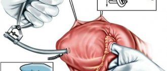
Mitral valve defects that affect only it are a significant rarity. Valve changes are often combined with other congenital heart defects, which relate either to valve rings in general (AVR) or to underdevelopment of the entire left half of the heart (LVH, i.e. hypoplastic left ventricular syndrome, aortic atresia). However, congenital valve defects with a normally developed left ventricle have been described: valve stenosis and valve insufficiency. Congenital mitral valve stenoses are extremely rare. They may be associated with improper formation of the valves, with a membrane located above the valves, with improper formation of the valves, creating two small holes instead of one large one. In all cases, the cause of heart failure in the form of an enlarged heart, stagnation of blood in the lungs, and enlarged liver can be suspected and identified using non-invasive diagnostic methods. Surgical treatment is possible, and its choice depends on the type of damage to the valve structures: from dissection of the narrowed sections of the valves to valve replacement.
Much more often, the mitral valve is affected in cases of endocarditis or rheumatism, and these, no longer congenital causes, should be excluded first.
Mitral valve prolapse
Congenital mitral valve insufficiency, which would necessitate early referral to cardiac surgery, is extremely rare. But one of the most common
Cardiological diagnoses have recently affected him directly.
This is mitral valve prolapse . Typically, the diagnosis is made in a completely asymptomatic child during a random examination on the basis of a slight murmur, which was previously called a “functional heart murmur” and was not paid much attention to. With the advent and implementation of echocardiography, the causes of this noise have become clearer. Most often, it is caused by inaccurate closure of the valve flaps with each other, as a result of which a stream of blood at the moment of contraction of the ventricle flows back and leaks into the atrium. This may be caused by the fact that one of the sections of the valve bends back under pressure, “prolapses” towards the atrium and allows this stream to pass through. This is prolapse. The degree of mitral valve prolapse can vary - from minor to severe regurgitation (backflow). This degree is clearly visible on echocardiograms. If your child is diagnosed with this, you should neither be afraid nor automatically consider him a “heart patient.” So far this is only an “echocardiographic” diagnosis and, if there are no signs of heart failure, then your child is practically healthy and can do everything that children of his age can do. You just need to be especially careful about colds, sore throats, and carious teeth so that they do not cause infection of the mitral valve, which is more likely to be affected in a child with prolapse than in other children.
A cardiologist will tell you about all this. How to get treatment at the Scientific Center named after. A.N. Bakuleva?
Online consultations
Degrees of mitral valve prolapse (classification).
Mitral valve prolapse is classified into primary and secondary (according to the origin of the disease), into prolapse of the anterior, posterior and both valve leaflets (according to location.
According to severity, 3 degrees of MVP are determined
- I degree - the valve leaflets “fall out” by 3–6 mm, the closure of the mitral valve leaflets is complete (for this reason, degree I MVP can be asymptomatic).
- Stage II - the valve leaflets “fall out” by 7–9 mm, the mitral valve leaflets do not close tightly, and there is a backflow of blood from the ventricle into the atrium (returgitation).
- III degree - valve flaps “fall out” over 9 mm.
In addition, doctors separately classify the severity of mitral regurgitation (reverse movement of blood from the left ventricle to the left atrium):
- I degree - reverse movement of blood occurs at the level of the valves;
- II degree - the wave of regurgitation reaches the middle of the left atrium;
- III degree - the regurgitation wave passes more than half of the atrium;
- IV degree - the wave of regurgitation reaches the opposite end of the atrium.
Why is valve prolapse dangerous and its types?
Basically, heart valve prolapse is not expressed by any symptoms, and is detected by chance, during examination for other reasons. But some patients may feel pain in the heart area with vegetative manifestations (especially women), dizziness, fainting and lightheadedness, increased fatigue, rapid heartbeat and interruptions in heart function, a feeling of incomplete breathing and shortness of breath, an unreasonable feeling of anxiety, frequent nosebleeds, as well as a slight increase in body temperature. The cause of these manifestations is poor blood clotting, which is caused by a violation of the structure of connective tissue fibers.
Heart valve prolapse usually has a favorable course and a person with this pathology, as a rule, does not need special treatment. But still, heart valve prolapse should not be treated as a harmless disease. In some cases, severe forms of prolapse can cause serious complications in the heart - increased arrhythmia (which can cause sudden death), thromboembolism and infective endocarditis. If these complications occur, urgent medical treatment or surgical intervention is necessary.
There are two types of heart valve prolapse: primary and secondary.
Primary prolapse
is congenital and is often inherited. It is caused by a genetic defect in the structure of the connective tissue of the valve leaflets and chordae tendineae and is called myxomatous degeneration. People with the pathology of congenital prolapse often exhibit the following signs: a long, thin face, tall, long arms and legs, thin and elastic skin, hypermobility (too mobile joints), squint with poor vision. But nevertheless, congenital prolapse does not cause significant disruption of blood circulation and treatment for this is not required.
Secondary prolapse
heart valves is acquired. It occurs with myocardial infarction, rheumatism, as a result of chest trauma and other reasons. In secondary prolapse, sagging of the valve leaflets can cause rupture of the chordae tendineae or inflammation.
Diagnosis of PMC.
Mitral valve prolapse in the vast majority of cases is detected using non-invasive examinations, the final diagnosis is made using echocardiography, and a number of other studies are also used:
- Interviewing the patient for complaints, collecting anamnesis.
- General blood analysis.
- Blood chemistry.
- Auscultatory examination. The doctor listens to the heart with a stethoscope and can detect a specific click and/or systolic murmur at the apex of the heart that is characteristic of the disease - these sounds decrease with the patient standing, sitting or lying down.
- Electrocardiography (ECG). On it, the doctor can see nonspecific changes in the ST segment and T wave in leads III and aVF, as well as “short PQ syndrome”) and supraventricular tachycardia.
- Echocardiography (ultrasound of the heart). Here the doctor can detect such signs of disease as sagging of the mitral valve leaflets (posterior or both at once) into the cavity of the left atrium in mid-systole, in late systole or throughout systole. Echocardiography is one of the most important examinations in diagnosing MVP in a patient.
- Chest X-ray. It may show signs of smoothed lordosis (but it may also be normal).
- MRI (chest).
After all the studies, the cardiologist prescribes the necessary treatment.
Mitral valve prolapse: treatment.
Treatment for MVP can only be prescribed by a cardiologist; it is required if a person has pain in the heart and there is an abnormal heart rhythm.
Factors influencing the need for treatment:
- degree of “fallout” of the valve leaflet;
- presence of returgitation;
- presence of complications.
In case of grade I MVP and the person does not have any complications, pain, or discomfort, treatment is not required. However, it is necessary to quit smoking and consume large quantities of coffee; It is important to constantly visit a doctor and get examined.
For grade I MVP with grade I regurgitation and degree II and III MVP (especially if cardiac arrhythmias and/or circulatory failure are detected), drug therapy is prescribed. In addition, in difficult cases, surgical treatment is prescribed.
It is also recommended for patients diagnosed with MVP:
- avoid excessive physical activity, avoid overwork; physical activity should be moderate;
- give up smoking, alcohol, coffee and strong tea;
- maintain a sleep schedule;
- eat a balanced diet.
PMC: prognosis and prevention.
In general, the prognosis for MVP is quite favorable, the main thing is constant clinical monitoring of the patient to determine the degree of mitral valve insufficiency. Often, mitral valve prolapse does not affect physical activity (among people with MVP there are professional athletes, dancers, etc.), however, there are signs, the identification of which is an “alarm bell” and leads to a ban on active anaerobic exercise:
- long QT syndrome;
- Marfan syndrome;
- history of loss of consciousness (without reason);
- left ventricular dysfunction;
- attacks of atrial tachycardia;
- severe mitral regurgitation.
All patients with MVP need to monitor their health, avoid frequent infections and be sure to treat them if they occur.
Prevention of MVP is the absence of adverse effects on the health of the pregnant woman and the fetus developing in the womb, timely diagnosis and treatment of diseases that damage the valvular apparatus of the heart.
Do not under any circumstances neglect the diagnosis of MVP and always strictly follow the doctor’s recommendations!