Minor anomalies of cardiac development (MARS for short) include a number of pathological changes observed in the anatomical structure of the heart and blood vessels in children. In some cases, MARS can lead to serious consequences for blood circulation, and therefore require constant monitoring and a serious approach during clinical examination.
Thanks to modern diagnostic approaches (echocardiography and ultrasound of the heart vessels), it is much easier for doctors to identify such abnormalities and take action than before.
This material will discuss the main pathologies that a medical specialist may encounter in the course of his professional activities.
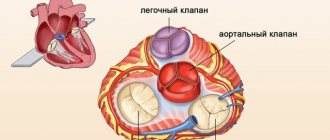
Mitral valve prolapse
Prolapse refers to an anatomical defect in the mitral heart valve. It is located between the left ventricle and the atrium. When the atrium contracts, it opens, which ensures the free flow of blood into the ventricle - and further, through the general blood flow. If a serious pathology of the mitral valve develops, its leaflets “bend”. In the worst case, the blood partially remains in the left atrium, which is fraught with unpleasant symptoms:
- heart pain;
- dizziness and shortness of breath;
- fainting;
- interruptions in heart function.
Typically, such symptoms are the exception to the rule, and minor mitral valve prolapse does not pose a threat to the life and health of the child. Severe prolapse is dangerous. It is manifested by severe deflection of the valves and more serious disturbances of cardiac activity (in the second and third degrees).
Heart aneurysm
A cardiac aneurysm is a thinned and protruding section of the heart wall that forms as a result of a myocardial infarction or other causes.
In most cases, cardiac aneurysm occurs in older people (especially men), but not always. In cardiology, a cardiac aneurysm is considered a very dangerous disease that can lead to immediate death.
Our heart is responsible for circulating blood in the body. Without it, the cells and tissues would not receive the necessary oxygen and essential nutrients, without which the cells would gradually begin to die.
Unfortunately, over time, disruptions in the functioning of the heart muscle begin to occur. Its walls, consisting of the endocardium, myocardium and epicardium, become thinner, therefore, the pressure on them increases.
As a result, where the walls are completely weakened, they begin to sag, that is, the outer side of the heart or the septum between its ventricles or atria protrudes, which leads to the formation of this disease.
The aneurysm is localized in those places where the maximum pressure is observed: in the wall of the left ventricle and in the area of the interventricular septum, which protrudes towards the right ventricle.
Its size varies from a few centimeters to large sizes (20 cm in diameter).
If an aneurysm ruptures, the consequences will be extremely sad.
However, in addition to its rupture, patients should be wary of heart failure, in which a large amount of circulating blood is lost due to the fact that it enters the cavity of the aneurysm and remains there, not taking part in the internal life of the body.
Also a very unpleasant complication can be called parietal blood clots, which form in the aortic cavity due to the fact that blood begins to stagnate there. Blood clots, leaking from the aorta, gradually clog the vessels, thereby causing the death of a section of an organ whose saturation was regulated by the clogged vessel.
Reasons for education
In 95% of cases, a cardiac aneurysm forms after a myocardial infarction. Almost all aneurysms are located in the area of the left ventricle.
Factors such as smoking, high blood pressure, tachycardia, constant heavy physical activity, heart failure, and recurrent heart attack can provoke an even greater increase in pressure on the heart walls.
Other factors influencing the formation of cardiac aneurysm include:
- Infectious diseases
They do not directly affect the formation of this disease, but can cause inflammation of the heart muscle, as a result of which many cells die and the heart walls become noticeably thinner.
Therefore, influenza, diphtheria, streptococcal and fungal infections, Coxsackie virus, etc. should be avoided.
- Congenital cardiac aneurysms
If the fetus develops any pathologies during the formation of the heart muscle, there is a high probability that at birth doctors will diagnose the baby with a heart aneurysm.
Perhaps such pathologies can be explained by the incompetent behavior of the mother during pregnancy: drinking alcoholic beverages, smoking, taking medications contraindicated for pregnant women, radiation exposure, occupational intoxication, etc.
- Aneurysms that occur after heart surgery
Sometimes cardiac aneurysms occur after heart surgery.
For example, during operations aimed at eliminating heart defects, sutures remain on the heart muscle. If these sutures do not heal well, or the patient experiences tachycardia or increased pressure in the ventricles, then most likely an aneurysm will soon form in the weakened areas.
- Injuries
Often this disease occurs after injuries that somehow affect the walls of the heart.
For example, with an open wound, a scar forms on the wall, at the site of which an aneurysm may occur.
Even with a closed injury, the appearance of a false cardiac aneurysm is quite likely. Typically, after such injuries, aneurysms occur almost immediately, so it is extremely important that specialists notice them and prevent them from rupturing.
- Toxic myocarditis
This is the name for the process of inflammation of the middle heart wall, which is caused by excessive consumption of alcoholic beverages, high concentrations of the thyroid hormone thyroxine in the body, intoxication with harmful chemical compounds, etc.
- Other reasons
During radiation treatment of organs in which malignant tumors have formed, gamma rays that are dangerous to health can reach the heart, which ultimately leads to an aneurysm. In addition, cardiac aneurysm is often observed in patients suffering from lupus erythematosus, dermatomyositis and idiopathic cardiosclerosis.
Varieties
Cardiac aneurysms are classified in different ways, allowing doctors to make diagnoses and begin treatment more quickly.
Depending on how long it takes for an aneurysm to occur, acute, subacute and chronic cardiac aneurysms are distinguished.
An acute aneurysm forms in the first two weeks after the patient has suffered a heart attack.
Subacute - after three to seven weeks, finally, chronic aneurysm occurs in the eighth week after the heart attack and grows very slowly.
A chronic aneurysm rarely ruptures, but blood clots often form in its cavity.
Cardiac aneurysms are small and wide ; they can be located in the anterior wall of the heart, at its apex, in the area of the intergastric septum, and extremely rarely in the posterior wall of the heart .
According to their shape , cardiac aneurysms are divided into:
- Diffuse aneurysms
They are distinguished by their small size and ability to grow. Blood clots rarely form in them, they almost never rupture, but such aneurysms provoke the development of arrhythmia in the patient.
- Fungiform aneurysms
The shape resembles a jug that has been turned over. They arise as a result of scars on the walls of the heart, often rupture, and blood clots can be seen in them.
- Saccular aneurysms
These aneurysms are large in size with a wide base, so blood flows into such aneurysms very well, as a result of which such aneurysms become very dangerous not only by ruptures, but also by the formation of blood clots.
- "Aneurysm within an aneurysm"
This is the name given to the most dangerous type of aneurysm, in which a new aneurysm forms in the walls of an existing saccular or diffuse aneurysm.
Finally, depending on the predominance of a particular tissue in the wall of the aneurysm , doctors talk about fibrous, muscular and fibromuscular aneurysm.
Fibrous aneurysm is considered the most unfavorable, because it is formed mainly from connective tissue after a myocardial infarction, as a result of which the heart walls very quickly stretch and become fragile.
A muscle aneurysm occurs due to congenital weakness of muscle fibers in some area of the middle wall of the heart, where the aneurysm forms.
A fibromuscular aneurysm contains both muscle and connective tissue and most often forms after a parietal infarction.
Symptoms of a cardiac aneurysm
Depending on where exactly the aneurysm is located, what its size and shape are, the symptoms of this disease can vary significantly. However, the leading signs of a cardiac aneurysm include:
- general weakness and apathy;
- frequent chest pain;
- tachycardia;
- shortness of breath, feeling of suffocation;
- increased sweating;
- a person can feel how hard his heart is beating;
- numbness of the limbs;
- pale skin;
- dry cough;
- dizziness, hallucinations.
Diagnosis and treatment
As you can see, there are no specific symptoms for a cardiac aneurysm, so it is quite difficult to identify.
Even for highly qualified cardiologists this is not an easy task.
After the initial examination, the patient is sent for electrocardiography, a general blood test, stress echocardiography and a special study - PET of the heart.
To understand how the valves function, echocardioscopy is performed.
For proposed surgical interventions, the doctor turns to cardiac scintigraphy, which allows one to obtain a good and clear image of the heart muscle.
Treatment of an aneurysm is carried out in two stages.
First, they turn to drug therapy, which allows you to maximally strengthen the heart walls and reduce the intensity of tissue and cell death. If the patient is old and cannot tolerate anesthesia, they are limited to only this stage.
The second stage involves surgery. It is necessary in the following cases:
- if the aneurysm grows rapidly;
- if drug treatment does not work;
- if there is progression of heart failure;
- if an aneurysm is diagnosed in a newborn baby;
- if the patient has suffered a second heart attack;
- if there is a high chance of aneurysm rupture or ventricular wall rupture.
The operation takes place under general anesthesia and lasts several hours. Unfortunately, even a perfectly performed operation does not exclude death in 5% of cases.
Prevention of cardiac aneurysm
Not every patient is able to undergo surgery. They need to know what to do to prevent the aneurysm from rupturing and causing a critical situation:
- you should stop smoking and drinking alcoholic beverages;
- you need to reduce physical activity: it is forbidden to run, walk quickly, or lift weights;
- it is important to control blood pressure;
- strictly monitor your weight and avoid extra pounds, as fat increases the load on the heart;
- lead a healthy lifestyle, eat right, walk in the fresh air, get enough sleep;
- avoid anxiety and nervous breakdowns;
- you need to give up coffee, spicy and sour foods, fried foods, fatty meats, etc.
Patent foramen ovale
This is the opening located between the two upper chambers of the heart (the left and right atria). It functions as a “door” during the development of the child in the womb. Through it, blood is discharged from the fetus from right to left. This is how blood flow exchanges between the small and large circles of blood circulation. An open foramen ovale in a child before birth plays a big role, “helping” the undeveloped lungs cope with blood flow. It is this that ensures the passage of blood to the left atrium and aorta.
When a child is born and his lungs, expanding, begin to work at full strength, the oval window “automatically” closes. A small depression remains in its place. However, there are cases when the oval window does not close after the baby is born. It is important to keep in mind that this anomaly does not belong to heart defects, and it can be detected by echocardiography. Usually, the bubble contrast technique is used for this, and if the baby’s oval window is not closed, air bubbles will move from right to left during examination.
Aneurysm of the interatrial septum
The wall between the left and right atrium can sometimes be curved. It protrudes to the side - to the right or to the left. Most often, the cause of its development in children is a hereditary factor. Intrauterine infections can also provoke the development of an aneurysm of the interatrial septum. At each age, different clinical manifestations of this anomaly can be distinguished:
- 1-3 years. Children lag behind in physical and mental development and often suffer from viral infections. During an objective examination, doctors often detect right ventricular hypertrophy with signs of pulmonary circulation overload;
- older age (12-15 years). Children and adolescents are stunted and do not tolerate physical activity well. Symptoms include: heart rhythm disturbances, weakness, pain, pale skin.
To avoid rupture of the aneurysm, it should be regularly monitored and modern diagnostic procedures performed.
False chords
In our heart there are so-called “true chords”, consisting of dense muscles. They are located in both ventricles of the heart and are attached by their endings to two valves - the triscupidal and mitral. Their function is to keep the valve leaflets in their natural position and prevent them from turning into the cavity of the heart when the ventricles contract.
The false chords of the left ventricle include anatomical formations that are not attached to the valve leaflets and are located in the cavity of the heart chamber. Pathological chords can be longitudinal and transverse, and when the heart contracts, they produce heart murmurs, sometimes reminiscent of the sound of a violin.
There are additional chords that do not cause serious pathological changes. False chords can be identified using echocardiography and ultrasound. Sometimes the child has no pronounced symptoms, except for the characteristic noise when listening to the heart, but if there are too many false chords and they are located transversely, this can lead to arrhythmias and serious circulatory disorders.
MARS are not classified as heart defects, but sometimes they can pose a real danger to the health and life of a child. That is why timely detection of such pathologies is very important for the work of a cardiologist, as well as any specialist involved in ultrasound and functional diagnostics.
Hello student
circulatory system of the heart
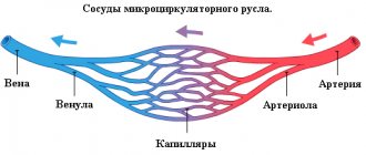
The activity of the heart, associated with large expenditures of energy, is ensured by a developed system of its own blood vessels, through which approximately 10% of all blood distilled in systole is pumped.
All superficial vessels are located subepicardially. Slightly convoluted vessels are surrounded by adipose tissue, the amount of which depends on the fatness of the animal. From all superficial vessels into the myocardium, branches extend at right angles, turning into a very dense capillary network. The capillaries of neighboring areas intersect with each other, but do not connect. Thus, the branches of the cardiac arteries are the terminal arteries responsible for the blood supply to a certain area, which, if the corresponding artery is blocked, does not receive blood (=infarction). ARTERIES
The blood supply to the muscles of the heart is carried out through two large arteries that arise from the aorta directly above the semilunar valve. In accordance with the topography of the initial sections, they are called coronary arteries, aa. coronariae
Left coronary artery, a. coronaria sinistra, after reaching the coronal sulcus, divides into two main branches. Its interventricular paraconal branch, ramus interventricularis paraccmalis, runs in the groove of the same name to the apex of the heart, and the circumferential, or circumflex, branch, ramus circumflexus, is located in the left section of the coronary sulcus.
From the interventricular paraconal branch in cats and dogs, proximal and distal collateral branches, rami collaterales proximalis et distalis, depart to the auricular surface of the left ventricle; in the cat, an angular branch, ramus angularis, still extends in front of these branches. Proximal and distal collateral branches supply blood to the cushion papillary muscle. In the cat, the interventricular paraconal branch additionally gives off into the wall of the right ventricle a branch of the conus arteriosus, ramus coni arteriosi, as well as several small branches into the areas of the interventricular septum along the paraconal interventricular groove. The blood supply to most of the interventricular septum, atrioventricular node and atrioventricular bundle is carried out through a branch of the interventricular septum, ramus septi interventricularis, which also supplies the mastoid muscles of the right ventricle.
Rice. 11. Base of a dog's heart after removal of the atria. Large arteries and veins are depicted separately, small ones together (according to Liicke, 1955) a. b, with valva aortae: a valvula semilunaris sinistra, b valvula semilunaris dextra, with valvula semilunaris septalis; d, e, f valva trunci pulmonalis: d valvula semilunaris sinistra, e valvula semilunaris dextra, f valvula semilunaris intermedia; g, h valva atrioventricularis sinistra s. bicuspidalis: g cuspis septalis, h cuspis parietalis; i, k, I valva atrioventricularis dextra s. tricuspidalis: i cuspis septalib, k cuspis parietalis, l cuspis annularis; m septum interatrialc; n conus arteriosus; o margo ventricularis dexter; p margo ventricularis sinister; q sulcus interventricularis paraconalis; r sulcus interventricularis subsinuosus
1 a. coronaria sinistra; 2 ramus interventricularis paraconalis; 3 ramus col lateral Is proxi-malb, 4 ramus circumflexus; 5 ramus proximalis ventnculi sinistri; 6 ramus marginis ventriculi sinistri; 7 ramus et v. distalis ventnculi sinistri; 8 ramus interventricularis subsinuosus; 9 a. coronaria dextra, ramus circumflexus: 10 ramus et v. coni arteriosi; 11 ramus et v. proximalis ventriculi dextri; 12 ramus et v nuirginis ventriculi dextri; 13 ramus et v. distalis ventriculi dextri; 14 ramus attrii dextri; 15 ostium sinus coronarius; 16 sinus coronarius; 17 v. cordis media; 18 v. cordis magna; 19 v. interventricularis paraconalis; 20 v. obliqua atria sinistri
Rice. 12. Auricular surface of the dog’s heart. Large arteries and veins are indicated separately, small ones together (according to Liicke, 1955)
auricula cordis dextra; b auricula cordis sinistra; with atrium sinistrum; d margo ventricularis dexter; e margo ventricularis sinister; f ventriculus dexter; g ventriculus sinister; h sulcus interventricularis paraconalis; i conus arteriosus; k sulcus coronarius; I apex cordis; m incisura apicis cordis 1 arcus aortae; 2 a subclavia sinistra; 3 truncus brachio-cephalicus; 4 truncus pulmonalis: 5 a. pulmonalis sinistra; 6 vv pulmonales; 7 v. cava cranialis; 8 a. coronaria sinistra, 9 her ramus circumflexus, 10 her ramus interventricularis paraconalis, 11 her ramus collaterals proximalis, 12 her ramus collateralis distalis; 13 ramus proximalis ventriculi sinistri, 14 ramus marginis ventriculi sinistri; 15 a.m. coronaria dextra, ramus circumflexus; 16 a.m. et v. coni arteriosi; 17 ramus et v. proximalis ventriculi dextri; 18 v. cordis magna; 19 v. interventricularis paraconalis; 20 v. proximalis atrii sinistri; 21 v. intermedia atria sinistri
Rice. 13. Atrial surface of the dog’s heart. Large arteries and veins are indicated separately, small ones together (according to Lucke, 1955)
a atrium sinistrum; b atrium dextrum; with sinus venarum cavarum; d ventriculus sinister; e ventriculus dexter; f margo ventricularis sinister; g margo ventricularis dexter; h sulcus interventricularis subsinuosus; i, k sulcus coronarius; l apex cordis; m incisura apicis cordis 1 arcus aortae; 2 a. subclavia sinistra; 3 truncus brachio-cephalicus; 4 a. pulmonalis dextra, 5 a pulmonalis sinistra; 6 w pulmonales dextrae, 7 vv. pulmonales; 8 v cava caudalis; 9 v. cava cranialis; 10 v. azvgos dextra; 11 sinus coronarius et a. coronaria sinistra, ramus circumflexus; 12 ramus interventricularis subsinuosus and v. cordis media, 13 ramus et v. distalis ventriculi sinistri; 14 v. ventriculi dextri; 15 ramus distalis atrii sinistri; 16 a.m. coronaria dextra, ramus circumflexus; 17 ramus et v. marginis ventriculi dextri; 18 ramus distalis ventriculi dextri; 19. 20 rami distales atrii dextri; 21 ramus intermedius attrii dextri
Rice. 14. Base of the cat's heart after removal of the atria (approximately 1.5 times magnification) (after Habermehl, 1959)
a, b, c valva aortae: a valvula semilunaris sinistra, b valvula semilunaris dextra, c valvula semilunaris septalis; d, e, f valva trunci pulmonalis: d valvula semilunaris sinistra, e valvula semilunaris dextra, f valvula semilunaris intermedia; g, h valva atrioventricularis sinistra s. bicuspidalis: g cuspis septalis, h cuspis parietalis; i, k, I valva atrioventricularis dextra s. tricuspidalis: i cuspis septalis, k cuspis parietalis, I cuspis angularis; m septum interatriale; n conus arteriosus;
o margo ventricularis dexter; p margo ventricularis sinister; q sulcus interventricularis paraconalis; r sulcus interventricularis subsinuosus 1 a. coronaria sinistra, 2 a. coronaria sinistra, ramus interventricularis paraconalis, 3 a. coronaria sinistra, ramus circumflexus; 4 ramus et v. angularis; 5 ramus et v proximalis ventriculi sinistri; 6 ramus et v. marginis ventriculi sinistri; 7 ramus et v. distalis ventriculi sinistri; 8 ramus proximalis atrii sinistri; 9 a. coronaria dextra, 9′ her ramus circumflexus; 10 a.m. et v. coni arteriosi; 11 ramus et v. proximalis ventriculi dextri; 12 ramus et v. marginis ventriculi dextri; 13 ramus et v distalis ventriculi dextri; 14 ramus distalis atrii dextri, 15 ramus mtermedius atrii dextri; 16 ramus proximalis atrii dextri; 17 sinus coronarius; 18 v. cordis magna; 19 v. interventricularis paraconalis; 20 v. obliqua atria sinistri; 21 v. cordis media; 22 a.m. coronaria sinistra, ramus interventricularis subsinuosus; 23 ramus distalis atrii sinistri; 24 ramus intermedius attrii sinistri
Rice. 15. Arteries and veins of the auricular surface of the cat’s heart (approximately 1.5 times magnification) (after Habermehl, 1959)
A auricula cordis dextra; B auricula cordis sinistra, partially removed;
With margo ventricularis dexter; D margo ventricularis sinister; E ventriculus dexter; F ventriculus sinister; With sulcus interventricularis paraconalis; H conus arteriosus; J sulcus coronarius; To apex cordis; L incisura apicis cordis
a arcus aortae; b truncus brachiocephalicus; ca subclavia sinistra; d truncus pulmonalis; e vv. pulmonales sinis-trae; f v. cava cranialis; g v. azvgos dextra
1 a. coronaria sinistra, 2 its ramus interventricularis paraconalis, 3 its ramus circumflexus; 4 ramus angularis et v. angularis; 5 ramus et v. proximalis ventriculi sinistri; 6 ramus et v. marginis ventriculi sinistri; 7 ramus et v. collatcralis proximalis; 8 ramus et v. collateralis distalis; 9 a. coronaria dextra, ramus coni arteriosi et v. coni arteriosi; 10 ramus et v proximalis ventriculi dextri; 11 ramus et v. coni arteriosi; 12 v. cordis magna; 13 v. interventricularis paraconalis
Rice. 16. Arteries and veins of the atrial surface of the cat’s heart (approximately 1.5 times magnification) (after Habermehl, 1959)
A atrium dex trum; In atrium sinistrum; C marge) ventricularis dexter; D margo ventricularis sinister; E ventriculus dexter; F ventriculus sinister; With sulcus inter-ventriculans subsinuosus. H sinus venarum cavarum; J auricula cordis dextra; To apex cordis a arcus aortae; b truncus brachiocephalicus; with a. subclavia sinistra; d branches of truncus pulmonalis; e vv. pulmonales; f v. cava caudalis. gv cava cranialis; hv azygos dextra
1 a. coronaria sinistra, ramus circumflexus, 2 a. coronaria sinistra, ramus interventricularis subsinuosus; 3 ramus et v. distalis ventriculi sinistri; 4 ramus et v marginis ventriculi sinistri; 5 ra mus distalis atrii sinistri; 6 a. coronaria dextra. ramus circumflexus; 7 ramus et v. marginis ventriculi dextri; 8 ramus distalis ventriculi dextri; 9 ramus intermedius atrii dextri; 10 ramus distalis atrii dextri; 11 v. semicircumflexa dextra; 12 sinus coronarius, 13 v. cordis media; 14 v. cordis magna; 15 v. obliqua atrii sinistri or ramus intermedius atrii sinistri; 16 v. collateralis dextra proximalis
The circumferential branch, ramus circumflexus, runs in the coronary sulcus towards the subsinus interventricular sulcus. In the initial part it is covered with the left ear. In the dog, the circumferential branch passes into the subsinus interventricular branch, ramus interventricularis subsinuosus, which extends to the apex of the heart.
Along the course of the circumferential branch, several smaller branches are separated from it into adjacent areas of the interventricular septum of the left ventricular wall. Three main branches - the proximal, marginal and distal branches of the left ventricle, ramus proximalis, ramus marginalis et ramus distalis ventriculi sinistri - provide blood supply to the wall of the left ventricle. Other vessels arising from the circumferential branch of the left coronary artery supply blood to the wall of the left atrium and partially to the interatrial septum. These are the proximal, intermediate and distal branches of the left atrium, rami proximales, intermedii et distales atrii sinistri.
The subsinus interventricular branch, ramus interventricularis subsinuosus, in dogs, as a rule, also arises from the left coronary artery. Through small vessels extending to the sides, it supplies blood to the area along the subsinus interventricular groove, and the stripe on the wall of the right ventricle is wider than on the wall of the left ventricle.
In cats, this vessel only in about half of all examined cases originated from the left coronary artery and in approximately 30% from the right coronary artery. In the first case we are talking about the left coronary, in the second about the average, or uniform, type of blood supply to the heart. In addition to these types of blood supply, a cat also has a third, in which blood enters through two subsinus interventricular branches, rami interventriculares subsinuosi, extending from the right and left coronary arteries.
Right coronary artery, a. coronaria dextra, in the dog it has only a circumferential branch, ramus circumflexus, which runs in the right coronary groove under the right ear and ends on the atrial surface, not reaching the subsinus interventricular groove. In cats, the same picture is observed with the left coronal type of blood supply. With an average type of blood supply, the right coronary artery continues in the form of a subsinus interventricular branch, ramus interventricularis subsinuosus, to the apex of the heart.
The circumferential branch of the right coronary artery in carnivores, along with small branches, gives off four large vessels to the wall of the right ventricle. These are the branch of the conus arteriosus, ramus coni arteriosi, and the proximal, marginal and distal branches of the right ventricle, ramus proximalis, ramus marginalis et ramus distalis ventriculi dextri.
With an average type of blood supply in a cat, the subsinus interventricular branch vascularizes the walls of both ventricles with small branches, and the branch of the interventricular septum, ramus septi interventricularis, also vascularizes the interventricular septum.
The proximal, intermediate and distal branches of the right atrium, rami proximales, intermedii et distales atrii dextri, which supply blood to the interatrial septum, extend into the right atrium from the right coronary artery.
VIENNS
For didactic reasons, the description of the cardiac veins, venae cordis, is given in the direction opposite to the blood flow. From the coronary sinus, sinus coronarius, which is a short large vessel for collecting most of the venous blood, the middle cardiac vein, v., first departs. cordis media. It runs along the subsinus interventricular groove and, dividing into several branches, reaches the apex of the heart, where it anastomoses with the branches of the paraconal interventricular vein, v. interventricularis paraconalis. Due to the fact that the middle cardiac vein runs parallel to the subsinous interventricular branch of the left coronary artery, it is also called the subsinous interventricular vein, v. interventricularis subsinuosa. A branch extends from it into the wall of the right ventricle. In a cat, this is the powerful right proximal collateral vein, v. collateralis proximalis dextra, in a dog - a much smaller vein of the right ventricle, v. ventriculi dextri. Other vessels arising from the coronary sinus at approximately the same level are the oblique vein of the left atrium, v. obliqua atrii sinistri, and distal vein of the left ventricle, v. distalis ventriculi sinistri. The first is in carnivores a rudiment of the left azygos vein, v. azygos sinistra, and collects blood from the wall of the left atrium. The distal vein of the left ventricle is connected to the same area as the branch of the same name of the left coronary artery.
The continuation of the coronary sinus is the great cardiac vein v. cordis is mean. It passes along with the circumferential branch of the left coronary artery in the coronary groove, then passes through its terminal, often paired branch - the paraconal interventricular vein, v. interventricularis paraconalis - in the groove of the same name and in the region of the apex of the heart, anastomoses with the terminal branches of the middle cardiac vein, v. cordis media. The branches of the great cardiac vein are: marginal vein of the left ventricle, v. maryinis ventriculi sinistri, proximal vein of the left ventricle, v. proximalis ventriculi sinistri, and the cat also has the angular vein, v. angularis.
From the paraconal interventricular vein depart: vein of the conus arteriosus, v. coni arteriosi, distal collateral vein, v. collateralis distalis, and vein of the interventricular septum, v. septi interventricularis.
Small veins of the heart, vv. cordis parvae, collect blood from areas of the wall of the right ventricle adjacent to the coronary sulcus and carry it directly to the right atrium. According to their topography, clogged veins can be divided into the vein of the conus arteriosus, V. coni arteriosi, the proximal vein of the right ventricle, v. proximalis ventriculi dextri, marginal vein of the right ventricle, v. marginis ventriculi dextri, and distal vein of the right ventricle, v. distalis ventriculi dextri. In the cat, the last three vessels unite into a common trunk, which can be designated as the right semicircular vein, v. semkircumflexa dextra.
Part of the venous blood from the endocardial area flows directly into the heart cavity through the smallest cardiac veins (thebesian veins), vv. cordis minimae (Thebesii). Their number is greatest in the right atrium, less in the left atrium, even less in the right ventricle and least in the left ventricle. Thebesian veins are very narrow tubules several millimeters long, extending from the muscle capillaries and flowing into the cavities of the heart through the openings of the smallest veins, foramina venarum minimarum. The smallest veins do not have valves.
Rice. 17. Arteries and veins of the interventricular septum of the cat’s heart. The wall of the right ventricle is cut off (magnification 1.5 times) (according to Habermehl, 1959)
A atrium dextrum; In auriculus dexter; With mm. papillares parvi; D, D' septum interventriculare. E m. papillaris subarteriosus; Apex cordis
a arcus aortae; b v. cava caudalis; with v. cava cranialis; d truncus pulmonalis
1 ramus circumflexus, a. coronaria dextra; 2 ramus interventricularis subsinuosus, a. coronaria dextra, 3 ramus interventricularis paraconalis, a. coronaria sinistra; 4 ramus septi interventricularis; 5, 6, 7 ramus interventricularis subsinuosus or paraconalis
Literature used: Anatomy of a dog and a cat (Coll, authors) / Transl. with him. E. Boldyreva, I. Kravets. - M.: “AQUARIUM BUK”, 2003. 580 pp., ill. color on
Download abstract: You do not have access to download files from our server. HOW TO DOWNLOAD HERE